 |
 |
| Fig. 1. Forming fluidic & air-stable supported lipid bilayers on the FluidicArray surface | Fig. 2. Fluorescence recovery after photobleaching (FRAP) for an SLB fromed on the FluidArray surface after air-drying and rehydration |
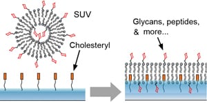
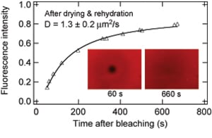
As drug delivery, therapy, and medical imaging are becoming increasingly cell-specific, there is a critical need for high fidelity and high-throughput screening methods for cell surface interactions. Membrane-mimicking surfaces, i.e., supported lipid bilayers (SLBs), formed from traditional methods do not possess sufficient robustness to meet this need. We have developed a groundbreaking technology to form fluidic and air-stable SLBs through tethered and dispersed cholesterol groups incorporated into the bottom leaflet (Fig. 1). The SLB remains fluidic after repeated exposure to air, drying, and rehydration (Fig. 2). Achieving air-stability allows one to easily fabricate SLB microarrays from direct robotic spotting of vesicle solutions. One can reconstitute peripheral as well as integral membrane components into the SLB to fabricate content microarrays in high throughput studies, e.g., the screening of drugs and nanomedicine targeting cell surface receptors. Read more in J. Am. Chem. Soc. 2008, 130, 6267; J. Am. Chem. Soc. 2009, 131, 13646; ACS Chem. Biol. 2014, 9, 1877.
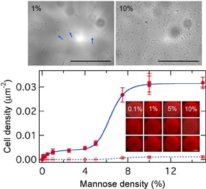
To demonstrate the application of the fluidic and air-stable SLB technology, we show in Fig. 3 the quantitative determination of multivalent binding curves for two E-coli strains on SLB microarrays with varying mannose densities. In this experiment, mannose-linked lipids are mixed into the lipid solution for robotic spotting on our surfaces to form fluidic mannose microarrays with precisely known surface mannose densities. The microarray is incubated with E-coli and the density of adhered E-coli cells on each spot is determined from microscopy. This experiment can yield a complete binding curve in 2-3 hours, as compared to days-weeks in traditional assays. Another application is the inhibition assay, as demontrated in Fig. 4 for the inhibition of E-coli adsorption by mannose-presenting nanoparticles.
 |
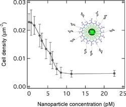 |
| Fig. 3. Multivalent binding curves of two E-coli strains on mannose density gradient microarrays (see inset). The upper shows two optical microscope images of E-coli cells adsorbed on SLB spots with 1% and 10% mannose, respectively | Fig. 4. Number density of adsorbed E-coli cells on the lipid membrane surface containing 7.5% mannose as a function of solution phase nanoparticle (inhibitor) concentration. |
Applications of the FluidArray® technology include a wide range of cell surface interactions, ranging from pathogen detection and characterization to the screening of nanomedicine. We supply FluidArray® series of coated surfaces (glass slides, coverslips, etc.) as well as associated reagents and protocols. We provide customer coating services for specific samples such as membrane based bio-sensors. We also provide high throughput screening (HTS) services for a wide range of cell surface interactions. Contact us with your R&D questions, problems, or ideas.
Copy Right © 2015 Athena Guo. All rights reserved.