The plasma membrane is a thin semi-permeable membrane that separates the interior of all cells from their extracellular environment. It is comprised of a phospholipid bilayer that is embedded throughout with proteins, such as ion channels, carrier proteins and receptor proteins, that are responsible for carrying out specific membrane. It provides structural support and protection for all cells, is responsible for maintaining ionic homeostasis, and is an integral component in cell adhesion, cell-cell communication, ion conductivity and cell signaling pathways.
Plasma Membrane

AAT Bioquest offers a wide range of plasma membrane labeling kits, fluorescent wheat germ agglutinin conjugates and lipophilic tracers – DiI, DiO, DiD and DiR tracers – for investigating lipid metabolism, imaging cell structures, and analyzing biophysical and cell signaling processes.
Live Cell Plasma Membrane Staining
AAT Bioquest Cell Navigator™ Plasma Membrane Staining kits are designed to rapidly and uniformly label the plasma membrane in living cells without the cell-type differences exhibited by lectins. These kits, which employ our novel Cellpaint™ plasma membrane stains, may be used as a segmentation tool for high-content screening (HCS) or to stain cellular plasma membranes for standard fluorescence microscopy. The fluorescence staining in cell membranes is well-preserved after fixation with formaldehyde, enabling multiplexing with other fluorescent conjugates or proteins.
Key Features of Cell Navigator™ Plasma Membrane Staining Kits
- Bright fluorescence with high signal-to-noise ratios
- High photostability for stable signal generation
- Suitable for staining plasma membranes in suspended or attached live cells
- Uniform staining of plasma membrane across a broad range of mammalian cell types
- Staining well-retained in plasma membrane after fixation
- Available in green, orange and red fluorescence to facilitate multicolor staining
| Cellpaint™ Orange | Cellpaint™ Green | Cellpaint™ Red |
 |
 |
 |
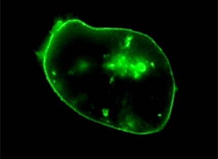
| Live HeLa cells were stained with Cellpaint™ Orange, then fixed in 4% formaldehyde and costained with Nuclear Green™ DCS1. | Live HL-60 cells were stained with Cellpaint™ Green. | Live HeLa cells were stained with Cellpaint™ Red. |
Table 1. Cell Navigator™ Plasma Membrane Staining kits for live cells.
| Product Name | Probe | Ex/Em (nm) | Filter Set | Unit Size | Cat No. |
| Cell Navigator™ Cell Plasma Membrane Staining Kit *Green Fluorescence* | Cellpaint™ Green | 497/505 | FITC | 500 tests | 22682 |
| Cell Navigator™ Cell Plasma Membrane Staining Kit *Orange Fluorescence* | Cellpaint™ Orange | 555/573 | TRITC | 500 tests | 22680 |
| Cell Navigator™ Cell Plasma Membrane Staining Kit *Red Fluorescence* | Cellpaint™ Red | 648/671 | Cy5 | 500 tests | 22681 |
Fluorescent WGA for Live and Fixed Cells
Wheat germ agglutinin (WGA) is a ∼36 kDa lectin used extensivly in cell biology. Its high affinity for N-acetyl-D-glucosamine and sialic acid residues on glycoconjugates is routinely exploited for labeling the cell membranes of mammalian cells, gram-positive bacteria and yeast bud scars, as well as, skeletal and cardiac sacrolemma and fibrotic scar tissue. When conjugated to AAT Bioquest iFluor™ dyes, fluorescent WGA conjugates exhibit superior brightness, photostability and water-solubility, outperforming Alexa Fluor® WGA conjugates and other spectrally similar conjugates. iFluor™ dye-labeled WGA are suitable for immunoassays, IHC, ELISA and Western Blot.
Key Features of iFluor™ WGA Conjugates
- Bright fluorescence with high signal-to-noise ratios and resistance to photobleaching and pH change
- Conjugates useful in live and fixed cells that have not been permeabilized
- Withstands fixations and permeabilization
- Can serve as a anterograde or retrograde neuronal tracer for neuronal mapping studies
- Available in a range of colors to support multiplex and colocalization studies with other fluorescent conjugates or proteins
| iFluor™ 488 WGA Conjugate | iFluor™ 647 WGA Conjugate | |
 |
 |
|
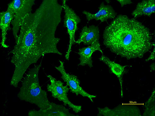
| Live HeLa cells were stained with iFluor™ 488 WGA conjugate (green) and Hoechst 33342 (blue). | Live HeLa cells were stained with iFluor™ 647 WGA conjugate (red) and Hoechst 33342 (blue). |
Table 2. iFluor™ wheat germ agglutinin conjugates for live and fixed cells.
| Product name | Permeability | Ex/Em (nm) | Filter Set | Unit Size | Cat No. |
| iFluor™ 488-Wheat Germ Agglutinin Conjugate | Membrane-impermeant | 491/516 | FITC | 1 mg | 25530 |
| iFluor™ 555-Wheat Germ Agglutinin Conjugate | Membrane-impermeant | 557/570 | Cy3/TRITC | 1 mg | 25539 |
| iFluor™ 594-Wheat Germ Agglutinin Conjugate | Membrane-impermeant | 588/604 | Texas Red | 1 mg | 25550 |
| iFluor™ 647-Wheat Germ Agglutinin Conjugate | Membrane-impermeant | 656/670 | Cy5 | 1 mg | 25559 |
Alexa Fluor® wheat germ agglutinin conjugates for live and fixed cells.
| Product name | Cell Permeability | Ex/Em (nm) | Filter Set | Unit Size | Cat No. |
| Wheat Germ Agglutinin, AF488 Labeled | Membrane-impermeant | 499/520 | FITC | 1 mg | 25500 |
| Wheat Germ Agglutinin, AF594 Labeled | Membrane-impermeant | 590/618 | Texas Red | 1 mg | 25509 |
Lipophilic Tracers – DiO, DiI, DiA, DiD, DiR and DiS
DiA, DiI, DiO, DiD and DiR are a family of dialkylcarbocyanines dyes used extensivly for labeling hydrophobic structures such as cell membranes, and as an anterograde or retrograde neuronal tracer in living and fixed cells and tissues. Once applied to cells, these dyes diffuse rapidly and laterally within the plasma membranes resulting in detailed uniform staining of the entire cell surface. Staining is specific as these dyes do not transfer via gap junctions or to unlabeled cells, unless the membrane of the labeled cell is disturbed. In addition to membrane staining and neuronal tracing, lipophilic carbocyanines have been successfully used in cytotoxicity assays, labeling lipoproteins, cell adhesion studies, tracing cell migration and fluorescence recovery after photobleaching (FRAP) assays.
Spectroscopic properties of dialkylcarbocyanines:
- Extremely high extinction coefficients
- Polarity-dependent fluorescence
- Short excited-state lifetimes
- DiO, DiI, DiD and DiR exhibit distinct green, orange, red and infrared fluorescence, respectively, to support multicolor imaging and flow cytometric analysis of live cells

Figure 1. Emission spectra of DiO, DiI, DiA, DiD and DiR.
Table 3. Lipophilic tracers for labeling cell membranes in live and fixed cells and tissues.
| Product Name | Ex/Em (nm) | Filter Set | Unit Size | Cat No. |
| DiOC2(3) iodide [3,3-Diethyloxacarbocyanine iodide] | 482/500 | FITC | 25 mg | 22038 |
| DiOC3(3) iodide [3,3-Dipropyloxacarbocyanine iodide] | 482/500 | FITC | 25 mg | 22039 |
| DiOC7(3) iodide [3,3-Diheptyloxacarbocyanine iodide] | 482/500 | FITC | 25 mg | 22040 |
| DiOC16(3) perchlorate [3,3-Dihexadecyloxacarbocyanine perchlorate] | 482/500 | FITC | 25 mg | 22042 |
| DiOC5(3) iodide [3,3-Dipentyloxacarbocyanine iodide] | 482/500 | FITC | 25 mg | 22045 |
| DiOC6(3) iodide [3,3-Dihexyloxacarbocyanine iodide] | 482/500 | FITC | 25 mg | 22046 |
| DiO perchlorate [3,3-Dioctadecyloxacarbocyanine perchlorate] | 482/500 | FITC | 25 mg | 22066 |
| DiIC12(3) perchlorate [1,1-Didodecyl-3,3,3,3-tetramethylindocarbocyanine perchlorate] | 549/563 | TRITC | 25 mg | 22035 |
| DiIC16(3) perchlorate [1,1-Dihexadecyl-3,3,3,3-tetramethylindocarbocyanine perchlorate] | 549/563 | TRITC | 25 mg | 22044 |
| DiIC12(3)-DS [1,1-Diododecyl-3,3,3,3-tetramethylindocarbocyanine-5,5-disulfonic acid] | 549/563 | TRITC | 5 mg | 22050 |
| DiIC18(3)-DS [1,1-Dioctadecyl-3,3,3,3-tetramethylindocarbocyanine-5,5-disulfonic acid] | 549/563 | TRITC | 5 mg | 22052 |
| DiSC2(3) [3,3-Diethylthiacarbocyanine iodide] | 560/571 | TRITC | 25 mg | 22073 |
| DiI iodide [1,1-Dioctadecyl-3,3,3,3- tetramethylindocarbocyanine iodide] | 549/563 | TRITC | 100 mg | 22101 |
| DiI perchlorate [1,1-Dioctadecyl-3,3,3,3-tetramethylindocarbocyanine perchlorate] | 549/563 | TRITC | 100 mg | 22102 |
| DiI triflate [1,1-Dioctadecyl-3,3,3,3-tetramethylindocarbocyanine triflate] | 549/563 | TRITC | 100 mg | 22103 |
| DiA [4-(4-(Dihexadecylamino)styryl)-N-methylpyridinium iodide] | 491/611 | Texas Red | 25 mg | 22030 |
| DiD labeling solution [1,1-Dioctadecyl-3,3,3,3-tetramethylindodicarbocyanine] | 645/663 | Cy5 | 10 mL | 22033 |
| DiIC12(5)-DS [1,1-Diododecyl-3,3,3,3-tetramethylindodicarbocyanine-5,5-disulfonic acid] | 650/670 | Cy5 | 5 mg | 22051 |
| DiIC18(5)-DS [1,1-Dioctadecyl-3,3,3,3-tetramethylindodicarbocyanine-5,5-disulfonic acid] | 652/668 | Cy5 | 5 mg | 22054 |
| DiIC1(5) iodide [1,1,3,3,3,3-Hexamethylindodicarbocyanine iodide] | 640/657 | Cy5 | 25 mg | 22056 |
| DiSC3(5) [3,3-Dipropylthiadicarbocyanine iodide] | 660/675 | Cy5 | 25 mg | 22076 |
| DiR iodide [1,1-dioctadecyl-3,3,3,3-tetramethylindotricarbocyanine iodide] | 754/778 | Cy7 | 25 mg | 22070 |
Product Ordering Information
Table 4. Ordering Info for Cell Nav. Cell Plamsa Membrane Products
| Cat# | Product Name | Unit Size |
| 22680 | Cell Navigator™ Cell Plasma Membrane Staining Kit *Orange Fluorescence* | 500 Tests |
| 22681 | Cell Navigator™ Cell Plasma Membrane Staining Kit *Red Fluorescence* | 500 Tests |
| 22682 | Cell Navigator™ Cell Plasma Membrane Staining Kit *Green Fluorescence* | 500 Tests |
Your first choice for scientific solutions
Find out moreSUPPORT
outstanding technical support
PRODUCT
we offer a full product guarantee
DELIVERY
we offer free delivery to UK universities and non profit organisations