Lysosomes are the “waste-disposal” system of the cell digesting unwanted materials and cellular debris in the cytoplasm. They are membrane-enclosed organelles that have an acidic interior (pH ∼4.8) and can vary in size from 0.1 to 1.2 µm. Lysosomes house various hydrolytic enzymes responsible for digesting biopolymers such as proteins, peptides, nucleic acids, carbohydrates and lipids. The synthesis of lysosomal hydrolases are controlled by nuclear genes, and mutations in these genes can cause an array of inherited metabolic disorders due to defects in lysosomal enzyme functionality. These metabolic disorders are collectively known as lysosomal storage diseases. Defects to lysosomal hydrolytic enzymes result in the accumulation of macromolecules or monomeric compounds contributing to abnormal signaling pathways which ultimately lead to pathogenic disorders. In addition to breaking down polymers, lysosomes can fuse with other organelles to allow lysosomal hydrolytic enzymes to digest the organelle’s contents and to clear out damaged structures, a mechanism known as autophagy.
Lysosomes
Lysosomal Labeling Dyes and Kits
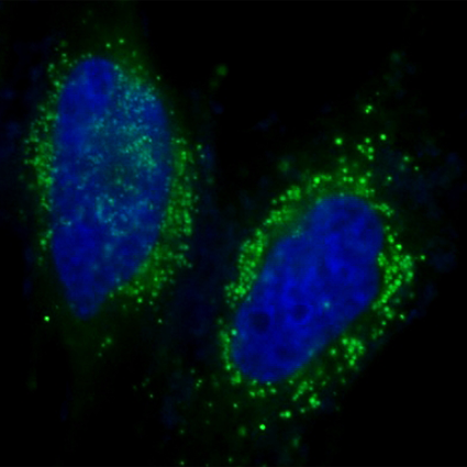
LysoBrite™ dyes are a series of cell-permeable fluorecent dyes for imaging lysosomal morphology and localization, and to investigate the biosynthesis and pathogenesis of lysosomes in live cells. At nanomolar concentrations, LysoBrite™ dyes selectively target and accumulate within lysosomes and other acidic organelles via the lysosomal pH gradient. Upon entering lysosomes, the pH-sensitive fluorescence of LysoBrite™ dyes significantly enhances. The resulting signal can be measured using either fluorescence imaging, high-content imaging, microplate fluorometry, or flow cytometry. LysoBrite™ Orange, LysoBrite™ Red, LysoBrite™ Deep Red, and LysoBrite™ NIR dyes have extremely high photostability as well as excellent cellular retention make them useful for a variety of studies, including cell adhesion, chemotaxis, multidrug resistance, cell viability, apoptosis and cytotoxicity.
LysoBrite™ Dyes for Live Cell Lysosomal Imaging
LysoBrite™ dyes are available in spectral characteristics ranging from blue to near-infrared to accommodate multiplexing with other fluorescent probes or after fixation for co-localization studies if using fixable dye LysoBrite™ Red DND-99 (Cat No. 22647). LysoBrite™ dyes are key components of Cell Navigator™ Lysosome Staining Kits made available separately here. Each kit provides sufficient materials for 500 tests.
Features of LysoBrite™ dyes:
- Selectively accumulates inside lysosomes and other acidic organelles via the lysosomal pH gradient
- pH-sensitive fluoresence for a more specific lysosomal staining
- Photostable signal generation for extended imaging windows
- Minimal cytotoxicity
- Can be imaged without washing
- Multiple wavelength options for multiplex analysis with other fluorescent probes
- Suitable for both proliferating and non-proliferating cells, as well as, cell in suspension and adherent cells

Table 1. Available LysoBrite™ dyes for labeling lysosomes in live cells.
| Product name | Sample Type | Fixable | Ex (nm) | Em (nm) | Filter Set | Unit Size | Cat No. | Assay Kit No. |
| LysoBrite™ Blue | Live Cells | No | 434 | 480 | DAPI | 500 Tests | 22642 | 22655 |
| LysoBrite™ VLG26 | Live Cells | No | 405 | 480 | Violet | 500 Tests | 22651 | |
| LysoBrite™ Green | Live Cells | No | 501 | 510 | FITC | 500 Tests | 22643 | 22656 |
| LysoBrite™ Orange | Live Cells | No | 543 | 565 | TRITC | 500 Tests | 22644 | 22657 |
| LysoBrite™ Red DND-99 | Live Cells | Yes | 573 | 592 | TRITC | 500 Tests | 22647 | |
| LysoBrite™ Red | Live Cells | No | 576 | 596 | TRITC | 500 Tests | 22645 | 22658 |
| LysoBrite™ Deep Red | Live Cells | No | 597 | 619 | Texas Red | 500 Tests | 22646 | 22659 |
| LysoBrite™ NIR | Live Cells | No | 636 | 651 | Cy5 | 500 Tests | 22641 | 22652 |
CytoFix™ Red Fixable Lysosomal Stain
The enhanced cellular retention of CytoFix™ Red lysosomal stain preserves its lysosomal localized fluorescence even after the pH gradient is lost during fixation. The fluorescence signal generated by this dye is well-retained in the lyososme for at least 1 week, making it well-suited for long-term lysosomal tracking. CytoFix™ Red can be used with GFP expressed cells, other fluorescent conjugates or other organelle stains for multicolor analysis. It can be used for both suspension and adherent cells and readily adapted for a wide variety of fluorescence platforms.
Features of CytoFix™ Red:
- High lysosomal staining efficiency
- Long retention after fixation (1 week or longer)
- Labeling protocol with minimal hands on time

Table 2. CytoFix™ Red fixable lysosomal stain
| Product name | Sample Type | Fixable | Ex (nm) | Em (nm) | Filter Set | Unit Size | Cat No. |
| CytoFix™ Red Lysosomal Stain | Live Cells | Yes | 540 | 590 | Cy3/TRITC | 500 Tests | 23210 |
Autophagy – Lysosomal Degradation Pathway
Autophagy is a natural and regulated lysosomal degradation pathway essential for cell viability, development and homeostasis. It is the destructive mechanism of cells that degrades and recycles unnecessary or dysfunctional cellular components. It achieves this through the sequestration of targeted cytoplasmic materials into double-membrane bound organelles known as autophagosomes. These autophagosomes later fuse with lysosomal organelles to have their contents digested and recycled by lysosomal enzymes. Autophagy serves as an adaptive mechanism to protect organisms against various pathologies. To investigate the role autophagy plays in cell homeostasis, imaging tools and assays have been developed to monitor autophagy functionality in response to cellular stress, microbial infection and disease. Cell Meter™ Autophagy Fluorescence Imaging Kits utilizes our proprietary Autophagy Super Blue™ probe, to selectively target and analyze autophagy activity.
 |
 |
| Control (DMEM Medium) |
Autophagy Treatment (1X HBSS buffer with 5% serum) |
Table 3. Cell Meter™ assay kits for measuring autophagy activity.
| Product name | Sample Type | Ex (nm) | Em (nm) | Filter Set | Unit Size | Cat No. |
| Cell Meter™ Autophagy Fluorescence Imaging Kit | Live Cells | 360 | 445 | DAPI | 200 Tests | 23001 |
| Cell Meter™ Autophagy Assay Kit *Blue Fluorescence* | Live Cells | 360 | 445 | DAPI | 200 Tests | 23000 |
| Cell Meter™ Autophagy Assay Kit *Green Fluorescence* | Live Cells | 485 | 525 | FITC | 200 Tests | 23002 |
| Cell Meter™ Mitochondrial Autophagy Imaging Kit *Red Fluorescence* | Live Cells | 540 | 590 | Cy3/TRITC | 100 Tests | 22998 |
Product Ordering Information
Table 4. Ordering Info for Lysosome Products
| Cat# | Product Name | Unit Size |
| 22641 | LysoBrite™ NIR | 500 Tests |
| 22642 | LysoBrite™ Blue | 500 Tests |
| 22643 | LysoBrite™ Green | 500 Tests |
| 22644 | LysoBrite™ Orange | 500 Tests |
| 22645 | LysoBrite™ Red | 500 Tests |
| 22646 | LysoBrite™ Deep Red | 500 Tests |
| 22647 | LysoBrite™ Red DND-99 | 500 Tests |
| 22651 | Cell Navigator™ Lysosome Staining Kit *Green Fluorescence with 405 nm Excitation* | 500 Tests |
| 22652 | Cell Navigator™ Lysosome Staining Kit *NIR Fluorescence* | 500 Tests |
| 22655 | Cell Navigator™ Lysosome Staining Kit *Blue Fluorescence* | 500 Tests |
| 22656 | Cell Navigator™ Lysosome Staining Kit *Green Fluorescence* | 500 Tests |
| 22657 | Cell Navigator™ Lysosome Staining Kit *Orange Fluorescence* | 500 Tests |
| 22658 | Cell Navigator™ Lysosome Staining Kit *Red Fluorescence* | 500 Tests |
| 22659 | Cell Navigator™ Lysosome Staining Kit *Deep Red Fluorescence* | 500 Tests |
| 23000 | Cell Meter™ Autophagy Assay Kit *Blue Fluorescence* | 200 Tests |
| 23001 | Cell Meter™ Autophagy Fluorescence Imaging Kit | 200 Tests |
| 23002 | Cell Meter™ Autophagy Assay Kit *Green Fluorescence* | 200 Tests |
Your first choice for scientific solutions
Find out moreSUPPORT
outstanding technical support
PRODUCT
we offer a full product guarantee
DELIVERY
we offer free delivery to UK universities and non profit organisations