ATS is known as the “saporin company” and initially focused on providing research tools for the Neuroscience research community.
Over the years these tools have been used in all areas of scientific research and drug development.
The Company’s current product line includes targeted toxins, antibodies and custom services designed to assist scientists in the study of system functions, cell functionality, diseases and disorders.

Advanced Targeting Systems pioneered the use of targeted conjugates for use in a technique called “Molecular Surgery.”
These products include specific lesioning agents for cholinergic basal forebrain neurons, noradrenergic and adrenergic neurons, macrophages and microglia and a pan-neuronal agent that strikes an epitope expressed on all neurons.
In addition, ATS makes “secondary conjugates” which allow users to convert their own targeting agents into specific cytotoxic tools and to screen antibodies for internalisation.


Product Categories

Products

Saporin belongs to the well-characterized family of ribosome-inactivating proteins and serves as a control

Targeted SAP conjugates are powerful and specific lesioning agents used in the technique known as Molecular Surgery.

The ribosome-inactivating protein, saporin (from the seeds of the plant, Saponaria officinalis) is bound to a targeting agent (anything that is recognized on the cell surface and internalized).

The targeted conjugate is administered to cells (in vitro or in vivo). The targeting agent seeks out and binds to its target on the cell surface.

The conjugate is internalized, saporin breaks away from the targeting agent, and inactivates the ribosomes which causes protein inhibition and, ultimately, cell death.

Cells that do not have the cell surface marker are not affected.

Products

Description Individual Kit w/controls
192-IgG-SAP IT-01 KIT-01
Anti-DBH-SAP IT-03 KIT-03
CTB-SAP IT-14 KIT-14
mu p75-SAP IT-16 KIT-16
CCK-SAP IT-31 KIT-31
Anti-ChAT-SAP IT-42 KIT-42
Anti-CD103-SAP IT-50 KIT-50
Bombesin-SAP IT-40 KIT-40
Bombesin-SAP IT-84 KIT-84
Anti-CD105-SAP IT-80 KIT-80
IB4-SAP IT-10 KIT-10
Chlorotoxin-SAP IT-86 KIT-86
NPY-SAP IT-28 KIT-28
Dermorphin-SAP / MOR-SAP IT-12 KIT-12
SSP-SAP IT-11 KIT-11
Dyno-SAP (Dynorphin-SAP) IT-68 KIT-68
NKB-SAP (NK3-SAP) IT-63 KIT-63
Melanopsin-SAP IT-44 KIT-44
Mac-1-SAP mouse/human IT-06 KIT-06
Orexin-B-SAP IT-20 KIT-20
Leptin-SAP IT-47 KIT-47
Anti-SERT-SAP IT-23 KIT-23
Anti-6 His-SAP IT-52 KIT-52
Ex4-SAP (GLP-1-SAP) IT-90 KIT-90
Nppb-SAP IT-69 KIT-69
FGF-SAP IT-38 KIT-38
Anti-DAT-SAP IT-25 KIT-25
Anti-YFP-SAP IT-66 KIT-66
NMB-SAP IT-70 KIT-70
GAT1-SAP IT-32 KIT-32
Anti-CD25-SAP human IT-24 KIT-24
Anti-vGAT-SAP IT-71 KIT-71
Anti-CD44-SAP IT-72 KIT-72
Anti-FLAG (M5)-SAP IT-59 KIT-59
Anti-V5-SAP IT-58 KIT-58
Neurotensin-SAP IT-56 KIT-56
Anti-GFP-SAP IT-53 IT-53
Anti-CD22-SAP human IT-37 KIT-37
ME20.4-SAP IT-15 KIT-15
Acetylated LDL-SAP IT-08 KIT-08
Anti-PD-L1-SAP IT-45 KIT-45
Anti-CD45.2-SAP IT-91 KIT-91
Hemokinin 1-SAP IT-76 KIT-76
Anti-CD117-SAP IT-83 KIT-83
Anti-CD133-SAP IT-82 KIT-82
MOA-SAP IT-92 KIT-92
Oxytocin-SAP IT-46 KIT-46
Galanin-SAP IT-34 KIT-34
Anti-CD25-SAP mouse IT-29 KIT-29
CRF-SAP IT-13 KIT-13

Products

Description Individual Kit
Streptavidin-ZAP IT-27 n/a
Fab-ZAP human IT-51 KIT-51
Fab-ZAP mouse IT-48 KIT-48
FabFc-ZAP human IT-65 KIT-65
Hum-ZAP IT-22 KIT-22
Fab-ZAP rat IT-55 KIT-55
MonoBiotin-ZAP BT-ZAP n/a
Mab-ZAP IT-04 KIT-04
Streptavidin-ZAP Kit n/a KIT-27-S
Fab-ZAP rabbit IT-57 KIT-57
FabFc-ZAP Hug-M IT-78 KIT-78
Rab-ZAP IT-05 KIT-05
Alpaca-ZAP IT-87 KIT-87
Goat-ZAP IT-36 KIT-36
Streptavidin-ZAP Antibody Kit n/a KIT-27-A
Biotin-Z Peptide Internalization Kit n/a KIT-27-ZB
FITC-Streptavidin-ZAP IT-85 n/a
Hug-M-ZAP IT-43 KIT-43
Anti-M-ZAP IT-30 KIT-30
Rat-ZAP IT-26 KIT-26
Biotin-Z Antibody Internalization Kit n/a KIT-27-Z
Streptavidin-ZAP Peptide Kit n/a KIT-27-B
FabFc-ZAP mouse IT-89 KIT-89
FabFc-ZAP rat IT-88 KIT-88
gPIG-ZAP IT-64 KIT-64
Chick-ZAP IT-62 KIT-62

ZAP conjugates are non-targeted saporin conjugates that “piggyback” on to your primary targeting agent (biotinylated material or antibody).

Biotin-Z Internalization Kit (for in vitro or in vivo use)

These kits convert biotinylated materials into targeted toxins and will allow you to evaluate the ability of your reagent to internalize upon binding to its receptor.


Target Recommended Product in vivo Recommended Product in vitro
biotinylated peptide Streptavidin-ZAP Peptide Kit (KIT-27-B) Biotin-Z Peptide Internalization Kit (KIT-27-ZB)
biotinylated antibody *choose host species of your material Streptavidin-ZAP Antibody Kit (KIT-27-A) goat, human, mouse, rat, rabbit, sheep Biotin-Z Antibody Internalization Kit (KIT-27-Z) goat, human, mouse, rat, rabbit, sheep
other biotinylated material Streptavidin-ZAP Kit (KIT-27-S) Streptavidin-ZAP Kit (KIT-27-S)

Streptavidin-ZAP (Cat. #IT-27) converts biotinylated materials into targeted toxins.

FITC-Streptavidin-ZAP (Cat. #IT-85) eliminate cells recognized by YOUR biotinylated targeting agent, FITC-labeled

MonoBiotin-ZAP (Cat. #BT-ZAP) eliminate cells recognized by YOUR streptavidinylated targeting agent

ZAP Antibody Internalization Kit (for in vitro use)

The Antibody Z-kit contains a Secondary Antibody conjugated to Saporin (ZAP), Control Conjugate, plus all the components needed to screen your primary antibody for internalization.


Target Recommended Products
alpaca/llama IgG (VHH domain) alpaca/llama IgG (IT-87, KIT-87)
alpaca/llama IgG (VHH domain) Chick-ZAP (IT-62, KIT-62)
goat IgG Goat-ZAP (IT-36, KIT-36)
guinea pig IgG gPIG-ZAP (IT-64, KIT-64)
human IgG Hum-ZAP (IT-22, KIT-22) Fab-ZAP human (IT-51, KIT-51) FabFc-ZAP human (IT-65, KIT-65)
human IgM Hug-M-ZAP (IT-43, KIT-43) FabFc-ZAP Hug-M (IT-78, KIT-78)
mouse IgG Mab-ZAP (IT-04, KIT-04) Fab-ZAP mouse (IT-48, KIT-48) FabFc-ZAP mouse (IT-89, KIT-89)
mouse IgM Anti-M-ZAP (IT-30, KIT-30)
rabbit IgG Rab-ZAP (IT-05, KIT-05) Fab-ZAP rabbit (IT-57, KIT-57)
rat IgG Rat-ZAP (IT-26, KIT-26) Fab-ZAP rat (IT-55, KIT-55) Fab-ZAP rat (IT-88, KIT-88)

made with bivalent antibodies that recognize the whole IgG

made with monovalent (Fab) antibodies that recognize the whole IgG, without bivalent capping

made with monovalent (Fab) antibodies that recognize Fc region only

pHast Conjugates are one of our pHastest tools for quantitative testing of your primary antibody’s specificity, binding, and internalization, providing results in 1 day.

Fluorescence intensity increases over time, indicating internalization of the pHast conjugate. Data generated using an IncuCyte SX5.

How It Works

The pHast conjugate binds to your primary antibody via a secondary antibody cross-linked to a pH-dependent fluorescent reporter. This fluorescent reporter will increase intensity as the pH of its surroundings becomes more acidic, as evident when exposed to the environment inside a cell. pHast conjugates are fluorescent plate reader compatible and can be used to illuminate your lead antibody candidates in a 96-well plate format.

Also Available:

Streptavidin-pHast (PH-03)
Streptavidin-pHast binds to your biotinylated protein and contains a pH-dependent fluorescent reporter (pHast).


Target Recommended Products made with whole IgG made with monovalent (Fab) made with monovalent Fc region (FabFc)
alpaca/llama IgG (VHH domain) Alpaca-pHast (PH-05)
chicken IgY Chick-pHast (PH-06)
goat IgG Goat-pHast (PH-07)
guinea pig IgG gPIG-pHast (PH-08)
human IgG Hum-pHast (PH-09) Fab-pHast human (PH-01) FabFc-pHast human (PH-10)
human IgM Hug-M-pHast (PH-11) FabFc-pHast Hug-M (PH-04)
mouse IgG Mab-pHast (PH-12) Fab-pHast mouse (PH-02) FabFc-pHast mouse (PH-13)
mouse IgM Anti-M-pHast (PH-14)
rabbit IgG Rab-pHast (PH-15) Fab-pHast rabbit (PH-16)
rat IgG Rat-pHast (PH-17) Fab-pHast rat (PH-18) FabFc-pHast rat (PH-19)

made with bivalent
antibodies that
recognize the whole IgG

made with monovalent
(Fab) antibodies that
recognize the whole IgG,
without bivalent capping

made with monovalent
(Fab) antibodies that
recognize Fc region only

Products

Description Catalog No.
Fab-pHast mouse PH-02
Fab-pHast human PH-01
FabFc-pHast human PH-10
FabFc-pHast HugM PH-04
FabFc-pHast rat PH-19
Fab-pHast rat PH-18
Rat-pHast PH-17
Fab-pHast rabbit PH-16
Rab-pHast PH-15
Anti-M-pHast PH-14
FabFc-pHast mouse PH-13
Mab-pHast PH-12
Hug-M-pHast PH-11
Hum-pHast PH-09
gPIG-pHast PH-08
Goat-pHast PH-07
Chick-pHast PH-06
Alpaca-pHast PH-05
Streptavidin-pHast PH-03

Products

Description Catalog No.
Blank-SAP IT-21
Rabbit IgG-SAP IT-35
BIgG-SAP Mouse IT-74
Goat IgG-SAP IT-19
Mouse IgG-SAP IT-18
Rat IgG-SAP IT-17
Human IgG-SAP IT-49
Blank-Streptavidin-SAP IT-27B
BIgG-SAP Human IT-77
Fab IgG-SAP IT-67
BIgG-SAP Sheep IT-93
BIgG-SAP Goat IT-81
BIgG-SAP Rabbit IT-75
BIgG-SAP Rat IT-73
Blank-CTA IT-61
Mouse IgM-SAP IT-41

Controls are a vital part of the scientific procedure; without them it is difficult to isolate the specific effects from the non-specific or artifactual. These control conjugates are the same molecular weight, consist of similar, comparable materials and are synthesized with the same protocols as the targeted conjugates. The difference is the cell-specific targeting agents are replaced with “blanks,” antibodies or peptides that have no specificity, and no ability to target cells.

Control for Targeted Toxins Control for Targeted Toxins using biotin-streptavidin technology
Goat Goat IgG-SAP (IT-19): preimmune goat IgG and saporin Fab IgG-SAP (IT-67): preimmune goat Fab IgG and saporin BIgG-SAP Goat (IT-81): biotinylated goat IgG and streptavidin-ZAP
Human Human IgG-SAP (IT-49): preimmune human IgG and saporin BIgG-SAP Human (IT-77): biotinylated human IgG and streptavidin-ZAP
Mouse Mouse IgG-SAP (IT-18): preimmune mouse IgG and saporin Mouse IgM-SAP (IT-41): preimmune mouse IgM and saporin BIgG-SAP Mouse (IT-74): biotinylated mouse IgG and streptavidin-ZAP
Rabbit Rabbit IgG-SAP (IT-35): preimmune rabbit IgG and saporin BIgG-SAP Rabbit (IT-75): biotinylated rabbit IgG and streptavidin-ZAP
Rat Rat IgG-SAP (IT-17): preimmune rat IgG and saporin BIgG-SAP Rat (IT-73): biotinylated rat IgG and streptavidin-ZAP
Sheep BIgG-SAP Sheep (IT-93): biotinylated sheep IgG and streptavidin-ZAP
Peptide Blank-SAP (IT-21): non-targeted peptide and saporin Blank-Streptavidin-SAP (IT-27B): non-targeted peptide and streptavidin-ZAP

ATS is pleased to announce Beta-release of a wide array of targeted toxins for use in eliminating specific cell types. This Beta-Testing Program will make new conjugates available to our customers sooner.

Description Catalog No.
CGRP-SAP BETA-011
Anti-OX1r-SAP BETA-007
Anti-CB1-SAP BETA-005
GnRH-SAP BETA-028
Vasopressin-SAP BETA-023
Anti-Human CXCR4-SAP BETA-018
Anti-CD38-SAP BETA-016
Anti-NET-SAP BETA-035
TIP39-SA BETA-030
Anti-Smoothened (SMO)-SAP BETA-033
ACTH-SA BETA-021
Deltorphin-SAP BETA-006
Azido-ZAP BETA-003
mAb35-SAP BETA-034
Amyloid Beta 42-SAP BETA-032
VIP-SAP BETA-027
Anti-CD24-SAP BETA-025
Anti-CD15-SAP BETA-024
αMSH-SAP BETA-022
Anti-MC4R-SA BETA-020
Anti-Cripto-SAP BETA-017
Methotrexate-SAP BETA-012
Nociceptin-SAP BETA-001

Each of the Beta products has had:

  1. Saporin activity confirmed,
  2. Peptide sequences published/confirmed, and/or
  3. Antibody binding specificity published/confirmed.

Beta Products now available as catalog items:

Anti-CD105-SAP (Cat. #IT-80) previously BETA-015

Anti-CD44-SAP (Cat. #IT-72) previously BETA-004

Anti-PD-L1-SAP (Cat. #IT-45) previously BETA-014

Chlorotoxin-SAP (Cat. #IT-86) previously BETA-010

Ex4-SAP (GLP-1-SAP) (Cat. #IT-90) previously BETA-009

MOA-SAP (Cat. #IT-92) previously BETA-013

Orexin-B-SAP (Cat. #IT-20) previously BETA-031

PACAP-SAP (Cat. #IT-84) previously BETA-029

Specific Product Protocols

AB-N34 Antibody to Choline Acetyltransferase (ChAT) Rabbit Polyclonal Immunostaining
AB-N39 Antibody to Melanopsin, affinity-purified Immunostaining
AB-T01 Anti-Conjugated Glutathione Rabbit Polyclonal ELISA
AB-T03 Anti-Conjugated 5-Hydroxytryptamine (Serotonin) Rabbit Polyclonal ELISA, Immunohistochemistry
AB-T06 Anti-Conjugated Noradrenaline Rabbit Polyclonal ELISA, Immunohistochemistry
AB-T07 Anti-Conjugated Dopamine Rabbit Polyclonal ELISA, Immunohistochemistry
AB-T08 Anti-Conjugated L-Glutamate Rabbit Polyclonal ELISA, Immunohistochemistry
AB-T10 Anti-Conjugated GABA (Gamma-Aminobutyric acid) Rabbit Polyclonal ELISA, Immunohistochemistry
AB-T11 Anti-Conjugated Dopamine Mouse Monoclonal ELISA, Immunohistochemistry
AB-T12 Anti-Conjugated L-Glutamate Mouse Monoclonal ELISA, Immunohistochemistry
AB-T16 Anti-Conjugated Histamine Mouse Monoclonal ELISA
AB-T023 Anti-Conjugated Glycine Rabbit Polyclonal ELISA, Immunohistochemistry
AB-T029 Anti-Conjugated Acetylcholine Mouse Monoclonal ELISA, Immunohistochemistry
AB-T030 Anti-Conjugated Tyrosine Rabbit Polyclonal ELISA
AB-T044 Anti-Conjugated Trans-Hydroxyproline Rabbit Polyclonal Immunohistochemistry
AB-T048 Anti-Conjugated D-Serine Rabbit Polyclonal ELISA, Immunohistochemistry
AB-T067 Anti-Conjugated L-Dihydroxyphenylalanine (L-DOPA) Rabbit Polyclonal ELISA, Immunohistochemistry
AB-T070 Anti-Conjugated Octopamine Rabbit Polyclonal ELISA, Immunohistochemistry
AB-T071 Anti-Conjugated Tyramine Rabbit Polyclonal ELISA, Immunohistochemistry
AB-T079 Anti-Melatonin Rabbit Polyclonal ELISA
AB-T080 Anti-Conjugated 5-Methoxytryptophan Rabbit Polyclonal ELISA, Immunohistochemistry
AB-T086 Anti-Conjugated Myristic Acid Rat Polyclonal ELISA
AB-T125 Anti-Conjugated NO-L-Cysteine Mouse Monoclonal ELISA, Immunohistochemistry, Western Blot
AB-T129 Anti-Conjugated Indole 3 Acetic Acid Rabbit Polyclonal ELISA
AB-T147 Anti-Conjugated Folic acid Rat Polyclonal ELISA, Immunohistochemistry
AB-T168 Anti-Conjugated Uric Acid Rabbit Polyclonal ELISA, Western blot
AB-T171 Anti-Conjugated L.Kynurenine Antibody ELISA, immunocytochemistry, immunohistochemistry, immunoblotting

Antibody Protocols

1. Coat maxisorp well plates (Nunc) with a solution of antigen (the dilutions for antigen range from  4-50 µg/ml, please see data sheet) in sodium carbonate buffer 0.05 M (pH 9.6) containing sodium metabisulfite (SMB) (Acros) 0.001 M, for sixteen hours at 4°C.

2. Saturate well plates with a solution of PBS (pH 7.3) containing 2.5 g/l of BSA (Acros), 0.05% Tween 20 (Acros) and SMB 0.001 M for one hour at 37°C.

3. Wash with PBS/0.5% Tween three times.

4. Dilute primary antibody as per antibody data sheet in PBS containing 2.5 g/l BSA, 10% of glycerol and SMB 0.001 M, 200 µl by well plate (incubating for 2 hours at 37°C).

5 . Wash with PBS/0.5% Tween three times.

6. 200 µl of peroxidase-labeled goat anti- “primary antibody host species” IgG (Sigma) diluted (1/10,000) in a solution of PBS containing 2.5 g/l BSA, 10% of glycerol, 0.5% Tween and SMB 0.001 M, will be applied by well plate (for one hour at 37°C).

7. Rinse well plates with PBS/0.5% Tween three times.

8. And finally, develop the peroxidase by incubating 200 µl by well plate of a citrate 0.1 M/phosphate, 0.2 M (pH 5) solution containing 0.4% of OPD (Sigma) and 0.03% of hydrogen peroxide (Acros) for ten minutes in the dark. After that, stop the reaction by the addition of 50 µl of 2 M HCl.

9. The optical density will be measured at 492 nm, to obtain the different values (IC50).

Perfusion protocol for Adult male Sprague Dawley (weight around 0.5 kg) :

1. The animals can be deeply anaesthetized, for example with urethane (0.5-1.5g/kg, intraperitoneal).

2. Heparinize and perfuse via the ascending aorta with 100 ml of cold physiologic saline (0.9% NaCl) and with the following fixative solution:

a) 300 ml of cold 4% paraformaldehyde and 2% glutaraldehyde in 0.1 M phosphate-buffer (PB), pH 7.2 (two minutes).
b) 600 ml of cold 4% paraformaldehyde and 2% glutaraldehyde in 0.1 M PB, pH 7.2 (ten minutes).
c) Dissect out the brains and place in a solution of 4% paraformaldehyde in 0.1 M PB, pH 7.2, at 4°C for twelve to sixteen hours.
d) Before the brains are cut on a freezing microtome, infuse the brain in increasing concentrations of sucrose (a first bath of 5% of sucrose in PBS until the brains sink), after that repeat the same process in a solution with a higher level of sucrose (10%, 20%, 25% and finally 30%).

3. Cut serial sections around 50 µm-thick. Keep at 4°C in PBS (0.1 M, pH 7.2) and process for
immunostaining.

Immunostaining protocol:

1. In order to avoid possible interference with endogenous peroxidase, treat free-floating sections with distilled water containing NH3 (20%), H2O2 (30%) and NaOH (1%) for 20 minutes (other method is using a solution with 33% of H2O2 and 66% of methanol).

2. Wash the sections for 20 minutes in 0.15 M phosphate-buffered saline (PBS) (pH 7.2).

3. Pre-incubate for 30 minutes in PBS containing 10% of normal horse serum and 0.3% of Triton X-100 (mixed solution).

4. Incubate at room temperature (1 hour 30 minutes) and overnight at 4°C mixing with a solution containing primary antibodies.

5. Wash for 30 minutes with PBS.

6. Incubate for 60 minutes at room temperature with biotinylated anti-rabbit IgG (Vector) diluted 1/200 in PBS.

7. Wash for 30 minutes with PBS.

8. Incubate sections for 1 hour with a 1/100 diluted avidin-biotin-peroxidase complex (Vectastain).

9. Wash for 30 minutes with PBS.

10. Wash with Tris-HCl buffer (pH 7.6)(10 minutes).

11. Develop the tissue-bound peroxidase with H2O2 using 3, 3′ diaminobenzidine as chromogen.

12. Rinse the sections with PBS and coverslip with PBS/Glycerol (1/1).

Custom Services

ATS has manufactured custom conjugates for over 20 years; providing consistent, reliable research tools for scientists all over the world. Check out these scientific publications utilizing custom conjugates.

ATS offers a variety of custom services including the following. Contact us to discuss your project.

Custom Conjugates are powerful tools that specifically destroy targeted cell types. This technique has important research applications in determining the function or contribution of the targeted cells in a biological system or for the removal of interfering cell types.

Custom Conjugates are powerful tools that specifically destroy targeted cell types. This technique has important research applications in determining the function or contribution of the targeted cells in a biological system or for the removal of interfering cell types. Through the use of custom conjugates, you can eliminate any cell type.

Requirements for Successful Targeting

  • A cell surface marker.
  • A molecule, such as an antibody or a ligand, that binds to that marker.

Your targeting agent is conjugated to Saporin, a highly active and potent ribosome-inactivating protein.

Getting Started

  • Select your targeting agent.
    As an example, for the specific and complete removal of mature T lymphocytes, use your own anti-transferrin receptor antibody; or obtain one from the ATCC.
  • Use the appropriate Secondary Conjugate to screen your targeting agent for specificity and internalization.
  • Contact us for a quote.
  • Prepare for stunning, powerful research results!

Examples of Uses

Examine the role of targeted neurons in behavior, in gene expression and to determine the effect of these neurons on other neurons in the brain.

Selective removal of subtypes of the hematopoietic system, which has well-characterized cell surface markers.

Research Applications

Custom conjugates are valuable tools with the potential for applications in many areas.

In vitro, custom conjugates can be used to selectively eliminate cells expressing a specific receptor/antigen. This could be very useful in various cloning and gene trapping strategies where the goal is to select for cells based on expression (or suppression of expression) of a particular gene.

In vivo, custom conjugates can be used to specifically eliminate selected types of neurons for studies of the function of those neurons and responsiveness of animals to a variety of manipulations, including:

  • Behavioral studies
  • Drug treatments
  • Physiological studies

Screening Targeting Agents

Use a Secondary Conjugate to test your antibody as a targeting reagent.

Proteins come in all shapes and sizes and don’t always contain a ready-to-conjugate binding site. This and other variables can cause you to look toward one-step kits, often leaving you with un-purified and un-verified conjugate.

Let the experts in the field help provide you with the most effective conjugate while retaining the functionality of each of the components.

Contact us now to get started discussing the strategy that best meets your needs.

Why biotinylate through Advanced Targeting Systems?

What you get with a “One-Step-Kit”

  • Un-purified conjugate
  • Un-verified conjugate
  • Estimated concentration
  • Reacts only with amine groups

VS

ATS Custom Biotinylation

  • Discuss options available for your protein
  • Removal of unbound biotin & protein
  • Calculation of ratio of moles biotin/moles protein
  • Concentration verified by BCA*
  • Zero hands-on time for you

*Dependent upon peptide, but typically available for most proteins such as antibodies.

Leonardo Ancheta,
Vice President and Director of Custom Conjugations at ATS, talks about important things to watch for to avoid compromising your next experiment.

Companion Product

Biotin-Z Internalization Kit

The streptavidin-biotin interaction has found extensive use as a research tool. The bond created between streptavidin and biotin is rapid and essentially non-reversible, unaffected by most extremes of pH, organic solvents, and denaturing reagents. A variety of molecules including lectins, proteins, and antibodies, can be biotinylated and reacted with streptavidin-labeled probes or other detection reagents for use in biological assays.

It’s easy to test your biotinylated targeting agent using the Biotin-Z Internalization Kit. Each kit contains all the components needed to allow your targeting agent to be screened quickly and economically to provide specificity, functional binding, internalization, and EC50 determination.

The key component of the Biotin-Z Internalization Kit is Streptavidin-ZAP; Streptavidin attached to Saporin, the most potent (and safest for use in the laboratory) of the plant ribosome-inactivating proteins. Streptavidin-ZAP “piggybacks” onto YOUR biotinylated material in order to evaluate the ability of the reagent to internalize upon binding to its receptor. Using Streptavidin-ZAP and biotinylated targeting agents, specific cytotoxins can be created JUST BY MIXING!

You can also test your biotinylated targeting agent in vivo. Streptavidin-ZAP can be mixed with your biotinylated targeting agent and delivered specifically to eliminate cells that recognize your targeting agent.

Your scientific applications demand a purified and verified conjugation.
The experts at ATS can make your next conjugation a solid success with “zero” hands-on time for you.


Data sheets accompany all biotinylations and include:

  • Protein Concentration Verified by Bicinchoninic Acid Assay (BCA assay)
  • Average chemical ratio of moles of biotin to moles of protein
  • Average effective molecular weight

pHast Protocols

100 mcg, 250 mcg, 1000 mcg

a tool to test antibody specificity, binding, and internalization with results in one (1) day

Fab-pHast in conjunction with your antibody can be analyzed via  fluorescent microscope, fluorescent plate reader, or flow cytometry. In order to  establish the EC50 of your antibody a fluorescent plate reader is recommended. A sample protocol for determining internalization via Fluorescent Plate reader

pHast Ab Internalization Assay

Parental HEK-293 cells, and HEK-293 cells transfected with the p75 receptor, were plated in a  96-well plate overnight. Titrated 192-IgG antibody (Cat. #AB-N43) was incubated at RT with 50 nM of Fab-pHast Mouse (Cat. #PH-02) for 20 minutes prior to addition to cells. Plates were  incubated overnight to allow maximum internalization, but a few hours is sufficient for detection.

Plates were read on a Spectra Max Gemini EM (Ex: 532nm/Em: 560nm). Data analysis was done by PRISM (GraphPad, San Diego).

Fab-pHast Internalization Protocol

1. Determine the number of cells needed for the planned number of plates.  Cells are plated in the center 60 wells in 90 μl of media per well.

2. Plate cells in a 96-well black, clear bottom plate or all black plate. The clear  bottom plate allows visualization of antibody internalization using a microscope. Cells are usually plated at 20,000 cells per well.

3. Transfer 100 μl of media into the wells around the edge of a 96-well plate.  These wells simply offer some protection from evaporation for the experimental wells.

4. Incubate plates for 20-24 hours before treatment.

5. It’s recommended to use a constant concentration of Fab-pHast at 50 nM and titrate your antibody. A good starting concentration of antibody is 10 nM. Optimization of concentration and dilutions will need to be established. High concentrations of unconjugated antibody may act as an inhibiter of fluorescent activity.

6. Make a stock solution of 500 nM of Fab-pHast, 10X the concentration that will be used in each well.

7. Add your desired concentrations of antibody to the Fab-pHast, 10X the desired concentration, and incubate at room temperature for 20 minutes.

8. The first and last experimental columns (2 and 11 in most plates) of cells are controls, only medium or Fab-pHast alone is added to these wells.

9. Add 10 μl of your 10X concentrations of Fab-pHast with titrated antibody to each experimental well.

10. Mix the plate gently on a plate mixer for 1-2 min, then incubate overnight to allow internalization. (Internalization can start to be detected in 1 hour, but maximal fluorescence occurs typically 19 hours after addition of the Fab-pHast conjugated antibodies).

11. To achieve a higher sensitivity, replace media with PBS before reading the plate.

12. Read the plate in a fluorescent plate reader at Ex: 532 nm/Em: 560 nm.

13. The absorbance of the sample wells is compared to the control wells to establish a curve.

14. The EC50 of your antibody can be derived from the curve, using PRISM software (GraphPad, San Diego).

Streptavidin-pHast [PH-03]
100 mcg, 250 mcg, 1000 mcg

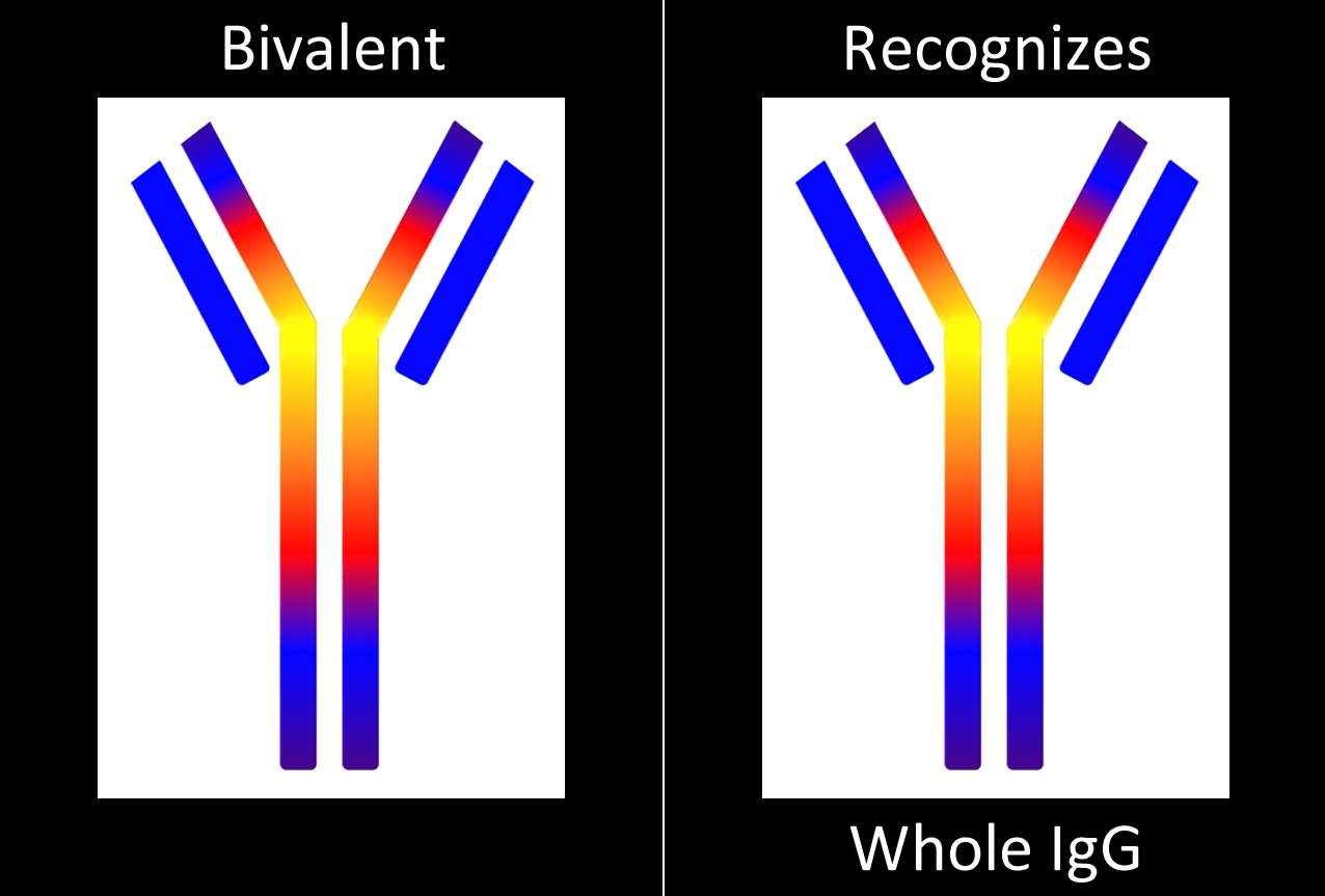
a tool to test biotinylated protein specificity, binding, and internalization with results in one (1) day

Streptavidin-pHast in conjunction with your biotinylated protein can be analyzed via fluorescent microscope, fluorescent plate reader, or flow cytometry. In order to establish the EC50 of your antibody a fluorescent plate reader is recommended.

A sample protocol for determining internalization via Fluorescent Plate reader

Streptavidin-pHast Internalization Protocol

1. Determine the number of cells needed for the planned number of plates. Cells are plated in the center 60 wells in 90 μl of media per well.

2. Plate cells in a 96-well black, clear bottom plate or all black plate. The clear bottom plate allows visualization of antibody internalization using a microscope. Cells are usually plated at 20,000 cells per well.

3. Transfer 100 μl of media into the wells around the edge of a 96-well plate. These wells simply offer some protection from evaporation for the experimental wells.

4. Incubate plates for 20-24 hours before treatment.

5. Mix an equimolar amount of Streptavidin-pHast to your biotinylated protein. Incubate at room temperature for 20 minutes. Optimization of concentration and dilutions will need to be established. High concentrations of unconjugated antibody may act as an inhibiter of fluorescent activity.

6. Serial dilute your desired concentrations of Streptavidin-pHast-Biotinylated-Protein in microcentrifuge tubes, 10X the desired concentration planned for the plate.

7. The first and last experimental columns (2 and 11 in most plates) of cells are controls, only medium or Streptavidin-pHast alone is added to these wells.

8. Add 10 μl of your 10X concentrations of Streptavidin-pHast-Biotinylated-Protein to each of your plates.

9. Mix the plate gently on a plate mixer for 1-2 min, then incubate overnight to allow internalization. (Internalization can start to be detected in 1 hour, but maximal fluorescence occurs typically 19 hours after addition of the Streptavidin-pHast conjugated proteins).

10. To achieve a higher sensitivity, replace media with PBS before reading the plate.

11. Read the plate in a fluorescent plate reader at Ex: 532 nm/Em: 560 nm.

12. The absorbance of the sample wells is compared to the control wells to establish a curve.

13. The EC50 of your antibody can be derived from the curve, using PRISM software
(GraphPad, San Diego).

Conjugate Protocols

CONCENTRATION CALCULATIONS
Convert molarity to mg/ml and mg/ml to molarity

Normally, our concentrations are given in units of milligrams per milliliter (mg/ml), but often data sheet quality assurance data are expressed in molarity (moles per liter, M) or fractions of that (e.g., micromolar (µM), 10-6 M, or nanomolar (nM), 10-9 M). Here is how to convert between these units.

FROM (mg/ml) TO molarity (M):

Divide the concentration (mg/ml) by the molecular weight. We will use the example of a typical immunotoxin that has a molecular weight of 210,000 grams per mole (or mg/mmole or kDa) (the molecular weight is usually found on the data sheet) and a common concentration is 1.0 mg/ml.

1.0 mg/ml


2.1 x 10⁵ mg/mmole

 = 0.48 x 10⁻⁵ mmole/ml
= 4.8 x 10⁻⁶ mmole/ml

In the first line, the mg units cancel each other, leaving units of mmole/ml that is equal to moles/liter or molar (M). Therefore, 0.48 x 10⁻⁵ mmole/ml = 0.48 x 10⁻⁵ M or 4.8 x 10⁻⁶ M. This, of course, can be expressed as 4.8 µM, or 4.8 micromolar.

In summary: concentration (grams per liter) ÷ molecular weight (grams per mole) = moles per liter.

FROM molarity (M) TO (mg/ml):

Multiply the molar concentration (M, or moles per liter) by the molecular weight. In an example of an immunotoxin at 1.0 nM concentration (1.0 nmoles per liter or 1.0 x 10⁻⁹ M or 1.0 x 10⁻⁹ moles per liter) again using as an example, a targeted toxin of molecular weight 210,000 grams per mole (or mg/mmole or kDa):

1.0 x 10⁻⁹ moles per liter x 2.1 x 10⁵ grams per mole  = 2.1 x 10⁻⁴ grams per liter
= 2.1 x 10⁻¹ µg/ml
= 0.21 µg/ml

In summary: molar concentration (moles per liter) x molecular weight (grams per mole) = grams per liter.

Product Literature

Technology Tutorial

How Molecular Surgery Works

Advanced Targeting Systems’ technology – Molecular Surgery – is a modification of one of the most widely used techniques in the neurosciences: lesioning of a region by surgical means and observation of the effect.

Saporin has no way of entering a cell on its own, except by bulk-phase endocytosis. To be effective, Saporin is bound to a Targeting Agent. A Targeting Agent binds to the cell surface and is internalized. Examples include: antibody, ligand, cytokine, growth factor, aptamer, etc. When the conjugate is internalized it escapes the endosomal compartment and inactivates the ribosomes. This shuts down protein synthesis and results in cell death.

Videos


ATS FAQ’s

Contact UsView All products Equimolar mixing of Streptavidin-ZAP and Biotinylated Molecule I was wondering if you could elaborate on why Read More...

Screening targeting agents and their cell surface biomarkers for high specificity and rapid internalization via cell death and fluorescence.

Contact Us View All Products Abstract Some of the most recent successes in the treatment of cancers or research into Read More...

Validated Antibodies targeting NGFR and Melanopsin

Contact Us View All Products NGFR Antibodies NGFR (mu p75) Rabbit Polyclonal [AB-N01] AB-N01 also known as mu p75, and Read More...

A Brief History of Saporin and its Contributions to Neuroscience

Contact UsView All Products ABSTRACT When investigating the origins of targeted toxins (a drug, therapy, or scientific tool directed to Read More...

First for Antibodies

Stratech Scientific have been successful in a recent antibodies tender from the Southern Universities Purchasing Consortium. In the award criterion Read More…