DYED POLYSTYRENE
Our non-functionalized microspheres are suitable for coating with antibody or other large proteins via adsorption. See TechNote 204, Adsorption to Microspheres, for a general adsorption protocol. Unless noted otherwise, these visibly dyed microspheres are supplied as a 5% solids suspension (w/v) and are available in these standard amounts: 0.5g, 1.0g, 1.5g, and 5.0g.
*Please note: If you are NOT ordering via website, please specify a lot number if multiple lots are available.
| Catalog Number |
Dye |
Nominal Diameter |
Specification Range |
Product Data Sheet |
| DSCR001 |
Crimson Red |
0.050µm |
0.040 – 0.060µm |
PDS 717.pdf |
| DSCB002 |
Cabo Blue |
0.200µm |
0.190 – 0.210µm |
PDS 717.pdf |
| DSCR002 |
Crimson Red |
0.200µm |
0.190 – 0.210µm |
PDS 717.pdf |
| DSSG002 |
Shamrock Green |
0.200µm |
0.190 – 0.210µm |
PDS 717.pdf |
| DSBK002 |
Basic Black |
0.200µm |
0.190 – 0.210µm |
PDS 717.pdf |
| DSCR003 |
Crimson Red |
0.300µm |
0.270 – 0.330µm |
PDS 717.pdf |
| DSCR004 |
Crimson Red |
0.400µm |
0.370 – 0.430µm |
PDS 717.pdf |
| DSCB005 |
Cabo Blue |
0.80µm |
0.770 – 0.830µm |
PDS 717.pdf |
| DSCR005 |
Crimson Red |
0.80µm |
0.770 – 0.830µm |
PDS 717.pdf |
| DSSG005 |
Shamrock Green |
0.80µm |
0.770 – 0.830µm |
PDS 717.pdf |
| DSBK005 |
Basic Black |
0.80µm |
0.770 – 0.830µm |
PDS 717.pdf |
| DSCR006 |
Crimson Red |
5.0µm |
4.80 – 5.20µm |
PDS 717.pdf |
References
Henderson, K., & Stewart, J. (2002). Factors influencing the measurement of oestrone sulphate by dipstick particle capture immunoassay. Journal of immunological methods, 270(1), 77-84.(0.3, 0.5, and 0.8µm dyed blue PS)
Henderson, K., & Stewart, J. (2000). A dipstick immunoassay to rapidly measure serum oestrone sulfate concentrations in horses. Reproduction, Fertility and Development, 12(4), 183-189.(0.31µm dyed blue PS)
Campbell, K., Fodey, T., Flint, J., Danks, C., Danaher, M., O’Keeffe, M., … & Elliott, C. (2007). Development and validation of a lateral flow device for the detection of nicarbazin contamination in poultry feeds. Journal of agricultural and food chemistry, 55(6), 2497-2503.(0.43µm dyed blue PS)
DYED CARBOXYL POLYSTYRENE
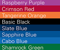
Biomolecules may be covalently immobilized to carboxyl-functionalized microspheres. A general coupling protocol is provided within TechNote 205, Covalent Coupling. Our dyed carboxyl microspheres are impregnated with vibrant dyes for optimal visualization. See our color palette for a representation of our standard colors. Unless noted otherwise, they are supplied as a 5% solids suspension (w/v) and are available in four standard volumes: 0.5g, 1.0g, 1.5g, and 5.0g.
*Please note: If you are NOT ordering via website, please specify a lot number if multiple lots are available.
| Catalog Number |
Dye |
Nominal Diameter |
Specification Range |
Product Data Sheet |
| DCCB001 |
Cabo Blue |
0.20µm |
0.190 – 0.210µm |
PDS 717.pdf |
| DCCR001 |
Crimson Red |
0.20µm |
0.190 – 0.210µm |
PDS 717.pdf |
| DCSG001 |
Shamrock Green |
0.20µm |
0.190 – 0.210µm |
PDS 717.pdf |
| DCBK001 |
Basic Black |
0.20µm |
0.190 – 0.210µm |
PDS 717.pdf |
| DCCB002 |
Cabo Blue |
0.50µm |
0.470 – 0.530µm |
PDS 717.pdf |
| DCCR002 |
Crimson Red |
0.50µm |
0.470 – 0.530µm |
PDS 717.pdf |
| DCCB004 |
Cabo Blue |
1.00µm |
0.95 – 1.05µm |
PDS 717.pdf |
| DCCR004 |
Crimson Red |
1.00µm |
0.95 – 1.05µm |
PDS 717.pdf |
| DCTA004 |
Tangerine Orange |
1.00µm |
0.95 – 1.05µm |
PDS 717.pdf |
| DCSG004 |
Shamrock Green |
1.00µm |
0.95 – 1.05µm |
PDS 717.pdf |
| DCCB005 |
Cabo Blue |
5.00µm |
4.80 – 5.20µm |
PDS 717.pdf |
| DCCR005 |
Crimson Red |
5.00µm |
4.80 – 5.20µm |
PDS 717.pdf |
REFERENCES
Terao, Y, Takeshita, K, Nishiyama, Y, Morishita, N, Matsumoto, T, & Morimatsu, F. (2015) Promising Nucleic Acid Lateral Flow Assay Plus PCR for Shiga Toxin–Producing Escherichia coli. Journ of food protection, 78(8), 1560-1568.
Sheng, W, Li, S, Liu, Y, Wang, J, Zhang, Y, & Wang, S. (2017). Visual and rapid lateral flow immunochromatographic assay for enrofloxacin using dyed polymer microspheres and quantum dots. Microchimica Acta, 184(11), 4313-4321.(Dyed Black carboxyl microspheres)
DYED PROTEIN-COATED POLYSTYRENE
At times, our inventory includes streptavidin-coated dyed microspheres supplied as ~1% solids (w/v) aqueous suspensions.
| Catalog Number |
Coating / Dye |
Nominal Diameter |
Specification Range |
| CDCR001 |
SA / Crimson Red |
0.20µm |
0.190 – 0.210µm |
| CDCB001 |
SA / Cabo Blue |
0.20µm |
0.190 – 0.210µm |