Visibly dyed microspheres are frequently used in lateral flow tests and “latex” agglutination tests. Our polystyrene microspheres are impregnated with vibrant dyes for optimal visualization. The colorpalette for a representation of our standard colors. Non-functionalized and functionalized versions are available to support adsorption and covalent binding strategies.
Dyed Polystyrene
Our non-functionalized microspheres are suitable for coating with antibody or other large proteins via adsorption. See TechNote 204, Adsorption to Microspheres, for a general adsorption protocol. Unless noted otherwise, these visibly dyed microspheres are supplied as a 5% solids suspension (w/v) and are available in these standard amounts: 0.5g, 1.0g, 1.5g, and 5.0g.
*Please note: If you are NOT ordering via website, please specify a lot number if multiple lots are available.
| Catalog Number | Dye | Nominal Diameter | Specification Range |
| DSCR001 | Crimson Red | 0.050µm | 0.040 – 0.060µm |
| DSCB002 | Cabo Blue | 0.200µm | 0.190 – 0.210µm |
| DSCR002 | Crimson Red | 0.200µm | 0.190 – 0.210µm |
| DSSG002 | Shamrock Green | 0.200µm | 0.190 – 0.210µm |
| DSBK002 | Basic Black | 0.200µm | 0.190 – 0.210µm |
| DSCR003 | Crimson Red | 0.300µm | 0.270 – 0.330µm |
| DSCR004 | Crimson Red | 0.400µm | 0.370 – 0.430µm |
| DSCB005 | Cabo Blue | 0.80µm | 0.770 – 0.830µm |
| DSCR005 | Crimson Red | 0.80µm | 0.770 – 0.830µm |
| DSSG005 | Shamrock Green | 0.80µm | 0.770 – 0.830µm |
| DSBK005 | Basic Black | 0.80µm | 0.770 – 0.830µm |
| DSCR006 | Crimson Red | 5.0µm | 4.80 – 5.20µm |
References
- Henderson, K., & Stewart, J. (2002). Factors influencing the measurement of oestrone sulphate by dipstick particle capture immunoassay. Journal of immunological methods, 270(1), 77-84.(0.3, 0.5, and 0.8µm dyed blue PS)
- Henderson, K., & Stewart, J. (2000). A dipstick immunoassay to rapidly measure serum oestrone sulfate concentrations in horses. Reproduction, Fertility and Development, 12(4), 183-189.(0.31µm dyed blue PS)
- Campbell, K., Fodey, T., Flint, J., Danks, C., Danaher, M., O’Keeffe, M., … & Elliott, C. (2007). Development and validation of a lateral flow device for the detection of nicarbazin contamination in poultry feeds. Journal of agricultural and food chemistry, 55(6), 2497-2503.(0.43µm dyed blue PS)
Biomolecules may be covalently immobilized to carboxyl-functionalized microspheres. A general coupling protocol is provided within TechNote 205, Covalent Coupling. Our dyed carboxyl microspheres are impregnated with vibrant dyes for optimal visualization. See our color palette for a representation of our standard colors. Unless noted otherwise, they are supplied as a 5% solids suspension (w/v) and are available in four standard volumes: 0.5g, 1.0g, 1.5g, and 5.0g.
*Please note: If you are NOT ordering via website, please specify a lot number if multiple lots are available.
| Catalog Number | Dye | Nominal Diameter | Specification Range |
| DCCB001 | Cabo Blue | 0.20µm | 0.190 – 0.210µm |
| DCCR001 | Crimson Red | 0.20µm | 0.190 – 0.210µm |
| DCSG001 | Shamrock Green | 0.20µm | 0.190 – 0.210µm |
| DCBK001 | Basic Black | 0.20µm | 0.190 – 0.210µm |
| DCCB002 | Cabo Blue | 0.50µm | 0.470 – 0.530µm |
| DCCR002 | Crimson Red | 0.50µm | 0.470 – 0.530µm |
| DCCB004 | Cabo Blue | 1.00µm | 0.95 – 1.05µm |
| DCCR004 | Crimson Red | 1.00µm | 0.95 – 1.05µm |
| DCTA004 | Tangerine Orange | 1.00µm | 0.95 – 1.05µm |
| DCSG004 | Shamrock Green | 1.00µm | 0.95 – 1.05µm |
| DCCB005 | Cabo Blue | 5.00µm | 4.80 – 5.20µm |
| DCCR005 | Crimson Red | 5.00µm | 4.80 – 5.20µm |
REFERENCES
- Terao, Y, Takeshita, K, Nishiyama, Y, Morishita, N, Matsumoto, T, & Morimatsu, F. (2015) Promising Nucleic Acid Lateral Flow Assay Plus PCR for Shiga Toxin–Producing Escherichia coli. Journ of food protection, 78(8), 1560-1568.
- Sheng, W, Li, S, Liu, Y, Wang, J, Zhang, Y, & Wang, S. (2017). Visual and rapid lateral flow immunochromatographic assay for enrofloxacin using dyed polymer microspheres and quantum dots. Microchimica Acta, 184(11), 4313-4321.(Dyed Black carboxyl microspheres)
REFERENCES
- Li S, Liu Y, Wang Y, Wang M, Liu C, & Wang Y. (2019) Rapid detection of Brucella spp. and elimination of carryover using multiple cross displacement amplification coupled with nanoparticles-based lateral flow biosensor. Frontiers in Cellular and Infection Microbiology, 9, 78. (Streptavidin Crimson Red nanoparticles [CP01R] for binding of biotinylated Brucella spp. amplicons in lateral flow biosensor.)

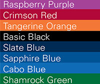
VISIBLE DYE COLOR PALETTE
The following color palette is provided to serve as a general reference only. Actual product hue may vary due to differences in microsphere composition and size, as well as the concentration of the suspension. Dyed spheres may also be coated (e.g. streptavidin) on a custom basis. Our custom capabilities include color matching, custom color formulation, or use of the client’s hydrophobic dye.
FLUORESCENT DYE SPECTRA
Here is a listing of our standard fluorescent dye spectra. See our standard fluorescent microspheres.


FLOW CYTOMETRY FLUOROCHOMES
See our Fluorescent Reference Standards.

Your first choice for scientific solutions
Find out moreSUPPORT
outstanding technical support
PRODUCT
we offer a full product guarantee
DELIVERY
we offer free delivery to UK universities and non profit organisations