Creative Diagnostics offer a wide range of high quality antibodies validated for use in multiplexed assays.
They have primary antibodies to multiple forms of virtually every protein in the proteome including cytokines and are a leading manufacturer whose speciality is the development of high sensitivity quantitative and reader based tests.
Creative Diagnostics is a manufacturer and supplier of antibodies, viral antigens, innovative diagnostic components and critical assay reagents. They provide contract biologic R&D and manufacturing services to the diagnostic manufacturers along with GMP biologics manufacturing for the biopharmaceutical market.
Creative Diagnostics is an evolving biotech company providing highly purified protein/ recombinant antigens worldwide. The protein/recombinant antigens are rigorously tested to meet the research and development demand for excellent quality, uncompromising biological activity at competitive prices.
Creative Diagnostics offers extensive expertise across expression systems for producing protein/recombinant antigens to support customs’ antibody generation. When you choose CD to develop protein/recombinant antigen for your antibody development, the end use application of your antibodies are considered during every phase of antigen production.
Bacterial Expression Systems (E. coli / Bacillus)
Creative Diagnostics provides rapid, high-level bacterial expression and purification of protein antigens to our customs. Our team utilizes both BacTEC? protein soluble technology and FoldEZ? protein refolding technology to achieve protein soluble expression and refolding. Besides, we generate OxiCyto? competent E. coli strains to enable the correct formation of disulfide bonds in proteins that require them for biological activity. Bacillus subtilis expression system, which is an ideal option for the expression of monomeric protein products, combined with our patented fermentation protocols allow us to offer fast process development and scale-up service to meet your antigen needs.
Yeast Expression Systems (P. pastoris / S. cerevisiae)
Yeast protein expression system is the most economical eukaryotic expression system for both secretion and intracellular expression. Our expertise in protein expression spans both P. pastoris and S. cerevisiae expression systems. Our YeastXceed? Technology considers promoters, optimal copies of vector per cell, inducibility, signal peptides, and cellular location, which are essential for high-level recombinant protein production. Many secretory proteins that bacterial expression systems yield as insoluble or as inactive inclusion bodies can be produced successfully by our yeast expression system using native or yeast secretion signals.
Baculovirus-insect Cell Expression Systems BacuFlex? Baculovirus/Insect Cell Expression Platform was developed by our in-house team of scientists for virus production and expression of proteins from baculovirus-infected insect cells in flexible scale. There are several advantages of insect cells over E. coli such as improved solubility, ability to incorporate post-translational modifications, and higher yields for secreted proteins.
Mammalian Expression Systems (CHO / 293)
Mammalian cells have the ability to perform most comprehensive post-translational modifications and to secrete glycoproteins that are correctly folded and contain complex antennary oligosaccharides with terminal sialic acid. Therefore, Mammalian antigen expression is the system of choice for many proteins that are difficult to produce and for antibody generation projects where detection of native antigen is required. Our Mammalian expression HostOptim? systems enable generation of high yielding production systems across different industry relevant platforms (e.g. CHO, HEK, Pichia pastoris).
Cell-free In Vitro Protein Expression
Site-specific incorporation of a biotinylated amino acid allows reduction of the required antigen material down to as little as 5-10 μg for an entire antibody generation project. Our team has developed cell-free expression systems in combination with site-specific biotinylation to meet the increasing demands for this purpose. Now, in only two hours, we can produce biotinylated or other modified proteins starting from plasmid or linear DNA.
Prokaryotic System: E.coli Lysates
Eukaryotic system: Rabbit Reticulocyte Lysates, Wheat Germ system.
As a leading supplier of highly purified protein/recombinant antigens, Creative Diagnostics is proud to offer the highest quality antigens supported by extensive research, development, and validation. We are committed to the highest standards of product performance, and our scientists are dedicated to the goal of accelerating your discovery.
Peptide Antigen Synthesis and Conjugation Services
In some cases, immunizing with a peptide antigen that corresponds to a specific region of the full length protein is an ideal strategy for developing certain epitope-specific antibodies (e.g. phospho-specific). Creative Diagnostics offer a series of services, concerning sequence design, gene synthesis and peptide synthesis, to generate an appropriate peptide antigen for the antibody generation process.
For peptide antigen generation, only the amino acid sequence of the target protein is needed. Our scientist can analyze the sequence and, with the use of sophisticated algorithms (or 3D structural models), predict regions that are predicted to correspond to exposed regions of the native protein. In addition, homologous regions can be avoided, thereby helping minimize cross reactivity when assaying with the antibody.
Because the molecular weight of the peptide is not large enough to stimulate an immune response, once a peptide sequence has been chosen, it should be coupled to a carrier protein before immunizations begin. Our team can assist you to select from the following list of common carrier proteins for the Peptide Conjugation:
Carrier Proteins:
KLH:
Keyhole lympet hemocyanin
CCH:
Concholepas concholepas hemocyanin
BSA:
Bovine serum albumin
cBSA:
Cationized bovine serum albumin
OVA:
Ovalbumin
THY:
Thyroglobulin
In order to isolate only those antibodies in the antiserum that bind with the peptide sequence, the antiserum need to be affinity purified against the peptide. This offers a significant advantage compared to immunizing with a full length protein since the resulting highly specific antibodies against the peptide sequence will approach a monoclonal antibody in specificity.
Antigen Retrieval Based Services
The primary goal of Creative Diagnostics Antigen retrieval (AR) services was always to meet the needs of clinical practice, specifically to facilitate the performance of IHC on FFPE tissues and extraction of antigens/proteins from FFPE tissue.
IHC:
Formalin-fixed, paraffin-embedded (FFPE) tissue sections must be treated to remove the paraffin and unmask the antigen epitopes in preparation for immunohistochemistry (IHC) staining. Two methods to break the protein cross-links formed by formalin fixation are heat-induced epitope retrieval (HIER) and proteolytic-induced epitope retrieval (PIER).
Unless the antigen retrieval method is stated on the antibody datasheet, the optimal method for each antigen must be found experimentally. Our scientists will help you to test several methods to find the retrieval method that gives optimal staining. Alternatively, we can combine HIER and PIER method to unmask antigens if other methods did not work. It is especially useful when performing double or triple labeling of two or more antigens simultaneously.
Extraction of Antigens/Proteins from FFPE tissue:Based on the principles of AR, Creative Diagnostics has developed a high-temperature heating protocol for antigens/protein extraction from FFPE tissue sections. Our method has improved both the protein extraction efficiency and the reversal of formaldehyde-induced protein modifications. The resulting preparation could be used either as the antigens for customs’ antibody generation, or as good materials for clinical and translational research (e.g. proteomics studies).
Welcome to discuss your antigen preparation options with our expert team. We will help you to develop the optimal antigen preparation procedure to reach the best balance between yield, purity and the cost.
We provide rat and mousemonoclonal antibodies construction services. In particular, our proprietary mouse immunization approach allows us to provide mouse monoclonal antibodies within 70 days.
To generate monoclonal antibodies that fit your specific purposes, we tailor our protocols in every major step of antibody production, including antigen preparation [peptide synthesis or protein expression in E.coli, yeast, insect or mammalian cells], animal immunization and hybridoma screening.
Monoclonal Antibody Production: One-Stop Services
Antigen Preparation
We express and purify your recombinant proteins with an appropriate E. coli, yeast, insect or mammalian cell expression system according to the specific purposes of the final antibodies.
Our proprietary chimeric protein expression method can produce a recombinant protein immunogen in a soluble form in bacterial cells that retains the specificity and immunogenicity of the original protein, and generate antibodies that bind to the protein surface. This approach highly increases the possibility of getting IHC-positive monoclonal antibodies, and antibodies that recognize the native protein and good for ELISA, IP, IHC, Immuno-fluorescence and Western Blotting.
We can design membrane mimc protein antigen (MMPA) using MPAT? platform. It remains structural characteristics such as multi-spanner extracellular domain and it is feasible to express in whole water-soluble form with high purity.
Alternatively, we perform peptide synthesis and conjugation.
Immunizations
We immunize animals with customized protocols that involve proper adjuvants, inoculation routes, dosage and timing. We can develop antibodies targeting impure native antigens or rare antigens of a minimum amount by adjusting the immunization strategies.
Our proprietary Magic? adjuvant can elicit stronger and faster immune response within only 21 days.
We use different formats of immunogens for animal immunizations depending on the specific purpose. For antibodies intended for immunohistochemical [or immunocytochemical] staining, in addition to the natural immunogens, e.g. recombinant protein, we may alternatively use denatured antigens in animal immunization and hybridoma screening.
Our fast track mouse immunization protocol allows us to provide monoclonal antibodies within 70 days.
Development of Hybridoma
We fuse splenocytes with proper myeloma cells using optimized PEG mediated fusion protocols.
We screen hybridoma clones with customized protocols according to the final applications of the requested antibodies, including but not limited to immunoprecipitation, immunoblotting, various ELISA and particularly, IHC methods.
Goal-oriented method to screen IHC positive monoclonal antibodies. We introduce our proprietary IHC-positive hybridoma screening protocols to provide you IHC suited hybridoma clones.
Our revolutionary platform Omni-Hybridoma? platform ensures that a large number of hybridoma clones can be selected after each cell fusion. It can eliminate the possibility of overgrowth of potentially valuable slow-growing clones by fast-growing clones.
We deliver your positive hybridoma clones in culture or in cryopreservation as well as the derived monoclonal antibodies.
Ascites Production or in Vitro Antibody Manufacturing
Inoculation of hybridoma cells in to relevant animals to produce ascites. We normally use immuno-sufficient mice to produce ascites. Immuno-deficient mice are available for ascites production for non-murine hybridoma cell lines or murine cell lines of distinct genetic backgrounds. Optimized methods are used to generate tumors and accumulate ascitic fluid. Using nude or SCID mice in ascites production will eliminate the contamination from endogenous mouse IgG.
Cell culture in protein-free medium. We use stationary flasks or 5-50liter bioreactors to manufacture monoclonal antibodies. We have an antibody fermentation capacity of over 300L.
Rat Monoclonal Antibody Production
We have established a unique platform to develop high-affinity monoclonal antibodies in rats.
Compared with commonly used mouse antibodies, rat antibodies do not have the background cross-reaction problems in immune-detection of antigens out of a mouse background, such as a mouse antigen from a mouse animal model. However, Rat monoclonal antibodies are much more difficult to develop compared with mouse monoclonal antibodies, due to the lack of a stable fusion partner cell line for rat hybridoma generation.
Creative Diagnostics has developed a proprietary hybridoma-generation platform for efficient development of high-affinity and high-specificity rat monoclonal antibodies. Our Rat Monoclonal Antibody platform allows us to offer the most comprehensive one-stop custom antibody development services in the industry.
Features of our rat hybridoma production services:
Picomolar affinity and high specificity.
Excellent results even for difficult antigens.
Ability to recognize subtle changes such as post-translational modifications.
Novel epitope recognition.
Cross-reactive antibodies recognizing human proteins and mouse orthologs.
Excellent IHC results with formalin-fixed paraffin-embedded tissue.
Ability to generate large panels of bioactive antibodies.
Rabbit Monoclonal Antibody Production
In contrast to rabbit hybridoma technology available in other leading companies, Creative Diagnostics has built up a unique rabbit monoclonal antibody production platform that is based on our unparalleled expertise in phage display antibody library technology. We generate rabbit antibody libraries and select high-affinity antigen-specific antibodies by phage display following Dr. Christoph Rader’s methodology. Usually, two or more rabbits of the b9 allotype are immunized with one antigen, total RNA from spleen lymphocytes and bone marrow cells are purified, and then the first-strand cDNAs are synthesized. cDNA from spleen and bone marrow samples derived from multiple groups of animals immunized by different antigens can be pooled to make a unified library for all the antigens. Chimeric rabbit/human Fab libraries are made in our pCDisplayphagemid vectors by fusing rabbit Vκ/Vλ to human Cκ and rabbit VH to human CH1 of human IgG1.
The rabbit antibody repertoire is an exceptional source for both polyclonal antibodies and monoclonal antibodies that combine high specificity with high avidity and affinity. In addition, rabbits, which belong to the order Lagomorpha (lagomorphs), are evolutionarily distant from mice and rats, which belong to the order Rodentia (rodents). As a consequence, epitopes conserved between rodent and human antigens that are invisible to rodent monoclonal antibodies (and also human monoclonal antibodies generated from transgenic mice with human immunoglobulin genes) can often be recognized by rabbit antibodies. Rabbit monoclonal antibodies have overcome this limitation, providing access to defined reagents of infinite supply from the rabbit antibody repertoire. Rabbit monoclonal antibodies generated by phage display offer additional advantages due to the fact that the phenotype (protein) and genotype (cDNA) are selected at the same time. Also, in comparison with rabbit hybridoma technology, our phage-display platform can produce much more antibody clones at a time.
It was well-known that rabbit Fab’s are toxic to in E. coli cells, in which phage display systems work. There are two approved approaches to avoid toxicity, insolubility and low expression problems of rabbit Fab in bacterial cells. The first is to make rabbit/human chimeric Fab libraries in which rabbit variable domains (Vλ, Vk, and VH) are linked with human constant domains (Ck and CH1). This approach has been well demonstrated by the work done by Drs. Carlos F. Barbas III and Christoph Rader. The other approach is to express the selected rabbit Fab [or scFv] antibodies in yeast Pichiapastoris, relying on yeast-display technology, which is current not available at our company.
Of note, most rabbit light chains have an extra disulfide bridge that links the variable and constant domains in addition to the two intra-chain disulfide bridges shared with mouse and human kappa light chains. By evaluating the impact of this increased disulfide bridge complexity on the generation and selection of chimeric rabbit/human Fab libraries, scientists have demonstrated that rabbits with mutant bas and wild-type parental b9 allotypes are excellent sources for rabbit monoclonal antibodies.
We have constructed a large number of immunized rabbit antibody libraries. In particular, we developed an in-frame-selection technique to exclude Fab library clones that have shortened antibody fragments and reading frame shifts, which are the greatest challenge in antibody library construction that are usually due to the errors in synthetic PCR Primers and PCR reactions used to amplify antibody genes.
Rabbit Antibody Library Screening
Selection of high affinity binders is mainly based on the in-solution phage display antibody library screening strategies we employ. We have two strategies: selection based on the equilibrium constant (Kd) and selection based on binding kinetics. In the first approach, sub-library phage is incubated with biotinylated antigen at controlled concentrations and bound phages are captured by immobilized NeutrAvidin. The second, selection based on binding kinetics is also termed off-rate (Koff) selection, in which phage population is allowed to saturate the labeled antigen before a large molar excess of unlabeled antigen is added to the mix for controlled periods of time. This allows the selection of mutant antibodies that have slower off-rates. Since a reduction in Koff usually results in a higher affinity, this selection approach singles out antibody variants with improved Kd. These methods allow selection of antibodies that have high affinity. In the end, the selected antibody fragments can be expressed to test their binding specificity and affinity.
Hamster Monoclonal Antibody Production
Creative Diagnostics has extensive experience in producing monoclonal antibodies in hamsters. The use of hamster as the host animal offers a unique species alternative in areas where mouse and rat antibodies may not work well. We can employ up to 10 hamsters for each antigen. Immune responses are elicited using immunogens such as transfected cell lines, DNA, recombinant proteins, native proteins, peptides or haptens conjugated to carrier molecules.
Fig.1 Outline of monoclonal antibody development in Hamster
We perform splenocyte fusion on one or more animals. We select and subclone the strongly positive clones from the parental cultures by 2 rounds of limiting dilution cloning. Stable clones will be expanded into 24-well plates and then into T-flasks for further characterization. At no additional cost, client could select one clone for in vitro production of approximately 1L of supernatant. Supernatants will be purified using Protein A/G column and pure antibodies will be deliverable. We also offer a full range of contract manufacturing services, including In vitro Antibody and Protein Production in bioreactors and large-scale Antibody Purification.
Sheep Monoclonal Antibody Production
Creative Diagnostics has accumulated considerable experience and expertise in the custom manufacture of high quality sheep monoclonal antibodies using our unique hybridoma technology.
Our sheep monoclonal antibody production service is based on the similar method to produce mouse monoclonal antibodies. The distinctive and key difference is that a sheep fusion helper is used to immortalize sheep B cells. The fusion helper is developed to establish our special hybridoma technology that can generate stable and productive cell lines.
Advantages of Sheep Monoclonal Antibodies:
Higher Specificity: As the antibody only binds to one specific region, it is possible to produce a highly specific antibody for the required target.
Broader range of epitope recognition.
Higher Affinity: Sheep monoclonal antibodies with extremely high affinity can be useful in assays for analytes at very low concentrations around10-10 to 10-14 molar.
Eliminated cross reactivity.
Polyclonal Antibody Production
You order, we deliver
As a professional producer in this field, Creative Diagnostics provides customers with the most competitive products and the most thoughtful services across the world. Low cost of the antibody production is our priority advantage. We have been committed to advancing technologies and streamlining management. Another factor makes us special is that we are capable of meeting the specific needs and requirements of customers. Horizontal and vertical cooperation established with different parties enables us to successfully accomplish whatever you are expecting in the most efficient way.
We are specialized in the following aspects:
? Bulk quantity polyclonal antibody production projects.
? The most complete collection of hosts eg. Mouse, rat, goat, rabbit, chicken, sheep, donkey, Guinea pig, Llama, Camel, horse, dogs, bovine, pig and primates.
? Custom phospho-specific polyclonal antibody service.
? A portfolio of purified immunoglobulin eg. Protein A, Protein G, ammonium sulfate and peptide affinity purification.
? Polyclonal packages: 28-days, 70-days and tailored ones.
What makes us special?
We have our own farm and are working with a team of skilled veterinarians and technicians in a modern specific pathogen free animal facility, we propose a complete palette of polyclonal antibody production options.
We use New Zealand White females rabbits, bred on our farm and adult laying leghorn chickens, Boer goats, and sheep. All of our animals are cared for by qualified personnel to ensure that you are receiving the highest quality service.
28-day fastest polyclonal antibody program and classical polyclonal antibody programs. Customized and flexible projects to meet your requirements.
You will be in contact directly with a project manager who will tell you how your project is going and give you updates with everything, for we actually are the manufacturer.
Our exclusive adjuvant used for 28-Day Speedy polyclonal antibody to make access to the fastest way to obtain high quality polyclonal antibodies.(This service is available for mouse, rat, Guinea pig, rabbit, goat and sheep)
One-stop solution will streamline the project as much as possible.
Immunogen Preparation → Animal Immunization → Antibody Development → Antibody Production → Product Evaluation.
Extended services:
Peptide Synthesis
Protein Conjugation, such as KLH, VOA, BSA
Elisa Screening
Serum Purification
Antibody Labeling
Antigen Expression and Purification with MPATTM Platform
Fab or F(ab’)2 Fragments Production
Chicken IgY Antibody Production
Creative Diagnostics provides the most cost-effective custom chicken IgY antibody production services in the industry. We offer standard 56 day chicken production packages which consist of 4 immunizations, resulting in the collection of 12-15 eggs per chicken (please see example project timeline below). We typically use 2 chickens for each project.
Creative Diagnostics provides free antigen design to assist in selection of the best possible antigen sequence for eliciting the desired antibody response. We use the latest in software with the proper algorithms to assess antigenicity, hydrophilicity, flexibility, secondary structure and aggregation potential.
Project Timeline:
Package 1
Customer Antigen to lgY Purified Antibody
Package 2
Customer Antigen to Affinity Purified Antibody
Package 3
Antigen Synthesis to lgY Purified Antibody
Package 4
Antigen Synthesis to Affinity Purified Antibody
Service Includes
Antigen design & synthesis
√
√
Immunization of 2 chickens
√
√
√
√
Prebleed
√
√
√
√
ELISA titre
√
√
√
√
lgY purification
√
√
Affinity purification
√
√
Time (weeks)
10-12
10-13
13-15
13-15
What You Receive
Full project report
√
√
√
√
ELISA data
√
√
√
√
Preimmune serum (5mL)
√
√
√
√
lgY purified antibody
√
√
Affinity purified antibody
√
√
Affinity resin (10mL)
√
√
Antigen (2-3mg)
√
√
How much antigen should you ship us?
We recommend immunization with, between 0.7 and 1 milligrams of purified recombinant protein or peptide conjugate per hen and prefer a concentration of at least 2 mg/ml. If you would like your IgY affinity purified, we need additional antigen at a concentration greater than 3 mg/ml to make an affinity column. The amount of immunogen required to make your affinity column depends on how much affinity purified IgY is needed for your project. Please contact us at info@stratech.co.uk and we’d be happy to discuss this with you.
Where do you ship your antigen?
Ship your purified recombinant protein or peptide conjugate to us at:
Creative Diagnstics
45-1 Ramsey Road
Shirley, NY 11967
As soon as we start the immunization, we will e-mail you an immunization/production schedule so you’ll know exactly when you can expect your custom IgY.
Industrial Quantities
For those customers requiring large scale production of IgY, we can meet your specific requirements. All laboratory work is performed using standard operating procedures to assure the highest quality of your custom product depending on your immunogen, how much IgY is required and over what time-frame, we will design a custom project that will suit your needs.
For bulk quantities of IgY, we usually recommend multiple hen immunizations and the pooling of their eggs for IgY purification. Following our standard immunization protocol (4 immunizations over 40 days), hens receive a booster shot every 70 days to maintain a high titre yolk.
Advantages of Chicken IgY Antibodies
Higher avidity: most mammalian proteins exhibit enhanced immunogenicity in chickens than in mammals due to phylogenetic distance, and thus raise antibodies of higher avidity; this also makes production of antibodies against conserved mammalian proteins more successful in chicken than in mammals.
Higher specificity: compared with mammalian IgG, chicken IgY has less cross reactivity with mammalian proteins other than the immunogen;
Lower background: IgY and IgG are structurally different in the Fc region; IgY does not bind to IgG Fc receptors and causes less false positive staining.
High yield: very low quantities of antigen are required to obtain high and long-lasting IgY titer in the egg yolk; chickens lay eggs regularly, providing a continual source of IgY antibody.
Anti-idiotype Antibody Production
Creative Diagnostics provides a full range of service in custom anti-idiotype antibodies (anti-IDs) to support your new monoclonal antibody drugs development. At Creative Diagnostics, our scientists utilize the proprietary technology to guarantee the high specificity and affinity of anti-idiotypic antibody, including magic adjuvant and immune mediators, super-sensitive hybridoma screening, and optimal clone selection processes. We have accomplished 95% success rate of anti-IDs antibody development for our customers.
The idiotype represents the complementarity determining regions (CDR) of an antibody, including the unique antigen binding site, and the combination of epitopes within the idiotype is unique for each antibody. When one antibody binds to one idiotope of another antibody it is referred to as an anti-idiotypic antibody (anti-ID). These highly specialized anti-idiotypic antibodies have become a powerful tool for antibody drug pharmacokinetics (PK), pharmacodynamics (PD) and immunogenicity studies.
Features of Our Anti-IDs Antibodies Service:
High specificity and affinity antibodies to your targets
Proven track record – 95% success rate of anti-IDs development
Generation of non-inhibitory and drug-target complex antibodies
Fab and F(ab)2 formats with a choice of detection tags
Conversion to full immunoglobulin with a choice of isotype
Antibodies sequenced for long-term security of supply
Readily integrated downstream antibody drug PK/PD and immunogenicity assay
Anti-idiotype Antibody Production Packages
Anti-idiotypic antibodies that recognize an antigen-combining site of an antibody can mimic the structure and/or function of certain nominal antigens. This feature makes them especially useful in unwanted immunogenicity assessment. For system suitability controls, the FDA recommends that a positive control antibody, either mono- or polyclonal, used at development and validation of immune assays for assessment of the immunogenicity of therapeutic protein products during clinical trials.
Generation of polyclonal anti-idiotypic antibody
Generation of monoclonal anti-idiotype antibody
Creative Diagnostics also provides immunogenicity testing of antibody drugs:
A number of various assay formats and platforms are available that can be employed for immunogenicity assessment.
Our services include but not limited to:
Type of assay
Binding Assays of Anti-IDs Antibodies
Enzyme-linked immunosobent assay (ELISA)
Electrochemiluminescence immunoassay (ECLIA)
Radioimmunoprecipitation assay (RIPA)
Surface plasmon resonance immunoassay (SPRIA)
Neutralization Assays of Anti-IDs Antibodies
In vitro cell – based bioassays
Competitive ligand – binding assays (CLBAs)
Both assay types can be used together for the complete characterization of the antibody response against the antibody drugs.
Polyclonal Anti-idiotypic Antibody Production
Creative Diagnostics has extensive experience in development a successful immunization protocol improving the generation of polyclonal anti-idiotype antibodies (anti-IDs). Polyclonal antibodies are a mixture of immunoglobulins that recognize different epitopes on a specific antigen. In addition to the traditional mouse and rabbit options, we also offer goat, chicken, sheep, donkey, Guinea pig, Llama, Camel, horse, dogs, bovine, primate or host animals specified by the clients.
Features and Benefits of Polyclonal Antibodies
Recognition of many epitopes on any single antigen
Easier detection of denatured proteins
Enhanced target protein signal amplification
Better management of antigen modifications
Fragmentation and purification of your antibody to obtain F(ab) & F(ab)2 fragment
Peptide Synthesis (up to 20 aa) & Conjugation (3-4 weeks)
5-10mg, purity > 90% by HPLC
Conjugation of peptide to carrier protein (KLH, BSA, OVA)
Immunization of conjugate into desired host species
√
Fab & F(ab)2 Fragmentation and purification (3-4 weeks)
Fab or F(ab)2 fragment generation with papain /pepsin
Fab or F(ab)2 purification from protein A /G
√
Services 1: 1 or 2 Rabbit, 63-days
3-5 immunizations
Test bleed day 35 & ELISA titer determination
Final bleed: 40 – 90 ml serum
Services 2: 1 Super-Fast rabbit, 28-days
4 immunizations
Final bleed: 40 – 90 ml serum
Services 3: PhosphoSpecific Antibodies
Peptide synthesis: phospho & non-phosphopeptide
Immunization of 2 rabbits
Purification: affinity purification & depletion
√
√
√
Antigen Affinity Purification
Antigens are covalently bound to agarose beads bysulfhydryls
Immuno-affinity purification of polyclonal antibodies
Common Applications of Polyclonal Anti-Idiotypic Antibodies
Preclinical research of therapeutic antibodies
Anti-drug Antibodies (ADA) for clinical development
Pharmacokinetic (PK) studies
Immune Response (IR) immunogenicity assays
Controls in ligand binding neutralizing assays
Controls in antibody blocking assays
The FDA recommends that the sponsor develops polyclonal anti-idiotypic antibodies as positive controls for antibody immunogenicity assays. Creative Diagnostics has a proven track record of achieving successful outcomes with higher complexity projects. Our ability to work closely with customers to clearly understand their end application, define their specific project goals and design a detailed project plan is the key to our high success rate delivering quality polyclonal anti-idiotype antibodies.
Monoclonal Anti-idiotypic Antibody Production
Monoclonal anti-idiotype antibodies are ideal for preclinical research (PK, Tox and immunogenicity study) and the generation of data to support the development of antibody drugs. Creative Diagnostics has developed high-quality monoclonal anti-idiotypic antibodies used in sandwich ELISA for quantification of drug antibodies in patient serum.
Our monoclonal anti-idiotype antibody solution is a highly customizable service based entirely on the customer needs. To complement the monoclonal anti-idiotype antibodies development, we offer following feature services:
Fast turnaround time
More than 95% success rate
Professional immunogenic design
Standard programs for affinity maturation
High-throughput cloning
High-throughput screening
Characterization assays
Monoclonal anti-idiotype antibody production packages
Item
Basic Package
Peptide Package
Fab & F(ab)2 Package
Experimental scheme design (1-2 days)
√
√
√
Peptide synthesis up to 20 aa, HPLC purity > 90% (3-4 weeks)
√
Peptide conjugation to carrier protein (KLH, BSA, OVA)
√
Fab & F(ab)2 fragmentation and purification (3-4 weeks)
√
Phase I: Immunization (4-6 weeks)
Immunization of 5 Balb/c mice
ELISA testing of serum
√
√
√
Phase II: Fusion (2-3 weeks)
Fusion of 1 mouse spleen with sp2/0
ELISA screening
√
√
√
Phase III: Sub-cloning (4-6 weeks)
2-3 sub-cloning steps
ELISA screening
√
√
√
Isotyping of final clones
√
√
√
Antibody production (3-4 weeks)
Ascetic fluid
Protein A/G purification
√
√
√
Our Guarantees:
Timely feedback
Personalized customization service
Competitive price
High specificity and affinity antibodies development
Please contact us to understand more custom monoclonal anti-Idiotypic antibodies production services from Creative Diagnostics to meet your specific requirements.
Anti-idiotype Antibody Assay Development Services
ELISA assays development
Monoclonal antibodies raised against the idiotype of ‘drug’ antibody are capable of uniquely identifying the drug’s presence and concentration in human serum or other tissues. Therefore, anti-idiotype antibodies are used in pharmacokinetic (PK) and pharmacodynamics (PD) studies. Typically we can develop capture and detection antibodies (Type 1-3) to be used in sandwich ELISA assays for the quantification of drug antibodies in patient serum or expression system supernatants.
Anti-drug Antibodies Assay
Anti-idiotype antibodies are typically used as positive control in Anti-Drug Antibodies Assay, which aims to determine whether a patient develops antibodies against the therapeutic antibody. For more information about Anti-Drug Antibodies Assay, please click the link: ADA assays
Neutralization Assay and Blocking assay
Often our customers develop anti-idiotype antibodies as controls for neutralization assays and blocking assay. Anti-Ids candidates are screened for the ability to neutralize or block specific ligand binding of an antibody/receptor. Creative Diagnostics has a proven track record of achieving successful outcomes with higher complexity projects.
Anti-Hapten Antibody Production
Anti-small molecule hapten antibodies enable the detection of small molecules using robust and rapid immune detection technologies, such as ELISA, LFIA and IHC/IF. Either raised against endogenous small molecules (amino acid metabolites, lipids, saccharids, nucleotides, steroids, …) or exogenous compounds (drug and pesticide residues, chemical pollutants,…), anti-hapten antibodies constitute attractive detection tools not only in the field of human health, for diagnostic and therapeutic applications, but also in areas such as food safety and environmental monitoring.
Combining 15 years of experience in the development of small molecule antibodies, Creative Diagnostics holds a specific expertise in the design of small molecule / protein carrier adducts. Based on our capacities in antigen design, we developed high quality small molecular monoclonal antibodies against a variety of targets.
Our advantages of custom small molecule mAbs services
A personalized strategy for antigen design, based on an in-depth analysis of the physico-chemical properties, immunogenicity and potential competitors of the small molecule target to address, and tailored to the client’s intended use of the antibody. Read More
Quality controls, 24 hours services, Go/No-Go decisions points at the end of each phase, for transparent projects with limited risk and reasonable costs.
Full ownership of the antibody-producing clone is granted to the client at the end of the study, together with at least 3mg of purified monoclonal immunoglobulin.
Summary of Project Proposed:
Phase
Summary
Description
Time
1
Synthesis of Immunogens
Small molecule conjugation to carrier proteins KLH for immunization. Small molecule conjugation to carrier proteins BSA for assay. Client provides small molecule to be conjugated.
1-2 weeks
2
Immunization
Immunization of 5 Balb/c mice with antigen (up to 4 times) and determination of antibody titer by ELISA. Animals with highest serum antibody titers are selected for fusion.
6-8 weeks
3
Fusion and Screening
Fusion of spleen cells from selected mice with myeloma cell line. ELISA screening.
Selection of clones producing specific antibody.
3-4 weeks
4
Subcloning and Generation
Subcloning and generation of monoclonals from 3 best chosen clones.
Expansion & storage of 3 vials each of the subclones.
6-7 weeks
5
Antibody Production
Production of antibodies on cell culture.
Protein G purification + ELISA validation.
2-3 weeks
Deliverables:
3 vials each of 3 antigen specific subclones (>90% clonality and single isotype), storage for 1 year.
3-5 mg of purified antibody.
ELISA data for a) mouse sera; b) 6x 96 well fusion plates; c) up to 96 selected positive wells from fusion plates (repeat validation); and d) subclones ELISA data.
Productivity and Clonality data for 3 chosen clones.
Mycoplasma test results & isotyping data for heavy & light chains.
Terms & Conditions:
Client to provide 5 – 10 mg of small molecule for conjugation to carriers.
Conjugation cost and quality not guaranteed until we know the molecule structure.
We accept payment by check, credit card, debit card, and bank transfer.
Payment required in 3 installments before the beginning of each phase of work.
All our services are guaranteed. Contact us for reference about our excellent services.
Creative Diagnostics also offers custom services for the detection of small molecules in biological samples.
Antigen design
Anti-small molecule hapten antibody production
Method development and validation for small molecule detection (ELISA, LFIA)
Membrane Protein Antibody Production
GPCRs represent a group of promising targets in science research and immune therapy. Unfortunately, they are multispan membrane proteins that are extremely difficult to generate antibodies using conventional approaches. The barriers are as followed.
1. Impossible to obtain a structurally intact protein as antigen;
2. Inaccessible to fold the discontinuous segments correctly;
3. Insufficient to elicit an immune response for a low level abundance.
Creative Diagnostics has developed proprietary methodologies MPATTM including conventional and novel approaches to solve these problems faced when generating antibodies against membrane proteins such as GPCRs. At least four feasible ways (Fig.1) access to you right now according to the structural property of membrane proteins. Our antibody platform also enables us to screen functional antibodies of membrane protein targets in vivo.
Fig.1 Approaches for membrane protein antibody generation. A: intact membrane protein; B: extracellular peptide as antigen; C: mimic of extracellular peptide; D: cell-based immunization; E: DNA immunization with special adjuvant.
While simple in theory, successful execution of this platform requires overall consideration and careful optimization according to specific membrane proteins. In fact, our platform is compatible with well-established antibody generation approaches such as cell fusion & screen.
Whatever membrane proteins you desired for antibody generation, just inform us and in most cases we can accommodate your request.
Phospho Antibody Production
The process of producing antibodies against a phospho-residue is more complicated than traditional antibody production using peptide immunogens. Phospho-specific antibodies are generated using peptides containing one or more phosphorylated amino acids. There are three residues which can be phosphorylated: Serine (S), Threonine (T) and Tyrosine (Y).
For phospho polyclonal antibody production:
To produce antiserum against Phospho-Peptides includes synthesis of phosphopeptides, conjugation and immunization of rabbits. In many cases, one would need to do affinity purification with a phospho-peptide column. Sometimes, one would also need to cross absorb the antibody with a non-phosphopeptide column in case there are some anti-non-phospho protein antibodies in the antiserum. In this case, synthesis of matching non-phospho peptides is required to make the negative-selection columns. Affinity purified, cross-absorbed polyclonal antibodies that are specific for the phosphor-peptides are usually required for downstream assays.
For phospho monoclonal antibody production:
In comparison with polyclonal antibody production, monoclonal antibody production against Phospho-Peptides is more straightforward; we just use the Phospho-Peptides to immunize the mice, and use Phospho-Peptides to screen for positive hybridoma clones. After that we use non-phospho-peptides to do negative selection. This negative selection is required [although widely forgotten] since peptide phosphorylation [or protein phosphorylation] is never 100% complete.
DNA Immunization Antibody Production
Creative Diagnostics offers proprietary genetic immunization based polyclonal and monoclonal antibody generation services. This unique antibody development approach involves direct immunization of host animals with plasmid DNA encoding the target protein of interest. The immunized hosts then produce the encoded protein and raise antibodies. Genetic immunization involves introducing the gene in the form of a cDNA directly into an animal which translates this cDNA into protein thus stimulating an immune response against the foreign protein. Protein purification is not necessary for this genetic immunization approach, which can save several months in time over recombinant protein generation followed by antibody production.
In order for genetic immunization to be successful, the cDNA-encoded protein must be secreted by the transfected cells in immunized animals or expressed on the surface of the transfected cells in order to get stimulation of an antibody response.
The foremost advantage of this antibody production approach is its high success rate in generation of high-affinity antibodies recognizing difficult-to-express proteins in their native confirmation, such as GPCRs, ion channels and other multiple membrane spanning proteins. For these proteins, recombinant protein fragments or peptides derived from their extracellular domains may raise antibodies workable in Western blotting but are extremely hard to produce high-affinity antibodies that can recognize their integral proteins in their native form. This point is important in raising antibodies for diagnostic use, in which recognition of the antigens in their native form can be required. For therapeutic antibodies, targeting the antigens in the native conformation with a high-affinity is of course required!
Of note, our technology allows guaranteed antibody development against 7-membrane-spanning GPCR proteins!
High affinity antibodies can result from genetic immunization because of low level of expressed proteins and constant presentation to the immune system; these tend to favor development of high affinity antibodies.
Usually polyclonal antibody development via genetic immunization is tried first since it is an economical way to see whether the protein-encoding plasmid/cDNA will raise the desired antibodies, e.g. antibodies recognizing an integral antigen in its native 3D conformation. If this is successful, monoclonal development is followed.
RIMMS Services
Affinity matured murine monoclonal antibody producing cell lines can be rapidly generated using repetitive, multiple site immunization strategy designated RIMMS. RIMMS capitalizes on rapid hypermutation and affinity maturation events which occur in B cell populations localized within secondary lymphatic tissue early in response to antigenic challenges. Along with using less antigen, this approach allows the investigator to obtain hybridomas rapidly.
The immunization sites used for RIMMS are proximal to easily accessible regional lymph nodes. RIMMS capitalizes on somatic fusion of immune B cells undergoing germinal center maturation in draining lymph nodes. Fusions can be performed as early as 7 days out to 14 days after the onset of immunization. Thus, RIMMS can be used to generate affinity matured murine hybridomas cell lines within one month period.
Creative Diagnostics have explored the feasibility of modifying immunization and fusion timelines used for developing monoclonal antibodies using RIMMS. We offer a full line of RIMMS services, including immunizations, somatic fusion, screening, and isolation of affinity-matured IgG-secreting hybridoma cell lines.
By using RIMMS, we have been able to expedite the isolation of affinity matured monoclonal antibodies to numerous antigens, including recombinant protein, conjugated synthetic peptides, and drug haptens. Our RIMMS service could be completed within one month, and it is an ideal choice if you want to make more diverse repertoire of antibodies or antibodies directed against conformational epitopes quickly.
Key Features of Services:
Require small quantities of antigen and fewer animals
Several different antigens could be developed (synthetic peptides, recombinant proteins, cellular extracts, conjugated compounds, DNA-based immunizations)
Produce affinity matured antibodies quickly (polyclonal and monoclonal)
Creative Diagnostics develops custom antibodies that work in your hands and in the end applications. Please do not hesitate to contact us if you need any free consultation and a detailed quotation of your project.
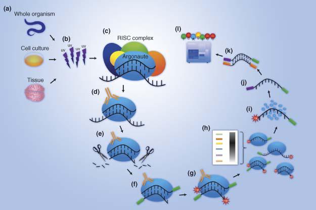
Quenchbody Generation Service
Antibody-based sensors have made outstanding contributions to the fields of molecular biology and biotechnology. Nowadays, a novel powerful fluorescent immunosensor strategy named Quenchbody (Q-body) has been applied to the detection of a range of antigens in a rapid, simple, and sensitive manner. Q-bodies, which are made by conjugation of fluorescent probes to antibodies, have merits such as no need of additional reagents and availability of sensitivity without need of long incubation.
Q-body works on the mechanism of antigen-dependent removal of quenching effect on popular fluorophores such as carboxyltetramethylrhodamine (TAMRA), which is incorporated to a specific position(s) of a single chain antibody (scFv) or Fab fragment. The primary reason of quenching is photoinduced electron transfer (PET) from conserved tryptophan (Trp) residues in the variable region, and secondarily, the other dye incorporated to the other site. Using this Q-body technology, just mixing a Q-body with antigen and measuring the fluorescence intensity enables antigen quantitation. Since no washing step is necessary, it is a remarkably simple and rapid quantification method, still allowing the use of several organic dyes with different colors. Q-bodies has been use to successfully quantify a range of biomolecules including small haptens, peptides, and larger proteins.
Ultra-Quenchbodies (UQ-bodies) refer to doubly labeled Fab Q-bodies, in which the same dye was incorporated to both the H and L chains, resulted in deeper quenching and a greater antigen-dependent release, due to H-dimer formation and Trp-mediated PET. Compared with single labeled Q-bodies, double-labeled UQ- bodies showed generally higher responses; Especially, a hetero double-labeled Fab has been successfully used to monitor changes in FRET that are dependent on a conformational change that occurs upon the binding of the antigen.
The services we provide:
Synthesis of Q-Bodies (scFv-type)
Generation of Ultra-Quenchbodies (Fab-type)
Generation of hetero double-labeled Fab-type Ultra-Quenchbodies
Generation of UQ-bodies using recombinant Fab fragments (E.coli)
CD’s Custom Technology Teams are experienced in performing both small-scale and large-scale antibody generations and purifications. Please feel free to contact us if you have any questions regarding our service.
Creative Diagnostics provides contract ELISA development kit services for the R&D and IVD community. We conduct ELISA kit development services for supporting regulatory approval submission. Creative Diagnostics will carry out the approval proposal and deliver the expected results and documents in a time and cost effective manner.
Through combined expertise in antibody generation and immunoassay development, we have produced a large number of diagnostic antibodies that cover a wide range of disease portfolio including infectious diseases, cardiovascular diseases, cancer and bone diseases, with osteoporosis in particular.
ELISA Formats:
Direct ELISA
directly labeling the antibody
relatively quick, and avoids potential problems of cross-reactivity
requires the labeling of every antibody, time-consuming and expensive
lack the additional signal amplification
Sandwich ELISA
measures the amount of antigen between two layers of antibodies
restricted to the quantitation of multivalent antigens
especially valuable when the concentration of antigens is low
reach detection sensitivity at sub-femtogram per ml level
Competitive ELISA
frequently used for the detection of small analyte antigens containing a single epitope
an inverse relationship between the signal obtained and the concentration of the analyte
Detection Methods:
Light absorbance
Fluorescence intensity or polarization
Fluorescence resonance energy transfer
Luminescence
Assay Milestones:
Why choose Creative Diagnostics?
There are a wide range of ELISA Kits for the detection of hundreds of different proteins and molecules including cytokines, growth factors, markers for infectious diseases, diabetes and tumor, drugs and small molecules, etc. in our website. You can select one directly.
We provide optimizing service. Our research team will optimize an existing kit by testing other antibodies or reagents based on your special request.
Start from scratch. Our staff scientists will work with you to avoid some of the early pitfalls of assay development by reviewing the purpose of your assay and the desired attributes before the process starts and follow rigorous guidelines for quality control from start to finish.
We focus on thorough optimization and validation of every aspect of assay development, including antibody specificity, assay sensitivity, reproducibility (intra- and inter-assay), cross-reactivity, standard curve range, assay accuracy (linearity and recovery) and kit stability.
Our featured small molecule design and rat IgG or chicken IgY monoclonal antibody production services can cover most of your needs.
CD Microplate Coating
Creative Diagnostics offers its vast expertise in microplate coating to many clients in the pharmaceutical, medical device and biotech industries. Creative Diagnostics has a highly trained staff operating its automated and validated microplate coating systems with a focus on quality control. Advantages include fast set-up, low minimums, expert quality control and professional technical support.
Expertise
Years of experience and advanced technological capabilities Creative Diagnostics combines the most advanced facilities and automated coating equipment with years of proven expertise in coating services. At Creative Diagnostics, our manufacturing processes and solutions are as flexible as your application requires.
Advantages
Creative Diagnostics coated plates are suitable for a wide range of applications, like immuno or nucleic acid hybridization assays. The plates are available in any lot size of the formats, have high affinity towards biotin and are stable over a wide range of pH, detergent concentration and ionic strength.
Services
Creative Diagnostics’s highly trained technical support staff can assist in the selection and qualification of the right coating surfaces, raw materials and coating process design. Below are some of the ways Creative Diagnostics can serve your organization:
Outsourcing: We can utilize existing coating process and protocol to coat microplates with our equipment.
Development: Our team can design an application-specific coating process for you or modify an existing coating protocol to suit your needs.
Planning: We can schedule manufacturing and delivery of your coated microplates on a regular basis.
Planning: We can schedule manufacturing and delivery of your coated microplates on a regular basis.
Creative Diagnostics performs in-process and QC release testing on each production batch to ensure accuracy and precision testing, stability testing, plate-to-plate and lot-to-lot correlation all pass specifications.
Additional highlight
Creative Diagnostics Services 96-well and 384-well microplate technology FlashPlate and non-scintillating microplate development Bar-coding technology for optimal control over product manufacturing, quality, and availability Versatile FlashPlate and Microplate Coating Services.
CD ELISA Matched Pair Development
Matched pairs are the basis of many sandwich ELISAs, either in kits or for in house assay set up. The name refers to sets of antibodies which are known to be capable of detecting different epitopes on the same protein antigen, so they can be used together for the capture and detection of a single antigen in a sandwich ELISA or related immunoassay. Matched pairs can consist of two monoclonals, two polyclonals, or a combination of both.
What we can do
Creative Diagnostics can develop the most suitable matched pairs for your ELISA assay. Projects can be initiated in conjunction with our custom hybridoma development services or antibodies can be supplied by customers for evaluation. We utilize the expertise of our own internal ELISA development team which supports a variety of ELISA products.
Antibody Testing:
ELISA assay/s are run to determine best antibody pair.
Plates are coated with antibody and antigen to be tested.
By the epitope mapping, each coating antibody is tested with antibody candidates.
A polyclonal Abs for the antigen of interest (if available) can be used as a coating or detection option.
A 5-7 point standard curve of the antigen is typically used.
A typical standard curve range is 6.8pg/ml – 5000pg/ml, plus a blank.
Best pair/s is/are determined based on signal intensity, sensitivity, background and S/N.
ELISA optimization
Optimizing capture antibody concentration
Optimizing the blocking buffer
Optimizing the standard diluent
Optimizing sample concentration
Optimizing the detection antibody concentration
Optimizing the enzyme conjugate concentration
Optimizing signal detection
Expertise
Ready to Use Antibody Pairs for ELISA
Creative Diagnostics provides a turnkey solution for quantitative measurement of your protein of interest. Leveraging our tested antibody pairs, Creative Diagnostics allows you to focus on more important parts of your research instead of the arduous task of searching and testing for the matching antibody components.
Search our collection of validated matched antibody pairs.
Antibody considerations
The antibodies used in ELISA assays can be monoclonal, polyclonal, or a combination of both. Each antibody type offers distinct advantages in the development of ELISAs, so it is important to appreciate the differences between them and how these can be used to advantage during ELISA development.
Monoclonal antibodies are homogeneous by definition, with specificity for a single epitope or small region of a protein. As a result, they are less likely to interact with closely-related proteins and are not generally expected to trigger non-specific signals in a given immunoassay.
Monoclonal antibodies can be used for all antibody-containing steps in all types of ELISAs. They are commonly used in sets as matched pairs in sandwich ELISAs, but can be used for capture or detection in conjunction with a polyclonal antibody to enhance signal or to provide a greater chance of capturing antigen from a complex solution.
Polyclonal antibodies are complex antibody pools which represent a collection of specificities to various epitopes found in a single antigen. Some epitopes predominate or there may be wide representation of the epitopes available in any given antigen. Polyclonals can very significantly from batch to batch, and must be tested and validated thoroughly.
As a result of their heterogeneity and the wide representation of epitopes present, polyclonal antibodies can be powerful tools for the thorough detection of an antigen, often yielding higher signal levels. It is also rare that they will fail to bind due to a single blocked antibody binding site, antigen configuration change, or misfolding. However, polyclonals are also more likely to share one or more epitopes with closely-related proteins, resulting in higher non-specific signal. One solution to reduce this problem is to use affinity purified or cross-absorbed polyclonal antibodies.
Sometimes the detection method for an ELISA is switched from direct to indirect detection, and thus from a monoclonal to a polyclonal in order to increase assay sensitivity due to higher levels of polyclonal antibody binding to the target antigen.
Creative Diagnostics always do our best to offer optimized ELISA reagent formats and services for your quantitation request.
Please feel free to contact us if you have any questions regarding our service.
Colloidal Gold Lateral Flow Strips Development
Creative Diagnostics offers extensive experience in the development of rapid, point-of-care, lateral-flow-format diagnostic assays. We can fully develop immunoassay test (or optimize an existing assay) according to your specifications. Once developed, we will ship the components to you, and all products associated with the project shall become the exclusive property of yours at the conclusion of the project. Full confidentiality is guaranteed.
Immunochromatography strip test, or namely lateral flow test, is a simple device intended to detect the presence or absence of the target analyte. Lateral flow test can operate as either competitive or sandwich assays. It’s a form of immunoassay in which the test sample flow along the PVDF membrane via capillary action.
Advantages of lateral flow tests
Simple to use, requires no highly technical personnel
One-step assay, no wash steps, short time to result in 5-10 minute
Portable, suitable for field testing
High sensitivity and specificity
Requires only a low sample volume and little or no samples/reagents preparations
Possibility of multiplexing
Low cost
High potential for commercialization
Markets
+ Medical diagnostics
+ Veterinary
+ Military
+ Pharmaceuticals
+ Environmental
+ Agriculture
+ Food safety
+ Blood banking
Sample Matrices
+ Whole blood, serum or plasma
+ Urine
+ Saliva
+ Other bodily fluids
+ Milk
+ Fuel
+ Food
+ Culture Media
Labeling Technologies
+ Gold nanoparticles
+ Latex
+ Quantum dots
+ Paramagnetic
+ Florescence
Expertise
As part of our full service solution, we have expertise in:
Sample preparation – including blood separation and experience in using analytes at various concentration
Antibody/antigen selection
Detector label selection – including gold, latex, paramagnetic and fluorescent particles
Numerous immunoassay techniques – such as sandwich, competitive and inhibition assays
Nitrocellulose membrane selection
Sample pad selection
Test strip architecture and chemistry
Reader solutions – improvements in reagents, component materials, and reader technologies along with manufacturing processes mean quantitative results are achievable.
Whether you require the complete outsourced service or just an initial feasibility analysis, we can provide customized development packages to accommodate most sample matrices, applications or budget. For customers looking for complete ‘turnkey’ assay development, a typical program will take from 10 to 14 months (assay specific), and follows our robust approach:
A Typical Project Plan
Advantages of our custom colloidal gold lateral flow strips development services
Full consultation is the key to a successful project. Each project is developed to exact requirements – no off-the shelf, one-size fits all packages.
We can manufacture high quality 40nm as well as 30 nm or 60 nm gold colloids depending on the design and application of your lateral flow test. (The quality of gold nanoparticles can have profound effects on the specificity, sensitivity and reproducibility of lateral flow assays. 40 nm gold nanoparticles are the most popular choice for lateral flow assays due to an optimal combination of high contrast color (absorption ~523 nm) and surface area for effective and efficient analyte testing.)
We make gold-antibody conjugates for minimum aggregation and best activity through optimizing parameters, methodology, conditions and so on. Expertise is required in conjugating antibodies to colloidal gold, and in assessing which components of the lateral flow strip (such as conjugate release pads and nitrocellulose membranes) are most suitable for a particular assay. These are steps that rely as much on experience as on technical ability.
The target could be small molecule. Small molecule design is our featured service; we have rich experience in developing small molecule antibodies and lateral flow strip products.
Perfect supply chain and strict quality controls designed to minimize lot-to-lot differences, ensure high sensitivity and provide complete quality documentation.
Chemiluminescent Immunoassay Development
Chemiluminescent Immunoassays (CLIAs) have revolutionized biological and chemical detection. Based on the detection of a protein that glows or emits light in a chemiluminescent reaction, CLIA is a faster assays technique that does not require stop reactions and has short incubation times. In addition, it has a much wider detection range than either RIAs or ELISAs and is also extremely sensitive under low analyte concentration conditions.
CDSimple? Chemiluminescent ELISA platform offers a seamless process through which we will develop high-quality assays for your application. Depending on your need, assays may be developed for basic research purposes or designed to include additional qualification and validation.
Key Benefits
Antibody matched pair screening experience
Extensive assay development experience
Facilitate the transition from RUO to IVD
Personalized assay development and support every step of the way
Delivery
Creative Diagnostics will carry out the approval proposal and deliver the expected results and documents in a time and cost effective manner. Our staff scientists follow rigorous guidelines for quality control with a focus on thorough optimization and validation of every aspect of assay development, including antibody specificity, assay sensitivity, reproducibility (intra- and inter-assay), cross-reactivity, standard curve range, assay accuracy (linearity and recovery) and kit stability.
Creative Diagnostics offers many antibody modification services including antibody fragmentation. Our experience in working with antibody enzymatic cleavage allows us to process immunoglobulins into their constitute components. In some assays, it is preferable to use only the antigen-binding portion of the antibody such as Fab or F(ab’)2 to prevent the constant (Fc) portion from binding to cell surface receptors.
Fragmentation will be achieved by papain, pepsin or ficin digestion to produce Fab or F(ab’)2 fragments depending on the final application, respectively. Generally, two step studies are included for antibody fragmentation – the first step is performed to optimize the cleavage conditions while the second step utilizes those conditions to produce the final product.
Advantages of Fab and F(ab’)2 Fragments
Lower immunogenicity than intact antibody for in vivo experiments
Reduced nonspecific binding resulting from Fc interactions
More efficient penetration of tissue sections for IHC
Better controlled binding to Protein A in IP and WB experiments
Our high-quality antibody fragmentation service rarely fails to give our clients better results in experiments and other downstream applications. Whatever antibodies you want for fragments generation, mouse IgG or human IgG and IgM, please contact us and we will try our best to meet your requirement.
Custom Antibody Labeling
We offer labeled antibodies using our catalogue antibody products and a broad range of intensely fluorescent dyes and labels including HRP, biotin, ALP, Alexa Fluor® dyes, DyLight® Fluor dyes, R-phycoerythrin (R-PE), at scales from less than 100 μg up to 1 g of IgG antibody.
Our Antibody Labeling Services:
Enzyme Labeling
Biotin Streptavidin Labeling
Fluorescence Labeling
Nanoparticles Conjugation
Oligonucleotide Conjugation
What’s Included?
Generation of conjugate at required scale from 100 ug to 1g
Antibody clean up prior to conjugation (removal of BSA or azide)
Antibody buffer exchanged into an optimized conjugation buffer
Performed by conjugation professionals
Evidence of successful conjugation provided
Benefits of This Service:
Agreed conjugation ratio and scale
Evidence of successful conjugation provided
Reproducible
Fast turnaround time
Full technical support
Antibody conjugates are used in a large number of immunoassay applications including:
*(R-)PE has three maximal asorbance, and all can be used. The optimal will depend on the application.
Nanoparticles Conjugation
Gold nanoparticles
Magnetic nanoparticles
Upconverting nanoparticles
Quantum dots
Oligonucleotide Conjugation
Custom Antibody Purification
Creative Diagnostics staff scientists have extensive antibody purification experience. We have developed robust proprietary purification procedures for antibodies from serum, ascites fluid, yolks and culture supernatants for use in early discovery research, preclinical trials and in vitro medical devices. All purified antibodies can be analyzed by electrophoresis, ELISA, western blot and HPLC to determine purity and integrity. Corresponding test data will be provided to the customer.
Available antibody purification services and methods include:
Antigen/Antibody Affinity Purification
Antigen/Antibody affinity can be used to obtain extremely pure specific antibodies from complex samples. Due to the highly specific purification process, the resulting antibody preparation tends to have lower background levels and lower non-specific binding rates. Our scientists have generated various affinity columns, like target-specific peptides, recombinant proteins and antibody-based column, and employed several key procedures to provide the best possible affinity-purified antibodies.
Protein A and Protein G Affinity Chromatography
Protein A and Protein G are the most common choices for antibody purification due to their ability to bind the constant (Fc) region of IgG from various species. Since Protein A and Protein G have differing binding efficiencies for IgG from different species, it is important to check this before deciding which method to use. Protein A/G, a recombinant fusion protein that combines IgG binding domains of both Protein A and Protein G, has the additive properties of Protein A and G.
ANTIBODY SOURCE
RECOMMENDED
Mouse ascites fluid
Protein G
Rat ascites fluid
Protein G
Mouse bioreactor tissue culture supernatant
Protein G
Rabbit monoclonal
Protein G
Raw rabbit serum
Protein A
Raw goat or sheep sera
Protein A
Protein L Affinity Chromatography
Protein L binds antibodies through kappa light chain interactions. Protein L binds a wider range of antibody classes than Protein A or G, including ScFv and Fab fragments. Protein L is extremely useful for purification of antibodies from culture supernatant because it does not bind bovine immunoglobulins, which are often present in the media as a serum supplement. Also, Protein L does not interfere with the antigen-binding site of the antibody, making it useful for immunoprecipitation assays, even using IgM.
IgY Purification from Egg Yolk
Chickens produce unique antibodies called IgY. Our team has developed multi-step purification, concerning PEG precipitation, ion exchange and Antigen/ Protein L affinity chromatography, to purify and enrich specific IgY antibodies from chicken egg yolk. Typically, we need to test the starting material by ELSIA assay and run SDS-PAGE to analysis the final purified antibodies.
Aseptic/Low Endotoxin Purification
Creative Diagnostics understand that each customer has unique requirements for their project. That’s why we are completely flexible with custom purification for list antibody products. We can custom provide 0.2 micron sterile-filtered, Low Endotoxin and Azide-Free antibodies for in vivo and in vitro assays. We are here to meet your needs.
Creative Diagnostics understand that each customer has unique requirements for their project. That’s why we are completely flexible with custom purification for list antibody products. We can custom provide 0.2 micron sterile-filtered, Low Endotoxin and Azide-Free antibodies for in vivo and in vitro assays. We are also committed to provide the highest quality and standards.
Functional antibodies can either mimic or interrupt the natural biologic effects associated with ligand-receptor interactions, or have a physiological effect on a target cell or molecule. They may be used in several functional assays, including depletion, activation, neutralization, or blocking experiments, both in vitro and in vivo. Functional grade antibodies are available free of preservatives and tested for low endotoxin content.
Low endotoxin purification
Endotoxins, toxic substance bound to the bacterial cell wall and released when the bacterium ruptures or disintegrates. Removal of endotoxin is one of the most difficult downstream processes during antibody purification, because endotoxin is extremely heat and pH stable. Our team has developed a low endotoxin purification process that serves to minimize endotoxin loads. Besides, our rapid test methods allow us to produce results right at the point of antibody purification.
Sodium azide removal
Sodium azide is a preservative used for inhibiting the growth of contaminants, such as bacteria or fungi. However, its presence in antibody solutions can affect the use of the antibody in cell culture assays as it is toxic to cells. It can also interfere with antibody conjugation and inhibits the activity of the enzyme horseradish peroxidase. Many CD antibody products contain sodium azide and this information is provided on individual datasheets. If the antibody is to be used for cell culture assays or conjugation, sodium azide removal from the antibody solution is recommended. Creative Diagnostics can provide sodium azide removal service to meet customer’s needs.
The following three procedures can be used to remove azide:
1. Dialysis
2. Desalting
3. Antibody purification kit
Custom Options
Bulk production from your hybridoma.
Custom purification from our list antibodies.
Custom concentrations available.
Custom endotoxin limits.
Flexible packaging.
Additional Service:
We also offer following services, which could be utilized dependently or independently, to support our customs’ antibody purification program.
HPLC purification
Size Exclusion/ Gel Filtration Chromatography
Ion Exchange
Hydrophobic Interaction
How it Works:
Working with Creative Diagnostics on antibody purification is easy: simply let us know some basic information, including:
Type of purification wanted
Protein being purified (ex: size, storage temperature, pH, toxicity)
One of our specialists will be in touch to confirm all of the information. Once the quote is approved, simply send in the protein to our laboratory or use our listed antibody products and within 2-3 weeks, the purified antibody will be sent to your lab.
Custom Hybridoma Optimization
Creative Diagnostics offers custom hybridoma optimization service. Our scientists have special experience in this field. Although hybridomas are theoretically immortal and produce antibodies indefinitely, there are several limitations in antibody production using hybridomas.
1. Hybridomas are artificial and unstable in practice
2. Hybridomas degenerate and reduce the monoclonality
3. Hybridomas diminish the antibodies production levels.
We are experts in solving these problems through hybridoma optimization such as Re-Cloning and Re-Fusion. This way you recover the best existing clone in terms of stability and productivity.
Re-Cloning: If your hybridoma has diminished in levels of antibody production, or the monoclonality is in question, Creative Diagnostics will subclone hybridomas and establish new monoclonal cell lines selected for maximum antibody productivity.
Re-Fusion: If your hybridoma has diminished in levels of antibody production, or no longer grows well, Creative Diagnostics will re-fuse hybridomas to at least two different myeloma cell lines and establish new monoclonal cell lines for you.
We will make our best effort to optimize the hybridomas you request and ensure the secreted antibodies remaining the same. If you have any demand in this service, just inform us and in most cases we can accommodate your request.
Custom Monoclonal Antibody Scale Up
Creative Diagnostics is a technology-based manufacturer specializing in monoclonal antibodies development and large-scale production. Due to strong technical background and numerous successful cases, Creative Diagnostics is perfectly qualified for monoclonal antibodies production in bulk. We are unparalleled in offering the best matched pairs of monoclonal antibodies in the industry. Both in vivo ascites production service and in vitro tissue culture are available for monoclonal antibody production at mg to gram scales.
Features & Benefits:
Rat monoclonal antibody using ascites production
A nude mice facility for ascities production can be used as an alternative method to get monoclonal antibodies which cannot be produced in vitro system
Size ranging from milligrams to grams
Serum-free to lower contamination of bovine immunoglobulin
A lower endotoxin level making possibility of antibodies application in vivo
The very competitive price in the industry
Full privacy on your project
Welcome to contact us. Specialized antibody team from Creative Diagnostics is committed to offering the best possible monoclonal antibodies to your satisfaction.
The ImmunoFISH technique, combined conventional double immunofluorescence with a standard FISH technique, provides a unique opportunity to complement molecular and biochemical analyses by assessing specific interactions/associations of nucleic acid sequences and proteins in individual cells. The major challenge is, on the one hand to preserve the epitope detected by the antibody as well as the 3D architecture of the nucleus, and on the other hand, to allow the penetration of the DNA/RNA probe to detect gene loci or chromosome territories.
Creative Diagnostics offers well-established procedure for simultaneous detection of protein modifications by immunofluorescence and nucleic acid sequences by DNA/RNA FISH. Our experience in this field has enabled us to offer a full line of customized immune-FISH service.
Furthermore, we have developed a technical platform to generate mAbs that could be used specifically with stand DNA/RNA FISH, which allows the end user to quantify not only the number of nucleic acid molecules per cell (and location), but also quantify the amount of the target protein.
GAF antibody (green), FISH (red), DAPI for DNA (blue) and the merge of the three are shown
Our modified ImmunoFISH technique is easy to perform and to analyze, requiring a short time to learn. Comprehensive consultation from our experienced staff is also available upon request. We are eager to learn of our customers’ specific ImmunoFISH requirement and to facilitate their research and product development.
Fluorescence in Situ Hybridization (FISH)
Fluorescence in situ hybridization (FISH) techniques allow specific nucleic acid sequences to be detected in morphologically preserved chromosomes, cells or tissue sections. In combination with immunocytochemistry, FISH can relate microscopic topological information to gene activity at the DNA, RNA, and protein level.
Creative Diagnostics offers a full line of FISH services, from standardized testing of validated assays to custom development of new assays. Our well-established standard operating procedure allows efficient optimization of labeling variety of fluorescent probes, sample preparation, hybridization conditions and imaging analysis. Drawing on many years of experience and in-depth knowledge, we guarantee that you will get the best FISH services from us for the ultimate success of your scientific research.
Creative Diagnostics offers a range of different FISH services including:
Metaphase and Interphase FISH (chromosomal assignment and clone ordering)
Fibre-FISH (Chromosome Painting)
RNA-FISH (cell-based gene expression assay)
M-FISH (multicolour karyotyping)
3D-FISH (on three-dimensionally preserved nuclei )
Flow-FISH (quantify the length of telomeres)
FISH on paraffin sections (analysis of archive material)
ImmunoFISH (combined FISH and Immunofluorescenc
With combined academic and industrial experiences, we are confident that we will achieve high quality work and guarantee you complete satisfaction. We are eager to learn of our customers’ specific FISH requirement and to facilitate their research and product development.
In Situ Hybridization (ISH)
In Situ Hybridization (ISH) refers to localizing a specific DNA or RNA sequence in whole embryos or tissues/tissue sections/cells using a labeled probe. The probe is either a labeled complementary DNA (oligoprobe) or a complementary RNA (riboprobe).
Creative Diagnostics offers customized ISH service, including probe design, tissue procurement and expertly interpreted gene expression studies. Our scientists have successfully developed a fixative that preserves nucleic acid best and allows the most beautiful ISH results. Besides, our scientists have undergone extensive ISH training to bring you highly sensitive nucleic acid detection in the sample.
Featured Services:
Multiplex ISH
Multiplex ISH can be performed whereby multiple probes are used to simultaneously localize different sequences in the same tissue section. Creative Diagnostics has more options of probe design, labeling and purification.
Dual ISH application: normal cell (left) compared with cancer cell (right)
MicroRNA ISH
Creative Diagnostics has extensive experience with microRNA ISH using our or client’ probes. Besides, we offer in situ hybridization screening of your microRNAs using enhanced microRNA detection probes.
Combination ISH and IHC – This service allow you to quickly visualize your target protein and transcript in its native setting.
Combined IHC labeling (brown) with ISH for target RNA (blue)
With combined academic and industrial experiences, we are confident that we will achieve high quality work and guarantee you complete satisfaction. We are eager to learn of our customers’ specific ISH requirement and to facilitate their research and product development.
Immunohistochemistry (IHC)
Immunohistochemistry (IHC) refers to the process of detecting antigens (e.g., proteins) in cells of a tissue section by using the principle of antibodies binding specifically to antigens in the tissue. In the most common instance, an antibody is conjugated to an enzyme, such as peroxidase, that can catalyze a color-producing reaction.
Creative Diagnostics has been providing high quality IHC service to our customers for years. We have established well-tested procedure to meet your research demands, which include different combinations of various tissue fixation processes, antigen retrieval, antibody titration options and eliminate background staining. Also, we have unparalleled bioinformatics support in performing IHC studies.
Creative Diagnostics offers a variety of IHC services:
Diagnostic IHC – For our veterinary diagnostic services, we offer a full complement of IHC markers to phenotype lymphomas and differentiate various soft tissue tumors.
Research IHC – We provide to screen the most appropriate antibody from our existing optimized antibodies. Besides, we are able to maximize the staining signals even for antigens with extremely low expression level using our proprietary technology.
Combination IHC and ISH – This service allow you to quickly visualize your target protein and transcript in its native setting.
Combined IHC labeling (brown) with ISH for target RNA (blue)
Antibody Validation – Using tissue array blocks, we can also validate your antibodies on a variety of tissues and species.
With combined academic and industrial experiences, we are confident that we will achieve high quality work and guarantee you complete satisfaction. We are eager to learn of our customers’ specific IHC requirement and to facilitate their research and product development.
Antibody-dependent enhancement (ADE) is a phenomenon by which non-neutralizing or sub-neutralizing antibodies bind to the virus and are then internalized by cells surface Fc Receptor, increasing virus entry. During the neutralizing process between SARS-CoV-2 and its antibody, the antibody Fab region will bind with receptor binding domain (RBD) on virus surface spike protein, blocking the virus entry. However, in ADE settings, the antibody Fc region interacts with cell surface Fc receptor, which leads to the entry of virus-antibody complex and causes ADE (Figure. 1).
Figure 1: ADE activity generation. Adapted from: Wu, F., Yan, R., Liu, M., Liu, Z., Wang, Y., Luan, D., … & Huang, J. (2020). Antibody-dependent enhancement (ADE) of SARS-CoV-2 infection in recovered COVID-19 patients: Studies based on cellular and structural biology analysis. medRxiv.
ADE plays an essential role in SARS-CoV-2 vaccine and antibody drug development. ADE has exhibited dependency on antibody concentration and its neutralization ability, which are important aspects involved in vaccine design. Moreover, high ADE activity tightly correlates with high risks of lung injury in COVID-19 settings, which can result in failed vaccine development. In immunized macaques, a modified vaccinia Ankara viral vector expressing the SARS-CoV S protein had elicited anti-S IgG response which enhanced pulmonary infiltration of inflammatory macrophages and resulted in more severe lung injury compared to unvaccinated animals. Another experiment also confirmed that peptide vaccines induced antibodies can enhance infection in vitro and resulted in severe lung pathology in vivo. Regarding its impact on antibody responses and high risks in lung injury, ADE assay is an irreplaceable part of SARS-CoV-2 vaccine investigation.
How our ADE assays are done
Effector Cell:
cells carrying the Fc Receptor (Raji cells, K562 cells, primary B cells etc.)
293T/ACE2 cells (ACE2 overexpression 293T cells) as the positive system control
Detection Method
SARS-CoV-2 or pseudotyped SARS-CoV-2 infects effector cells after incubation with the serial dilution of test antibody or serum.
ADE is measured by comparing the percentage of infected cells at different serum/antibody concentrations with untreated/non-enhancing controls.
In the live virus-based ADE assay (BLS3), infection is quantified by immunostaining and flow cytometry analysis. In the Reporter (eg: GFP or Luciferase) viral particles (RVPs) based ADE assay, infection is quantified by flow cytometry analysis or luciferase assay system.
Untreated and uninfected controls are included in each plate to control for assay variability across plates. The assay can be internally controlled using known concentrations of purified enhancing and isotype antibodies.
The assay is optimized in a 96-well plate format to reduce the volume of testing material required, which in turn allows for parallel neutralization tests and/or multiple replicates/repeats for increased robustness.
Positive control antibody or serum with ADE activity
Deliverable
Project Report
Timeline
4-5 weeks
Example Schematic of the ADE assay based on Pseudotyped Luciferase rSARS-CoV-2 Spike (CD Cat # COV-PS01)
We also provide follow on services – read our SARS-CoV-2 Pseudovirus Neutralization Assay pages for more information.
We provide a full serial of SARS-CoV-2 and SARS-CoV-2 Mutation pseudovirus with GFP or luciferase reporter gene as well as cell lines carrying the Fc Receptor or ACE2. See below Related Products for more information.
SARS-CoV-2 Pseudovirus Neutralization Assay
Overview of Pseudovirus Neutralization Assay
Pseudovirus refers to a retrovirus that can integrate the envelope glycoprotein of another virus to form a virus with an exogenous viral envelope, and the genome retains the characteristics of the retrovirus itself.
Some viruses, such as the new coronavirus SARS-CoV-2 (2019-nCoV), SARS-CoV, Ebola virus, H5N1, etc., are highly infectious and highly pathogenic, which have brought great difficulty and danger to the screening of neutralizing antibodies. Compared with natural virus, the pseudovirus can only infect cells in a single round, has broad host range, high titer, and is not easily inactivated by serum complement.
In the pseudovirus luciferase assay (PVLA), the inhibition of viral entry into cells by NtAb is correlated to the decreased levels of luciferase signals in the cells. This method is superior to the conventional assay because of its simplicity, higher sensitivity and accuracy, suitability for high-throughput experiments. In addition, no live virus is used during the test. Therefore, this method could be used as an alternative for safely conducting serologic studies in a rapid response in assessing the threat posed by SARS-CoV-2.
At present, we have successfully developed a SARS-CoV-2 pseudovirus with HIV lentiviral vector. The Luciferase luminescence value reaches 106 RLU after the pseudovirus infection, which can meet the requirement for SARS-CoV-2 neutralization assay and screening of neutralization antibodies or serum.
Calorimetry Services
Isothermal Titration Calorimetry (ITC)
ITC is a powerful analytical tool which measures the binding affinity and thermodynamics between any two biomolecules. Binding constants in the range of 103 to 108 M-1 can be accurately measured.
ITC is considered the “gold standard” assay for binding, and has many advantages:
Universal assay – directly measures heat change associated with binding
Label-free – uses native materials
True in-solution technique
Requires minimal assay development
Has no molecular weight limitations
Non-optical
Versatile, can be used with any biomolecule – proteins, nucleic acids, small molecule drugs, lipids, etc. Can be used with a wide range of biological buffers, ionic strengths, pH’s In a single ITC experiment, one can determine:
Binding affinity – Kd
Directly measure sub-millimolar to nanomolar
Can extend affinity range to picomolar using competitive ITC method
Number of binding sites
Multiple and different binding sites
Enthalpy and entropy of binding
Differential Scanning Calorimetry (DSC)
DSC continuously measures heat capacity of a system as a function of temperature allowing for simultaneous determination of enthalpy, entropy and Gibbs energy for a thermal transition. This information has proven to be extremely important in the characterization of protein folding and oligonucleotide conformation. Differential Scanning Calorimetry (DSC) is a powerful analytical tool which directly measures the change in heat associated with the unfolding of a protein, lipid, or nucleic acid. In DSC, as the biomolecule is heated at a constant rate, a detectable heat change associated with thermal denaturation can be accurately measured. DSC experiments are:
Label-free and use native materials
True in-solution technique
Easy to perform-require minimal assay development
Conducted using a wide range of biological buffers, ionic strengths and pH’s
Universal assay-measures the heat change associated with denaturation
Non-optical-unaffected by colored or turbid samples
Versatile-can be used with proteins, nucleic acids, lipids and other biomolecules
In a single DSC experiment, one can determine:
Transition midpoint (Tm)
Enthalpy and heat capacity change associated with unfolding
Biomolecular Interaction Analysis
Biomolecular Interaction Analysis, Biacore
This biosensor technology is based on the principle of surface plasmon resonance (SPR). This phenomenon occurs when polarized light, under conditions of total internal reflection, strikes an electrically conducting gold layer at the interface between media of different refractive index: the glass of a sensor surface (high refractive index) and a buffer (low refractive index). SPR is a powerful technique to detect the binding activity between all types of molecular interactions. This includes drugs and targets, antibodies and antigens or any pair of interacting molecules including protein-protein, protein-nucleic acid and protein-lipid interactions. Interactions are measured in real time allowing relative comparisons between different molecules or complete determination of kinetic parameters. Typically, a protein is bound to a ‘chip’ and analyte containing interacting components is examined. SPR does not require any type of labeling of the test molecules. Real time kinetics data is accomplished on the Unit BIAcore 3000 instrument. Quantitative information, such as kinetic parameters and equilibrium constants for complex formation can be obtained within a range of 100mM-1pM are analyzed with the BIAcore evaluation software. Biacore systems characterize molecules in terms of:
specificity of their interactions
on and off rates constants (kinetics)
binding strength (affinity)
All routine procedures for preparation of chips, as well as immobilization, programming and initial running of the sample are performed by the CD staff.
Epitope Mapping
Epitope mapping has become more and more vital in both vaccine and antibody drug development. Knowledge on epitope of an antibody will largely facilitate your drug design and patent application. Creative Diagnostics is now able to offer a comprehensive and modular antibody epitope and seromarker profiling service workflow. It ranges from high throughput screening, using thousands of custom peptides, and parallel verification of the peptide hits for hundreds of samples to the validation of results by robust custom peptide ELISA. The assay modules can be combined or utilized individually according to your requirements.
Just send your sample to CD together with the sample submission form (see link below) and our experienced scientists will work with you on the optimal screening strategy for your project and help to design the peptide sequences. We will then produce tailored CDStarTM peptide microarrays and/or custom peptide ELISA plates specifically optimized for your requirements and perform screening and control experiments using your samples. The peptide screening platform is compatible with antibodies, sera, whole blood and any other biological fluids that contain antibodies. Subsequent to the assay, CD’s biologists and computer scientists will perform data evaluation and analysis. Experiments and results are presented to you as a detailed report.
Key Features
Comprehensive support and consultation for optimal project setup including free microarray design services
Tailored CDStarTMpeptide microarrays based on your samples
Up to 25,000 different peptides as duplicates fully adjusted to your project needs
Full immunoassay validation service based on control and patient sera or serum pools
Only 4-6 weeks from project launch to reporting
Comprehensive service
This is a comprehensive epitope mapping service. Just send us your protein/antibody sample and we’ll perform all the necessary functions from sample assay to data analysis. Our Epitope Mapping comprehensive service includes: assistance with your sequence designs, synthesis of a custom epitope mapping – peptide microarray, all on-chip binding reactions, detection of the bound antibody using standard immunological detection techniques, array image scan, and data analysis. We offer additional services for in-depth binding data analysis and curve analysis of quantitative measurements.
High throughput microarray format
Epitope mapping on a peptide microarray offers the opportunity to study thousands (up to tens of thousands of peptides/chip) of specific binding sequences in a single experiment. One experiment using our 4K epitope mapping – peptide microarray is equivalent to 41 experiments using a 96-well plate. A 30K microarray format is also available.
Custom microarray content
Peptides are synthesized on-chip, not pre-synthesized and spotted. There is no up-front cost to pre-synthesize a peptide library. We can synthesize a completely custom one-of-a-kind peptide microarray specifically for your experiment delivering results that cannot be achieved with an off-the-shelf assay. With the ability to program new sequence designs on the fly, you can quickly revise microarray design and content to keep experiments moving forward based on previous results.
Quantitative results
Microfluidic epitope mapping – peptide microarrays using our CDStar? technology produce more uniform spots and enhanced binding kinetics to deliver a more reliable reading of the assay signals. Well-designed positive and negative controls as well as multiple replicates of each peptide sequence contribute to data confidence.
Marker validation
Parallel verification of the peptide hits for hundreds of samples by robust custom peptide ELISA was also performed to validate the epitope mapping results.
Anti-Drug Antibodies Assay
Immunogenicity testing is an essential step in therapeutic protein drug development process. Anti-Drug antibodies (ADA) may lead to many clinical problems including allergic reactions, altered pharmacokinetic, reduced efficacy, etc.
Clinical problems with immunogenicity:
Clinical Concerns
Clinical Outcome
Safety
Hypersensitivity reactions;
Deficiency syndrome
Efficacy
Enhancing or decreasing efficacy by:
changing half-life;
changing biodistribution
Pharmacokinetics(PK)
Changes to PK;
Changes to PD
Others
Despite generation of antibodies, no discernable impact
Thus, perform an immunogenicity testing to evaluate the ADA level induced by therapeutic protein drug is very necessary in both clinical studies and post marketing clinical monitoring. The evaluation of ADA is a multi-step analytical approach including a broad range of different assay formats and methodologies.
A complete analytical assessment of ADAs includes the following steps: screening assay, confirmatory assay, titration assay and neutralization assay.
Screening Assay——Detect unique ADAs in patient samples
Confirmatory Assay——Confirm the drug specificity of ADAs
Titration Assay——ADAs Characterization
Neutralization Assay——ADAs Characterization
Creative Diagnostics provides complete, highly-sensitive, highly-selective ADA Assay Kit Development Service which is suitable in various product types:
Peptide
Recombinant protein
Therapy mAb
Bi-specific antibody
ADC
Biosimilar
PEGylated protein
CAR-T
The common methods we offer for ADA screening and confirmatory assay are listed as follow:
Format
Advantage
Disadvantage
Bridging/Direct ELISA
Low cost, high flux and wide application
Limited sensitivity
MSD
Multi-factor detection, high flux, high sensitivity
High cost, Limited availability
For neutralization assay, cell-based assays we take are able to produce results that are more biologically relevant or have physiological relevance, which is a regulatory authorities’ preferred method to detect NAb presence. Our extensive cross-platform experience allows to develop cell proliferation, gene expression, gene reporter, antibody-dependent cell-mediated cytotoxicity (ADCC), and complement dependent cytotoxicity (CDC) assays with high specificity, sensitivity, and precision.
Creative Diagnostics has years of experience in designing, optimizing, and validating ADA assays for biotech and pharma companies. Our veteran scientists are able to design scientific and appropriate assay format and optimum proposal for different products.
Our team can provide all the following services at all stages of the immunogenicity assay development process:
Development of specific antibodies
Reagent preparation
Development and optimization of ADA screening and confirmatory assay
Titration and isotyping
Neutralization assay
Features of our Services:
Full-scale ADA assays with high sensitivity
Competitive price with high quality
High-efficiency with short turnaround time
Professional technical team with scientific program
Our assay kit development is surely up to the FDA standard and we can tailor our protocols in every major step of the assay development to fit your specific purposes. We are glad to make it accessible to all kinds of research and industrial clients. If you are interested in the service we provide, please feel free to contact us for more information. We are looking forward to working with you.
Related service:
Antibody Custom Services suitable for ADA assay
Options
Antibody types
Time
Epitope
Characteristics
Remark
1
Affinity purified rabbit pAb
10-12 weeks
Multiple
Good specificity; High sensitivity; Low cost; Short-time; Batch variation
Recommended
2
Mouse mAb
26-29 weeks
Single
High specificity; Minimal batch variation; Lower sensitivity; High cost; Long-time
Multiple mAbs are recommended
3
Rabbit mAb
25-37 weeks
Single
High specificity; Minimal batch variation; Lower sensitivity; High cost; Long-time
Multiple mAbs are recommended
……
Custom appropriate antibodies to fit you need
Reference
Food and Drug Administration (2016). Assay Development and Validation for Immunogenicity Testing of Therapeutic Protein Products (Guidance for Industry).
Food and Drug Administration (2014). Immunogenicity Assessment for Therapeutic Protein Products (Guidance for Industry).
Biological Service
Biological Service
With an extensive versatile technical platform, coupled to over 500 m2 of independent laboratory space equipped and maintained to the highest standard, CD is at your disposal to aid your research projects and optimize your research programs.
Platform for preparation of biological samples
From biopsies, body fluids, cells etc
Preparation of PBMC, CBMC, TAM, ATM, NSC, MSC
Selection of cell subpopulations by sorting
Sample processing (extraction and purification of RNA, DNA, controls)
Quality control
Immunological analysis
Evaluation of immune responses by ELISA: Determination of markers in blood, serum, plasma, or culture supernatants
Titration by ELISA
Analysis of cell subpopulations by flow cytometry
Protein analysis including Western-blotting, IP, Co-IP, GST pull-down, ChIP-seq, EMSA, MS etc…
Quality control of antibodies by HPLC (SEC)
Cell test
Monitoring of cell proliferation in response to growth factors, cytokines or mitogens
Study of cell growth inhibition in response to antibodies or inhibitory mediators
Tests of cellular cytotoxicity
Tests of cell apoptosis status
The expertise of our multidisciplinary team and the quality of our services will definitely reduce your cost of functioning and save your invaluable time!
Immunoprecipitation (IP) is an assay used to purify an antigen from a sample mixture or remove antigens from a complex mixture. For this purpose, purified specific antibody is covalently immobilized to beaded agarose or other affinity support by any one of several efficient conjugation chemistries for preparing an antibody affinity column.
Creative Diagnostics can provide following services:
Sample preparation – lysis and quantitation
Antibody conjugation – polyclonal or monoclonal antibody conjugation to a solid matrix
Mild and irreversible coupling – which will retain biological activity of antibodies and allow their proper orientation
Purification – the resulting antigen/protein preparation tends to have lower background levels and lower non-specific binding rates
Report – provide WB picture and the experiment data
Creative Diagnostics provides professional immunology experiment technical service with experienced technicians and perfect equipment. We provide customers with reliable, high-quality experimental data and the complete test report.
Welcome to discuss your IP experiment options with our expert team. We will help you to develop the optimal procedure to reach the best balance between yield, purity and the cost.
Cross-Linking and Immunoprecipitation (CLIP)
Cross-Linking and Immunoprecipitation (CLIP) is an antibody-based technique used to study RNA-protein interactions related to RNA immunoprecipitation (RIP), but differs from RIP in the use of UV radiation to cross-link RNA and the binding proteins. Unlike DNA-protein crosslinking which is done with formaldehyde, CLIP uses ultraviolet (UV) light and the crosslinking is irreversible. UV crosslinks are also more specific and only link proteins to RNAs that are in very close proximity. Further, UV crosslinks do not form between two proteins. For these reasons, UV has become the standard in RNA research. The RNA-RBP complexes are then immunoprecipitated. Due to the irreversibility of the crosslinks, the next step is digestion with a proteinase.
Creative Diagnostics can serve full line of CLIP-Seq service. We combine UV cross-linking and immunoprecipitation with high-throughput sequencing to identify binding sites of RNA-binding proteins. CLIP-seq depends on cross-linking induced mutation sites (CIMS) to localized protein-RNA binding sites. Because CIMS are reproducible, high sequencing depths allow CIMS to be differentiated from technical errors.
Besides CLIP-Seq, we also offer following services to meet your research demands.
PAR-CLIP: Photoactivatable-ribonucleoside-enhanced crosslinking and immunoprecipitation attempts to solve some of the problems of CLIP, namely, efficiency of crosslinking and resolution of RBP binding sites.
iCLIP: Individual-nucleotide resolution CLIP is a refinement of CLIP that allows single-nucleotide resolution of RBP binding sites.
miCLIP: Methylation individual-nucleotide-resolution crosslinking and immunoprecipitation is a specialized version of iCLIP designed to determine which RNA nucleotides are methylated by the RNA methyltransferase Nsun2.
Creative Diagnostics offers its vast expertise in CLIP to many clients in the pharmaceutical, medical device and biotech industries. We are eager to learn of our customers’ specific CLIP requirement and to facilitate their research and product development.
RNA Immunoprecipitation (RIP)
RNA immunoprecipitation (RIP) is similar to chromatin immunoprecipitation (ChIP), but instead of DNA, RIP targets RNA-protein interaction. RIP precipitates a specific RNA binding protein (RBP) and associated RNAs which can be analyzed by real-time PCR, microarrays or sequencing. In addition to immunoprecipitation method, researchers sometimes fuse a tag at either the C-terminal or N-terminal of the protein of interest, and the tag antibody thus can be used as IP antibody. This approach is economical because it allows the same tag antibody to be used for different protein detections and avoids generating different antibodies for individual protein target.
Creative Diagnostics provide RIP service together with microarray (RIP-chip/rRIP-chip) and sequencing (RIP-seq) to identify RNAs that are bound by the RBP of interest.
RIP-chip/rRIP-chip
RIP-Chip is RIP method coupled to reverse transcription and a microarray. It can be used to identify subsets of RNAs that have related functions or potentially co-regulated, as well as proteins that are associated with them in RNP complexes. A noteworthy special application of RIP-chip is the recombinant RIP-chip (rRIP-chip). We use recombinant RBPs of interest to probe an isolated total RNA sample and immunoselect the messenger-ribonucleoproteins using a specific anti-RBP antibody.
RIP-seq
iRIP-seq is to sequence the pull-downed RNAs using high-throughput sequencing rather than analyzing them with a microarray. RIP-seq was recently developed to discover genome-wide RNA transcripts that interact with a protein or protein complex. RIP-seq is similar to both RNA-seq and ChIP-seq, but presents unique properties. We provide a full line of RIP-seq service to meet your research need.
As a global biotech CRO, we provide service to clients all cross the continents. Please let us know about your specific RIP requirement and how we can facilitate your research and product development.
Chromatin Immunoprecipitation (ChIP)
Chromatin Immunoprecipitation (ChIP) is a tool that allowed us to determine the location of DNA binding sites on the genome for a given protein of interest. In some cases, it is usually more of a qualitative than a quantitative approach. ChIP also aims to determine the specific location in the genome that various histone modifications are associated with, indicating the target of the histone modifiers.
Creative Diagnostics can serve both standard ChIP assay and modified ChIP procedure to meet your research demands. Besides, we provide full line of downstream analysis for most methods, including PCR/qPCR-based assays, amplification, cloning, sequencing, library preparation and microarray analysis.
(1) N-ChIP: Native chromatin is used as the substrate, which means that proteins are not cross-linked to the DNA. Fragmentation of the chromatin is achieved by micrococcal nuclease digestion, resulting in a nucleosome based resolution. N-ChIP is restricted to proteins that are very tightly associated with chromatin, typically limiting this type of ChIP to histones and their modifications.
(2) X-ChIP: Cross-linking is typically achieved using formaldehyde and chromatin is fragmented by sonication, creating random fragments. As the proteins are cross-linked to the DNA a broad range of chromatin associated factors can be analyzed using this technique.
(3) Biotinylation-ChIP: This modified ChIP procedure is to solve the problem that the cross-linking is never complete. The system is based on co-expression of the target protein fused to a short biotin acceptor domain together with the biotinylating enzyme BirA from Escherichia coli. The superior strength of the biotin–avidin interaction allows one to employ more stringent washing conditions in the ChIP protocol, resulting in a better signal/noise ratio.
Services:
Prepare chromatin samples.
Perform ChIP with a ChIP-qualified antibody.
ChIP-Seq service.
ChIP-on-chip service.
Analyze & deliver the data.
(Service 3 and 4 are optional)
Creative Diagnostics offers its vast expertise in ChIP to many clients in the pharmaceutical, medical device and biotech industries. We are eager to learn of our customers’ specific ChIP requirement and to facilitate their research and product development.
Tandem Affinity Purification (TAP) / MS
Tandem affinity purification (TAP) method is a tool that allows rapid purification protein complex under native conditions, even when expressed at their natural level. The method involves the fusion of the TAP tag to the protein and two-step affinity purification process. An advantage of this method is that there can be real determination of protein partners quantitatively in vivo without prior knowledge of complex composition. Its simplicity, high yield, and wide applicability make it a very useful procedure for protein purification and proteome exploration.
For tandem affinity purification service provided by Creative Diagnostics, only the information of the interested protein and the specific cell line is needed. Our scientists can help you to create fusion protein with a designed tag, and perform purification to reduce the amount of non-specific binding. This service can also be used in combination with mass spectrometry service in a large-scale approach to systematically analyze the proteomics.
Detection Options:
1D/2D Electrophoresis – determination the molecular weight and isoelectric point of immunoprecipitated proteins
2D Blue Native / SDS-PAGE – analysis of the subunits of membrane protein complexes
Western Blot analysis – detection of specific proteins in immunoprecipitated sample
Shotgun proteomics – identification and quantification of peptides from digested proteins using tandem mass spectrometry
LC-MS-MS – separation, identification and quantification substances in immunoprecipitated mixtures
Co-immunoprecipitation (Co-IP)/MS
Co-immunoprecipitation (Co-IP) is one of the most commonly used methods for determining protein-protein interactions. It relies on the ability of an antibody to stably and specifically bind complexes containing a bait protein. The antibody provides a means of immobilizing these complexes on a solid matrix. In this way, it is possible to detect the unknown members of proteins or ligands that are bound to the specific protein in natural condition.
Creative Diagnostics can perform your Co-IP experiments using the latest rapid liquid-phase reaction kinetics and magnetic (or resin) separation, which insure proper antibody orientation for optimal antigen binding with patented surface chemistry and save on antibody usage. The presence of target proteins in the bait complexes could be determined by leading protein / proteomics methods.
Detection Options:
1D/2D Electrophoresis – determination the molecular weight and isoelectric point of immunoprecipitated proteins
2D Blue Native / SDS-PAGE – analysis of the subunits of membrane protein complexes
Western Blot analysis – detection of specific proteins in immunoprecipitated sample
Shotgun proteomics – identification and quantification of peptides from digested proteins using tandem mass spectrometry
LC-MS-MS – separation, identification and quantification substances in immunoprecipitated mixtures
Because of the biochemical diversity of protein-protein interactions, it is not possible to describe a single set of conditions that will work for every immunoprecipitation experiment. Instead, our scientists can provide both practical starting conditions and potential modifications to the procedure so that best conditions for your Co-immunoprecipitations are optimized.
Welcome to discuss your Co-immunoprecipitations experiment with our expert team. We will help you to develop the optimal Co-immunoprecipitations procedure to reach the best results.
Exosome Research
What are exosomes? Extracellular vesicles (EVs) 30 – 200 nm in size were once thought to be natural cellular debris; Read More...
Monoclonal Antibodies
At Stratech, we specialise in providing a comprehensive range of monoclonal antibodies (mAbs) for scientific research. We partner with state-of-the-art Read More...
WNT Pathways
What is the Wnt Pathway? The Wnt pathway is a highly conserved signally pathway regulated by a series of Wnt Read More...
Research Solutions for Coronavirus – Creative Diagnostics
Contact usPlace an Order SARS-CoV-2 Immunoassay Kits Creative Diagnostics now offers immunoassay kits for direct detection of the new coronavirus Read More...
First for Antibodies
Stratech Scientific have been successful in a recent antibodies tender from the Southern Universities Purchasing Consortium. In the award criterion Read More…

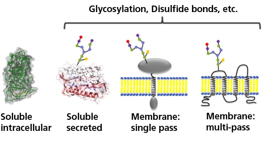
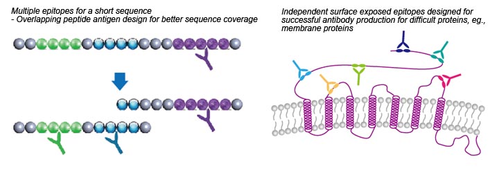
.jpg)
.jpg)
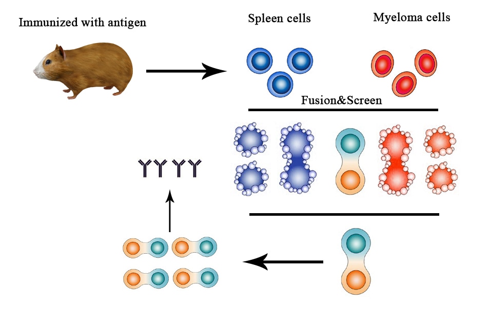
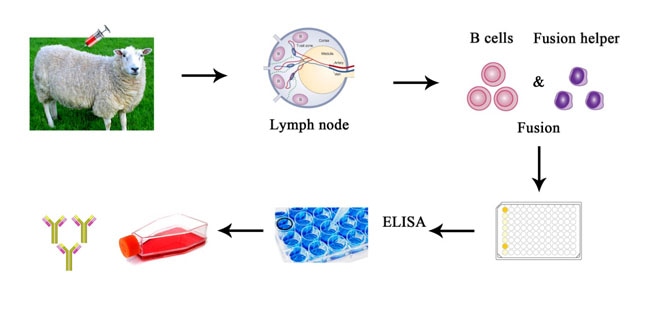

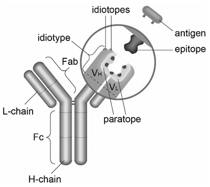
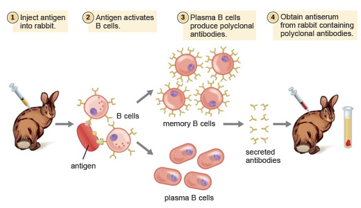
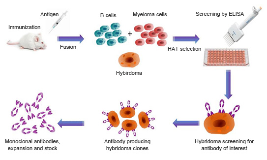
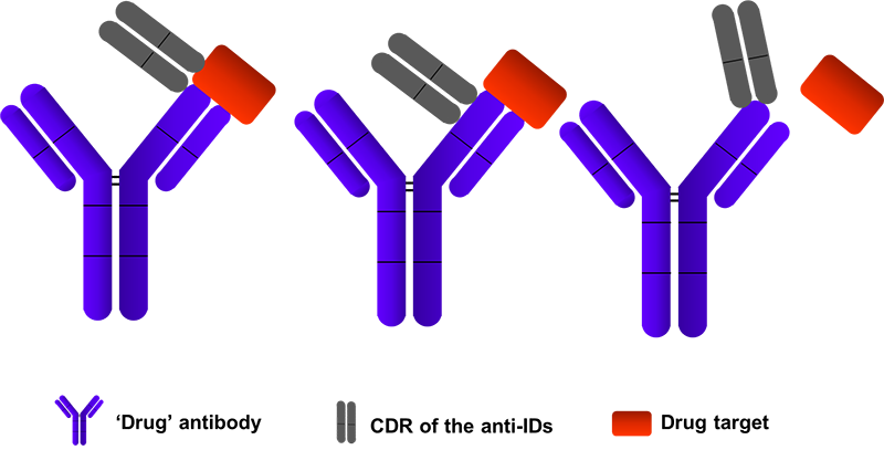

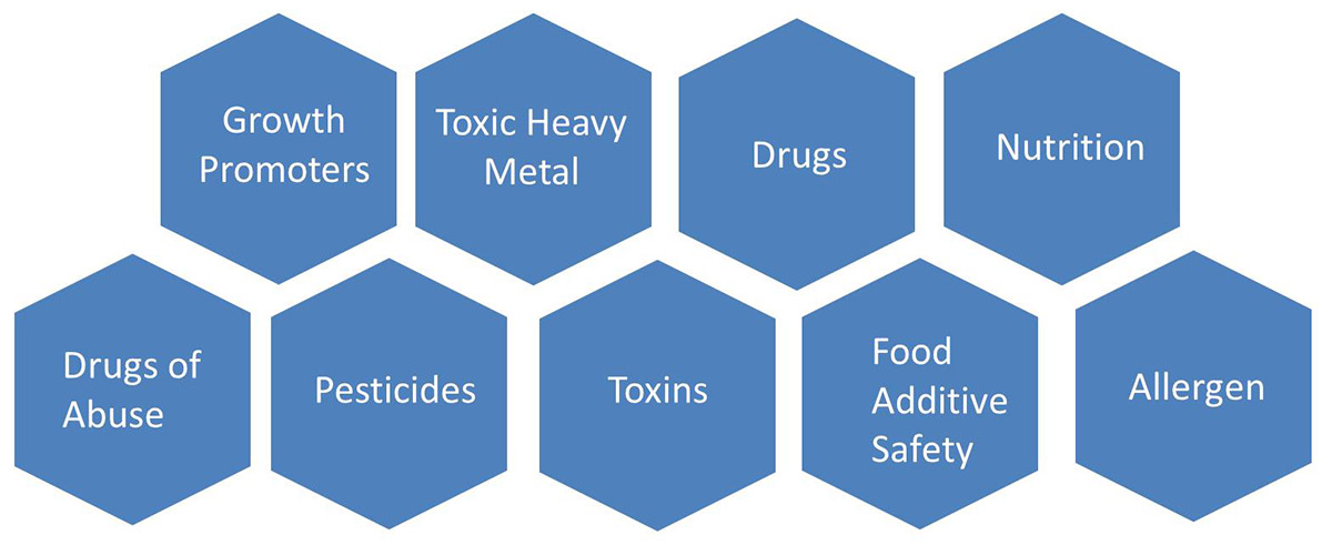
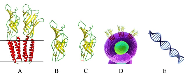
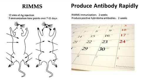
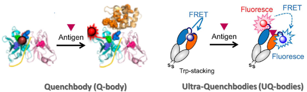
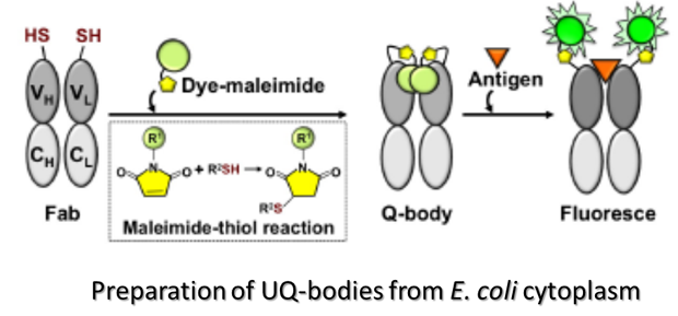
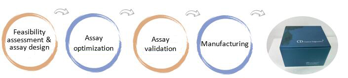
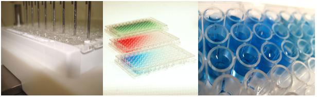
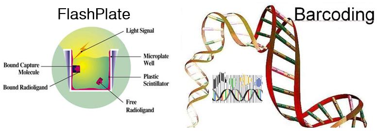
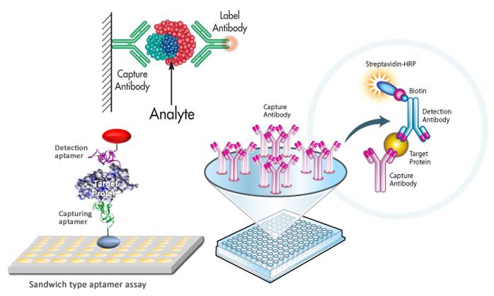
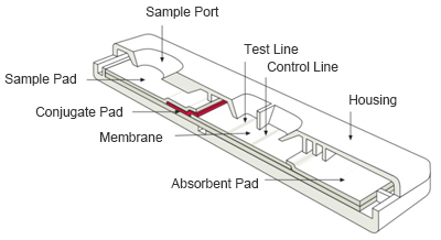
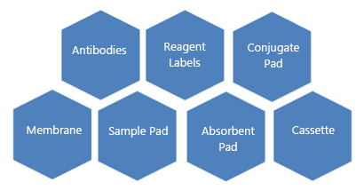
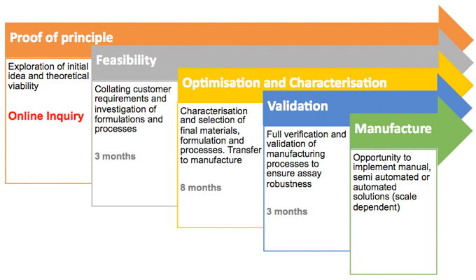
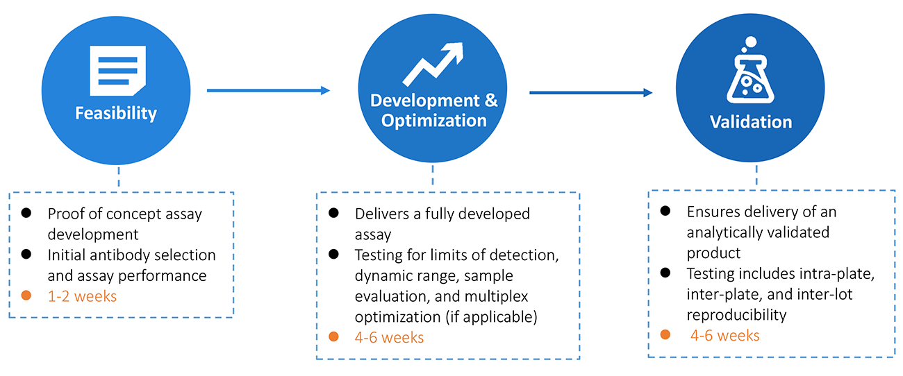
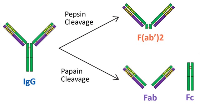
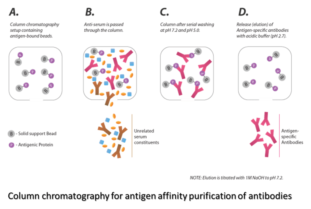
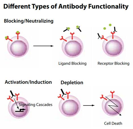
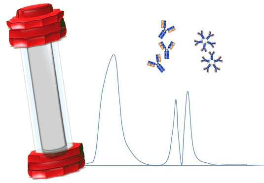
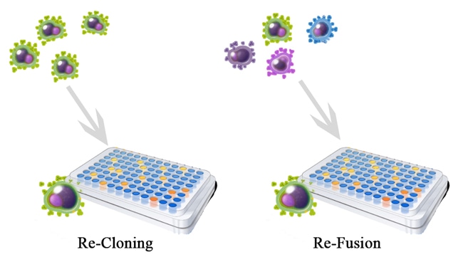
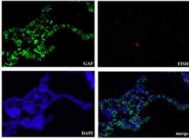
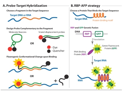
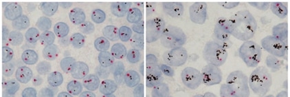

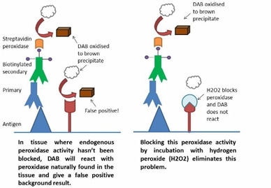
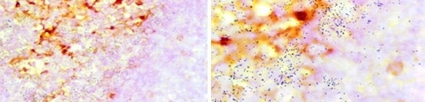
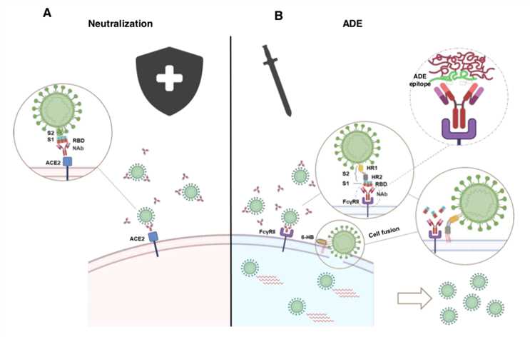 Figure 1: ADE activity generation. Adapted from: Wu, F., Yan, R., Liu, M., Liu, Z., Wang, Y., Luan, D., … & Huang, J. (2020). Antibody-dependent enhancement (ADE) of SARS-CoV-2 infection in recovered COVID-19 patients: Studies based on cellular and structural biology analysis. medRxiv.
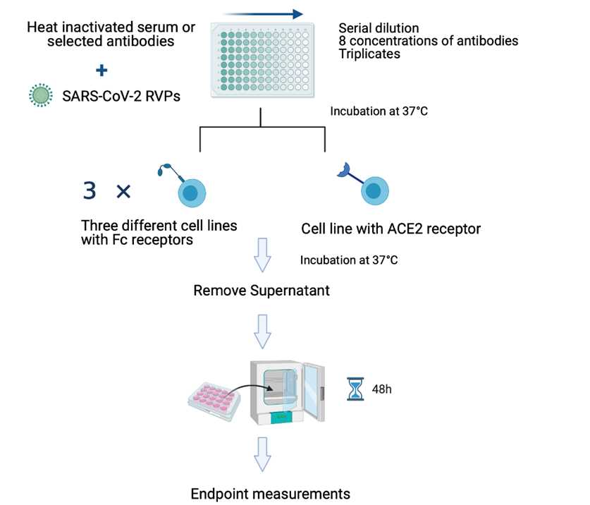
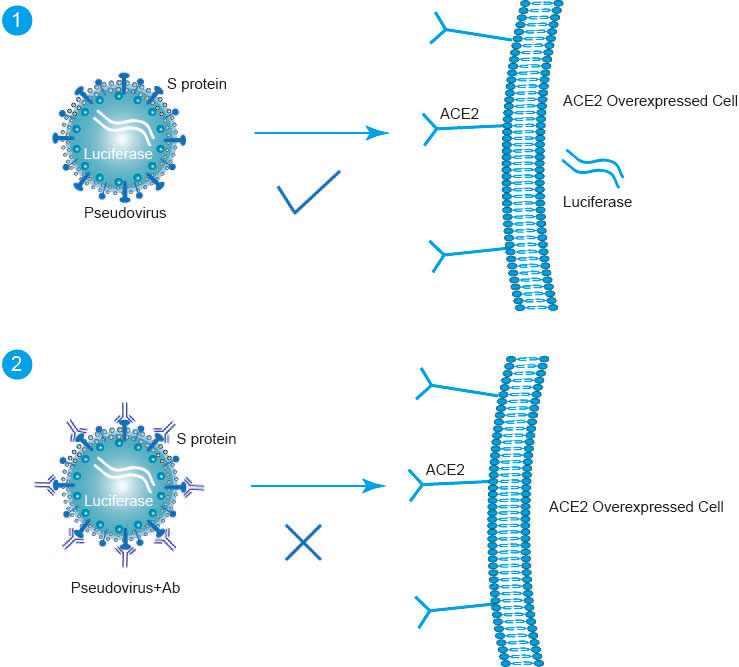 Fig1. Pseudotype-Based Neutralization Assays Principle