What is G protein-Coupled Receptor?
Transmembrane proteins (TP) is a type of membrane protein that spans one side of the membrane to the other. Transmembrane proteins perform many important cellular biological functions in the body. It is involved in the exchange of matter, energy and signals inside and outside the cell membrane. For example, transmembrane proteins can be receptors for a variety of signaling molecules, hormones, and other substrates, and can activate intra-membrane signaling pathways.
G protein-coupled receptor (GPCR) is a class of transmembrane proteins that detect extracellular molecules and activate internal signal transduction pathways that ultimately activate cellular responses.
G protein-coupled receptors are also known as seven alpha-helices transmembrane segment receptors (7TM receptors). They mediate signals involving visual control, renal function, tumorigenesis, immune response, and inflammation.
Classification of G protein-Coupled Receptor (GPCR)
GPCRs can be divided into six categories based on sequence homology:
Class A: Rhodopsin-like receptor
Class B: Secretin receptor family
Class C: Metabolic glutamate receptor
Class D: Fungal mating pheromone receptor
Class E: Cyclic adenosine receptor
Class F: Frizzled/Smoothened family
Class A accounts for nearly 85% of the GPCR gene and is currently the largest.
In a new classification system, namely GRAFS classification system, GPCR is divided into five categories: glutamate, Rhodopsin, Adhesion, Frizzled/Taste2, Secretin).
G protein-Coupled Receptor (GPCR) ligand
The ligand binds to the G-protein-coupled receptor, activates the G-protein, and transmits the signal to the cell, eventually activating the cell reaction. More than 100 ligands are known to achieve transmembrane signaling through G-protein coupled receptors, including: Biogenic amines (epinephrine, norepinephrine, histamine, 5-hydroxytryptamine); Peptide hormones (bradykinin, luteinizing hormone, parathyroid hormone); Odor molecules; photons.
Physiological effects of G protein-Coupled Receptor (GPCR)
Vision: Photosensitive G-protein coupled receptor rhodopsin can converts electromagnetic radiation into cellular signals and initiates downstream signaling processes.
Taste: GPCRs can mediate the release of bitter, umami and sweet substances by gustducin.
Olfactory: Olfactory epithelium in the nasal cavity and olfactory receptors on the vomeronasal can sense odor molecules and pheromones.
Physiological effects of GPCRs also include: behavioral and mood regulation; regulation of the immune system; regulation of the autonomic nervous system; detection and regulation of cell density.
G protein-Coupled Receptor (GPCR)and drug development
Major human diseases such as cancer, AIDS, heart disease, diabetes, Alzheimer’s disease, Parkinson’s disease, dwarfism, dwarfism, color blindness, retinitis pigmentosa and asthma are closely related to GPCRs. Therefore, GPCR is also a “star” in the field of drug development.
Currently, about 40% of marketed drugs are designed based on GPCRs. At least one-third of small molecule drugs in the world’s pharmaceutical market are currently activators or antagonists of GPCRs. Twenty percent of the top 50 best-selling drugs currently on the market are reportedly G-receptor-related drugs.
For more details about GPCRs, for example, the structure, history, etc., please read the article: G protein-Coupled Receptor – Targets for Drug Therapy.
The important role of GPCR in the drug target protein determines its importance in the life sciences. CUSABIO can provide the products you need for your research. Currently, we could product 183 GPCR-related products: 70 protein products, 89 antibody products, 24 ELISA products.
Hot Products


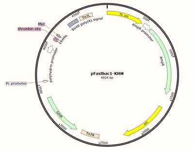
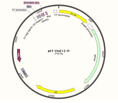 Vector characteristics:
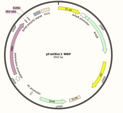
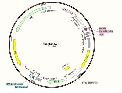

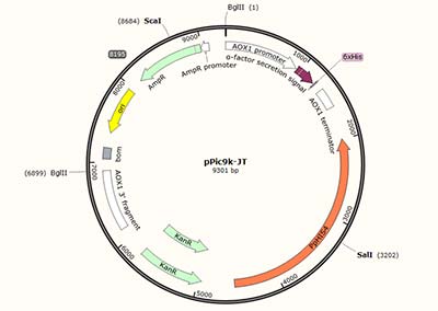
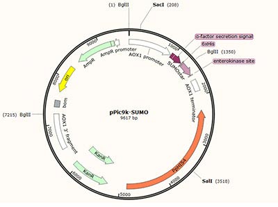
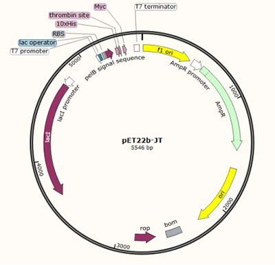 Vector characteristics:
 Vector characteristics: