- Target
This method is a guide to use the Elabscience® RTU antibody for IHC correctly with BOND™MAX (Leica) for immunohistochemical operation and obtain the optimal results.
- Limits
This method is suitable for the automatic immunohistochemical staining experiment on BOND™ MAX (Leica) instrument with the Elabscience® RTU antibody for IHC.
- Experimental Steps
1) Reagents and Consumables
1.1 Transparent dewaxing solution.
1.2 Absolute ethanol.
1.3 95% ethanol.
1.4 PBS Buffer.
1.5 Tween-20.
1.6 Immunohistochemical oil pen.
1.7 Bond™ Dewax Solution. Cat:AR9222, Leica.
1.8 Bond™ Wash Solution 10 x concentrate. Cat: AR9590, Leica.
1.9 Bond™ Epitope Retrieval Solution 1. Cat: AR9961, Leica.
1.10 Bond™ Epitope Retrieval Solution 2. Cat: AR9640, Leica.
(Please choose the antigen repair solutions for antigen repair according to the Elabscience® Antibody manual)
1.11 Bond™ Polymer Refine Detection. Cat: DS9800, Leica.
1.12 Bond™ Titration kit: (The Bond Titration Kit contains Bond Titration Container Inserts and Bond Titration Containers)Hematoxylin Staining Solution. Cat: OPT9049, Leica.
1.13 Bond™ Aspirating Probe Cleaning. Cat: CS9100, Leica.
1.14 Bond™ Printer Ribbon & Labels Cxi (6Pack) Cat: S21.4610.A, Leica.
1.15 Bond™ Universal Covertiles. Cat: S21.2001, Leica.
1.16 Neutral Balsam.
2) Instruments
2.1 Automatic Immunohistochemical Staining System: BOND™ MAX, Leica.
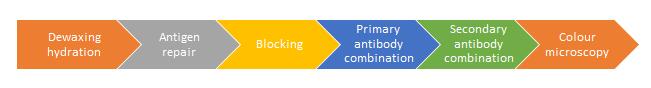
3) Staining Procedure
Ensure the type and quantity of tissue sections and reagents, set the “Reagent Setting” step, the “Slide Setting” step, the “Program Setting” step and other related settings through the software, determine the staining operation procedure, print the label, paste the label on the corresponding tissue slices, put the labeled slices and reagents into the staining machine for quality inspection, and enter the staining after passing the quality inspection. The procedure is as follows:
3.1. Bake and Dewax Protocol
3.1.1 Heat the slice to 60°C and bake for 30 min.
3.1.2 Heat the slice to 72°C, add 150 µL Bond Dewax Solution on it and incubate for 30 s, then discard the solution.
3.1.3 Maintained at 72°C, add 150 µL Bond Dewax Solution to wash the slice, then discard the solution.
3.1.4 Cool the slice to RT, add 150 µL Bond Dewax Solution to wash the slice, then discard the solution.
3.1.5 Add 150 μL alcohol to wash the slice and discard the alcohol at RT, wash for 3 times.
3.1.6 Add 150 µL Bond Wash Solution and wash the liquid at RT, wash for twice.
3.1.7 Add 150 µL Bond Wash Solution and incubate for 5 min at RT, then discard the solution.
3.2. HIER 20 min with ER1/ER2 Protocol
3.2.1 Add 150 µL ER1 or ER2 to wash the slice at RT.
3.2.2 Add 150 µL ER1 or ER2 at RT then heat to 100°C and incubate for 20 min.
3.2.3 Cool the slice to RT, and incubate for 20 min.
3.2.4 Heat the slice to 35°C, add 150 µL Bond Wash Solution to wash the slice, then discard the solution.
3.2.5 Cool the slice to RT, add 150 µL Bond Wash Solution and incubate for 3 min, then discard the solution.
3.3. IHC Protocol
3.3.1 Add 150 µL Peroxide Block on the slice and incubate for 5 min at RT.
3.3.2 Add 150 µL Bond Wash Solution to wash the slice then discard the solution, wash for 3 times.
3.3.3 Add 150 µL primary antibody from Elabsciecne and incubate for 30 min at RT.
3.3.4 Add 150 µL Bond Wash Solution to wash the slice then discard the solution, wash for 3 times.
3.3.5 Add 150 µL Post Primary to the slice and incubate for 8 min at RT, then discard the Post primary.
3.3.6 Add 150 µL Bond Wash Solution to wash the slice then discard the solution, wash for 3 times.
3.3.7 Add 150 µL Polymer to the slice and incubate for 8 min at RT, then discard the Polymer.
3.3.8 Add 150 µL Bond Wash Solution to wash the slice then discard the solution, wash for twice.
3.3.9 Add 150 µL Deionized Water to wash the slice then discard the Deionized Water.
3.3.10 Add 150 µL Mixed DAB Refine to the slice and incubate for 10 min at RT, then discard the Mixed DAB Refine.
3.3.11 Add 150 µL Bond Wash Solution to wash the slice then discard the solution, wash for 3 times.
3.3.12 Add 150 µL Hematoxylin to the slice and incubate for 5 min at RT, then discard the Hematoxylin.
3.3.13 Add 150 µL Deionized Water to wash the slice.
3.3.14 Add 150 µL Bond Wash Solution to wash the slice.
3.3.15 Add 150 µL Deionized Water to wash the slice.
3.4. Dehydration and Transparency
3.4.1 At the end of the staining procedure, take the slice and discard the Deionized Water.
3.4.2 Immerse the slice in 75% ethanol for 30 s/time, 2 times
3.4.3 Immerse the slices in 95% ethanol for 30 s/time, 2 times.
3.4.4 Immerse the slices in absolute ethanol for 30 s/time, 2 times.
3.4.5 Immerse the slices in transparent dewaxing solution for 5 min
Tips: Drain every time when changing the solution (about 5 s), and then put it into the next solution.
3.5. Seal the Slices
3.5.1 Take out the slices and drain them (about 5 s), blow them dry with cold air by blower.
3.5.2 Add 1~2 drops of neutral balsam to the slice (depending on the number and size of the slice), and cover the slide.
3.5.3 Check the slice to make sure that the slice tissue is completely covered with neutral balsam, and there is no big bubble in the tissue (discharge big bubble by squeezing the cover glass).
3.5.4 Place slices in fume hood to dry.
4). Observe the slice by microscope and judge the result.
- Notices
1) This method is for Elabscience® RTU Antibody for IHC applicable on the Autostainer BOND MAX with matched immunohistochemical staining system. Due to the different kinds / targets of Antibody for IHC, the antigen repair methods may differ, please read the manual carefully and strictly follow the manual.
2) This method recommends the varieties / Models / manufacturers of the important related reagents in the process of immunohistochemistry experiment. If users change the corresponding reagents / raw materials, you need to evaluate the equivalence of the alternative varieties and seek the optimal operating procedures.
3) This method is for Elabscience® RTU Antibody for IHC applicable on the Autostainer BOND MAX with matched immunohistochemical staining system. If the user changes the immunohistochemical staining system, please evaluate the equivalence of the alternative system and find the optimal operation procedure.