Recombinant Mouse Monoclonal Antibody to Glypican-3 (GPC3) (Hepatocellular Carcinoma Marker)(Clone : rGPC3/863)
Catalogue Number: 12-1003-ABO
12-1003 Image
12-1003 Image
12-1003 Image
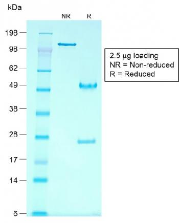
Figure 2: SDS-PAGE Analysis of Purified Glypican-3 Mouse Recombinant Monoclonal Antibody (rGPC3/863).
12-1003 Image
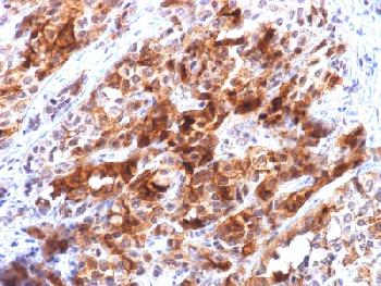
Figure 1: Formalin-fixed, paraffin-embedded human Hepatocellular Carcinoma stained with Glypican-3 Mouse Recombinant Monoclonal Antibody (rGPC3/863).
| Manufacturer: | Abeomics |
| Type: | Recombinant Monoclonal |
| Alias: | DGSX, Glypican proteoglycan 3, GPC3, GTR2-2, Heparan sulphate proteoglycan, Intestinal protein OCI-5, MXR7, OCI-5, SDYS, Secreted glypican-3, SGBS1 |
| Shipping Condition: | Blue Ice |
| Unit(s): | 100 ug, 20 ug |
| Host name: | |
| Clone: | rGPC3/863 |
| Isotype: | IgG1, kappa |
| Immunogen: | Recombinant full-length human GPC3 protein |
| Application: | FACS, IF, IHC |
Description
Description: Glypican-3 (GPC3) is a glycosylphospatidyl inositol-anchored membrane protein, which may also be found in a secreted form. Anti-GPC3 has been identified as a useful tumor marker for the diagnosis of hepatocellular carcinoma (HCC), hepatoblastoma, melanoma, testicular germ cell tumors, and Wilm s tumor. In patients with HCC, GPC3 is overexpressed in neoplastic liver tissue and elevated in serum, but is undetectable in normal liver, benign liver, and the serum of healthy donors. GPC3 expression is also found to be higher in HCC liver tissue than in cirrhotic liver or liver with focal lesions such as dysplastic nodules and areas of hepatic adenoma (HA) with malignant transformation. In the context of testicular germ cell tumors, GPC3 expression is up regulated in certain histologic subtypes, specifically yolk sac tumors and choriocarcinoma. A high level of GPC3 expression is also found in some types of embryonal tumors, such as Wilm s tumor and hepatoblastoma, with a low or undetectable expression in normal adjacent tissue. In patients with thyroid cancer, expression of GPC3 is dramatically enhanced in certain types of cancers: 100% in follicular carcinoma and 70% in papillary carcinoma. Expression of GPC3 in follicular carcinoma is significantly higher than that of follicular adenoma. In contrast, GPC3 is not expressed in anaplastic carcinoma.
Additional Text
Gene Name
GPC3
Gene ID
2719
Uniprot ID
P51654
Purification
Purified
Application Notes
Flow Cytometry (1-2µg/million cells); Immunofluorescence (1-2µg/ml); Immunohistochemistry (Formalin-fixed) (1-2µg/ml for 30 minutes at RT)(Staining of formalin-fixed tissues requires heating tissue sections in 10mM Tris with 1mM EDTA, pH 9.0, for 45 min at 95°C followed by cooling at RT for 20 minutes)
Antibody Clonality
Monoclonal, Recombinant Monoclonal
SUPPORT
outstanding technical support
PRODUCT
we offer a full product guarantee
DELIVERY
we offer free delivery to UK universities and non profit organisations