Anti-CD1a / HTA1 (Mature Langerhans Cells Marker) Recombinant Mouse Monoclonal Antibody (Clone:rC1A/711)
Catalogue Number: 12-1098-ABO
12-1098 Image
12-1098 Image
12-1098 Image
12-1098 Image
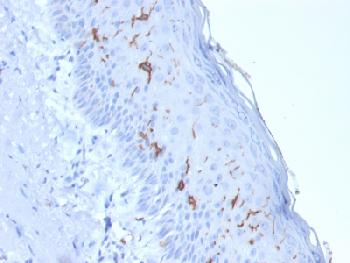
Figure 1: Formalin-fixed, paraffin-embedded human Skin stained with CD1a Mouse Recombinant Monoclonal Antibody (rC1A/711).
12-1098 Image
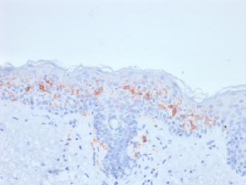
Figure 2: Formalin-fixed, paraffin-embedded human Skin stained with CD1a Mouse Recombinant Monoclonal Antibody (rC1A/711).
12-1098 Image
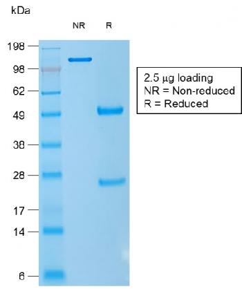
Figure 3:SDS-PAGE Analysis of Purified CD1a Mouse Recombinant Monoclonal Antibody (rC1A/711).
| Manufacturer: | Abeomics |
| Shelf Life: | 24 months |
| Type: | Recombinant Monoclonal |
| Alias: | Cortical thymocyte antigen CD1A, Epidermal dendritic cell marker CD1a antibody, FCB6, HTA1, T cell surface antigen T6 / Leu 6, T-Cell Surface Glycoprotein CD1A |
| Shipping Condition: | Blue Ice |
| Unit(s): | 100 ug, 20 ug |
| Host name: | Mouse |
| Clone: | rC1A/711 |
| Isotype: | IgG1, kappa |
| Immunogen: | Recombinant full-length human CD1a protein |
| Application: | IHC |
Description
Description: At least five CD1 genes (CD1a, b, c, d, and e) are identified. CD1 proteins have been demonstrated to restrict T cell response to non-peptide lipid and glycolipid antigens and play a role in non-classical antigen presentation. CD1a is a non-polymorphic MHC Class 1 related cell surface glycoprotein, expressed in association with Beta-2 microglobulin. Anti-CD1a labels Langerhans cell histiocytosis (Histiocytosis X), extranodal histiocytic sarcoma, a subset of T-lymphoblastic lymphoma/leukemia, and interdigitating dendritic cell sarcoma of the lymph node. When combined with antibodies against TTF-1 and CD5, anti-CD1a is useful in distinguishing between pulmonary and thymic neoplasms since CD1a is consistently expressed in thymic lymphocytes in both typical and atypical thymomas, but only focally in 1/6 of thymic carcinomas and not in lymphocytes in pulmonary neoplasms. Anti-CD1a is reported to be a new marker for perivascular epithelial cell tumor (PEComa).
Additional Text
Gene Name
CD1A
Purification
Protein A/G Purified
Gene ID
909
Uniprot ID
P06126
Application Notes
Immunohistochemistry (Formalin-fixed) (1-2µg/ml for 30 min at RT)(Staining of formalin-fixed tissues requires heating tissue sections in 10mM Tris with 1mM EDTA, pH 9.0, for 45 min at 95°C followed by cooling at RT for 20 minutes)
Antibody Clonality
Monoclonal, Recombinant Monoclonal
SUPPORT
outstanding technical support
PRODUCT
we offer a full product guarantee
DELIVERY
we offer free delivery to UK universities and non profit organisations