Foxp3/Transcription Factor Staining Kit
Catalogue Number: 17-1014-ABO
17-1014 Image
17-1014 Image
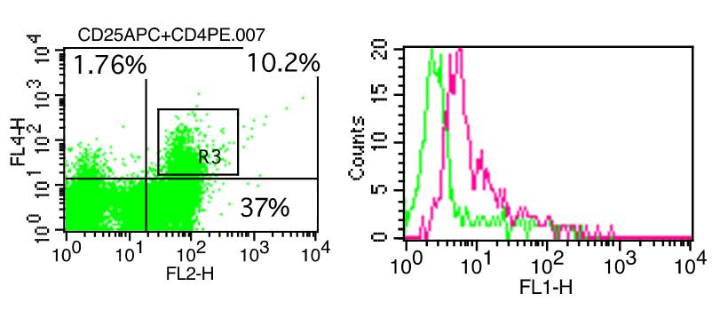
Figure-1: Human PBMC were stained with FoxP3/Transcription Factor staining kit (17-1014). Anti-CD4 PE and anti-CD25 APC positive cells were gated, further analyzed in anti-FoxP3 FITC (10-4074F) using 2 µg antibodies. Green represents FITC conjugated Isotype control (ABEOMICS). Red represents anti-FoxP3 FITC.
| Manufacturer: | Abeomics |
| Type: | Flow Cytometry Kit |
| Shipping Condition: | Blue Ice |
| Storage Condition: | 2-8°C |
| Unit(s): | 1 kit |
| Application: | FACS |
Description
Description: The FoxP3 / Transcription Factor Staining Kit has optimized for staining with antibodies to transcription factors and nuclear proteins, such as FoxP3. This kit can be used for staining cytokines and chemokines.
Additional Text
Application Notes
Protocol: FoxP3 FITC staining with CD4 PE and CD25 APC antibodies. 1. Determine number of cells required for staining. Each sample contains 0.5 -1 x 10^6 cells in 50µl of media or staining buffer. The following controls are needed for the experiment. Unstained cells (no antibodies were added), cells with isotype control and cells with secondary antibody (if secondary antibody was used). 2. Centrifuge cells at 1000 RPM for 10 minutes and decant supernatant. 3. Resuspend cells with appropriate volume of staining buffer. 4. Aliquote 1x10^6 cells in 50 µl to the desired number of flow tubes. Dilute anti-CD4 PE, anti-CD25 APC cell surface antibodies in 50 µl of 1X staining buffer. Add diluted antibodies into 50 µl of cell suspension. Mix antibodies in cells suspension thoroughly. 5. Incubate in ice for 30 minutes in ice pritected from light. Wash cells in 2-3 ml of 1X staining buffer. Centrifuge 1000 RPM for 10 minutes. Decant supernatant carefully. 6. Add 1ml of freshly prepared 1X fixation/permeabilization buffer. Mix well. 7. Incubate in ice for 30 minutes protected from light. 8. Whsh cells in 3-4 ml of 1X permeabilization buffer. Centrifuge at 1000 RPM for 10 minutes. Decant supernatant carefully. 9. Add 2% mouse serum to the cells in 50 µl of 1X permeabilization buffer (for blocking non-specific binding). Incubate 10 minutes in ice. (Optional) 10. Add fluorochrome conjugated Anti-FoxP3 antibody or isotype control in 50 µl of 1X permeabilization buffer. (Without washing blocking step). 11. Incubate in ice for 30 minutes, protected from light. Wash cells 3-4 ml of 1X permeabilization buffer. Centrifuge 1000 RPM for 10 minutes. Decant supernatant carefully. 11. After decanting, add 300-400 µl of staining buffer to each tube. If not analyzing same day, samples can be stored over night in dark at 4 degrees C. Samples can be analyzed in Flow Cytometer according to the manufacturer protocol.
SUPPORT
outstanding technical support
PRODUCT
we offer a full product guarantee
DELIVERY
we offer free delivery to UK universities and non profit organisations