Phosphothreonine Antibody
Catalogue Number: ASM10404-A700-ABG
ASM10404-A700 Image
ASM10404-A700 Image
ASM10404-A700 Image
ASM10404-A700 Image
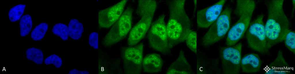
Immunocytochemistry/Immunofluorescence analysis using Rabbit Anti-Phosphothreonine Polyclonal Antibody (ASM10404). Tissue: HeLa Cells. Species: Human. Fixation: 2% Formaldehyde for 20 min at RT. Primary Antibody: Rabbit Anti-Phosphothreonine Polyclonal Antibody (ASM10404) at 1:60 for 12 hours at 4°C. Secondary Antibody: FITC Goat Anti-Rabbit (green) at 1:200 for 2 hours at RT. Counterstain: DAPI (blue) nuclear stain at 1:40000 for 2 hours at RT. Localization: Cytoplasm. Nucleus. Magnification: 100x. (A) DAPI (blue) nuclear stain. (B) Anti-Phosphothreonine Antibody. (C) Composite.
ASM10404-A700 Image
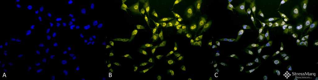
Immunocytochemistry/Immunofluorescence analysis using Rabbit Anti-Phosphothreonine Polyclonal Antibody (ASM10404). Tissue: HeLa Cells. Species: Human. Fixation: 2% Formaldehyde for 20 min at RT. Primary Antibody: Rabbit Anti-Phosphothreonine Polyclonal Antibody (ASM10404) at 1:60 for 12 hours at 4°C. Secondary Antibody: R-PE Goat Anti-Rabbit (yellow) at 1:200 for 2 hours at RT. Counterstain: DAPI (blue) nuclear stain at 1:40000 for 2 hours at RT. Localization: Cytoplasm. Nucleus. Magnification: 20x. (A) DAPI (blue) nuclear stain. (B) Anti-Phosphothreonine Antibody. (C) Composite.
ASM10404-A700 Image
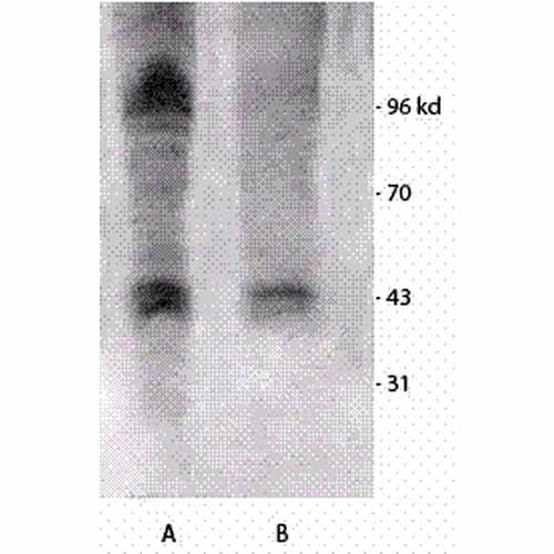
Western blot analysis of Mouse brain cell lysates showing detection of Phosphothreonine protein using Rabbit Anti-Phosphothreonine Polyclonal Antibody (ASM10404). Primary Antibody: Rabbit Anti-Phosphothreonine Polyclonal Antibody (ASM10404) at 1:1000. Left: Treated with Vanadium, Right: Non-treated.
| Manufacturer: | Abcepta |
| Type: | Primary Antibody |
| Shipping Condition: | Blue Ice |
| Unit(s): | each |
| Host name: | |
| Clone: | |
| Isotype: | |
| Immunogen: | Phosphothreonine conjugated to KLH |
| Application: | WB |
Additional Text
Purification
Protein A purified
Storage Note
-20C
SUPPORT
outstanding technical support
PRODUCT
we offer a full product guarantee
DELIVERY
we offer free delivery to UK universities and non profit organisations