AHA1 Antibody
Catalogue Number: ASM10442-A700-ABG
ASM10442-A700 Image
ASM10442-A700 Image
ASM10442-A700 Image
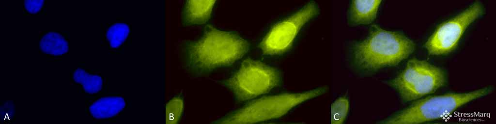
Immunocytochemistry/Immunofluorescence analysis using Rabbit Anti-AHA1 Polyclonal Antibody (ASM10442). Tissue: Heat Shocked HeLa Cells. Species: Human. Fixation: 2% Formaldehyde for 20 min at RT. Primary Antibody: Rabbit Anti-AHA1 Polyclonal Antibody (ASM10442) at 1:60 for 12 hours at 4°C. Secondary Antibody: R-PE Goat Anti-Rabbit (yellow) at 1:200 for 2 hours at RT. Counterstain: DAPI (blue) nuclear stain at 1:40000 for 2 hours at RT. Localization: Cytoplasm. Endoplasmic reticulum. Magnification: 100x. (A) DAPI (blue) nuclear stain. (B) Anti-AHA1 Antibody. (C) Composite. Heat Shocked at 42°C for 1h.
ASM10442-A700 Image
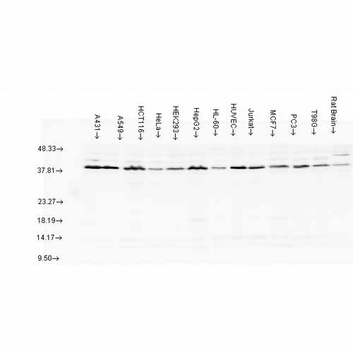
Western blot analysis of multiple Cell line lysates showing detection of AHA1 protein using Rabbit Anti-AHA1 Polyclonal Antibody (ASM10442). Load: 15 µg protein. Block: 1.5% BSA for 30 minutes at RT. Primary Antibody: Rabbit Anti-AHA1 Polyclonal Antibody (ASM10442) at 1:1000 for 2 hours at RT. Secondary Antibody: Donkey Anti-Rabbit IgG: HRP for 1 hour at RT.
| Manufacturer: | Abcepta |
| Type: | Primary Antibody |
| Shipping Condition: | Blue Ice |
| Unit(s): | each |
| Host name: | |
| Clone: | |
| Isotype: | |
| Immunogen: | Mouse Aha1 |
| Application: | WB |
Additional Text
Gene Name
AHSA1
Uniprot ID
Q8BK64
Gene ID
217737
Purification
Protein A purified
Accession Number
NP_666148.1
Storage Note
-20C
SUPPORT
outstanding technical support
PRODUCT
we offer a full product guarantee
DELIVERY
we offer free delivery to UK universities and non profit organisations