WIPI2 Antibody
Catalogue Number: ASM10525-A680-ABG
ASM10525-A680 Image
ASM10525-A680 Image
ASM10525-A680 Image
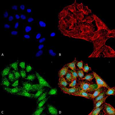
Immunocytochemistry/Immunofluorescence analysis using Rabbit Anti-WIPI2 Polyclonal Antibody (ASM10525). Tissue: HeLa Cells (Human Cervical Cancer). Species: Human. Fixation: 4% Formaldehyde for 15 min at RT. Primary Antibody: Rabbit Anti-WIPI2 Polyclonal Antibody (ASM10525) at 1:100 for 60 min at RT. Secondary Antibody: Goat Anti-Rabbit ATTO 488 at 1:200 for 60 min at RT. Counterstain: Phalloidin Texas Red F-Actin stain; DAPI (blue) nuclear stain at 1:1000, 1:5000 for 60 min at RT, 5 min at RT. Localization: Cytoplasm . Magnification: 60X. (A) DAPI (blue) nuclear stain (B) Phalloidin Texas Red F-Actin stain (C) WIPI2 Antibody (D) Composite.
ASM10525-A680 Image
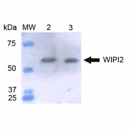
Western blot analysis of Human HeLa and 293Trap cell lysates showing detection of ~49.4 kDa WIPI2 protein using Rabbit Anti-WIPI2 Polyclonal Antibody (ASM10525). Lane 1: Molecular Weight Ladder (MW). Lane 2: HeLa cell lysates. Lane 3: 293Trap cell lysates. Load: 15 µg. Block: 5% Skim Milk in 1X TBST. Primary Antibody: Rabbit Anti-WIPI2 Polyclonal Antibody (ASM10525) at 1:1000 for 2 hours at RT. Secondary Antibody: Goat Anti-Rabbit IgG: HRP at 1:1000 for 60 min at RT. Color Development: ECL solution for 6 min in RT. Predicted/Observed Size: ~49.4 kDa.
| Manufacturer: | Abcepta |
| Type: | Primary Antibody |
| Shipping Condition: | Blue Ice |
| Unit(s): | each |
| Host name: | |
| Clone: | |
| Isotype: | |
| Immunogen: | Synthetic peptide from the C-terminal of Human WIPI2 |
| Application: | WB |
Additional Text
Gene Name
WIPI2
Gene ID
26100
Uniprot ID
Q9Y4P8
Accession Number
NP_001028690.1
Purification
Affinity Purified
Storage Note
-20C