Extracellular Drug Conjugates Therapeutically Exploit Protein Proximity

Pharmaceutical company Centrose, founded by James R. Prudent, Ph.D., developed a new class of antibody drug conjugates called extracellular drug conjugates. Nature Publishing Group published the research as a open access paper in its Molecular Therapy journal.1 Apart from the interesting and important development of a new class of antibody drug conjugate (ADC), the research also showed how important linker length 2 is to the potency and specificity of the EDC.

How Extracellular Drug Conjugates Work

Extracellular drug conjugates (EDCs) are designed similarly to ADCs. That is, EDCs consist of a monoclonal antibody (mAb), a linker, and a cytotoxic agent. Antibody drug conjugates require internalization into a diseased cell where the cytotoxic agent can then be released to act on its target.3 The cytotoxic agent may require intracellular modification or degradation to act on its target molecule.

By contrast, extracellular drug conjugates require no internalization. Rather, EDCs target cell surface proteins that are expressed on a target (i.e., cancerous) cell. Moreover, the cytotoxic agent that is linked to the mAb does not target the same protein targeted by the mAb. Rather, the cytotoxic agent kills the targeted cells by affecting a protein or enzyme that is different from the protein or enzyme bound by the mAb but that is closely associated with the target protein or enzyme.

In this particular study, the research team observed that some proteins that are overexpressed on the surface of cancer cells are closely associated with the sodium potassium ATPase1 (NKA, and also known as the sodium potassium pump; see Figure 1). Cardiac glycosides such as digoxin or ouabain inhibit the NKA. Cells affected by these or other cardiac glycosides swell, then undergo necrotic cell death.1,4,5 The team reasoned that by (1) combining antibodies to proteins that are both (a) overexpressed on the cell surface of cancer cells and (b) closely associated with the NKA (2) with cardiac glycosides that strongly inhibit the NKA (3) would create cancer therapeutic antibody drug conjugates that were localized to the extracellular space, hence, Extracellular Drug Conjugates.

Creating the Extracellular Drug Conjugates in this Study

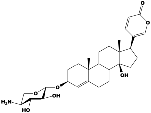
Through prior testing (not in this paper) the team found that the cardiac glycoside scillarenin β-L-aminoxyloside (Figure 1) highly inhibited the NKA. This was chosen as the drug for conjugation to the antibody.
The team also developed or acquired nine (9) mAbs. For directly testing the EDCs, the mAbs had to meet one of the following three criteria:

  1. was a marker for metastatic cancer commonly known to associate with the NKA;
  2. was cancer related and thought to associate with the NKA; or
  3. was found by the current study to associate with the NKA and was a current cancer antibody drug target.
Figure 1: Scillarenin β-L-aminoxyloside, the cardiac glycoside used in this study as the cytotoxic agent for the extracellular drug conjugates.

As controls, the research team selected mAbs to proteins that were expressed on the cell surface but did not associate with the NKA or that were not expressed on any cell surface.

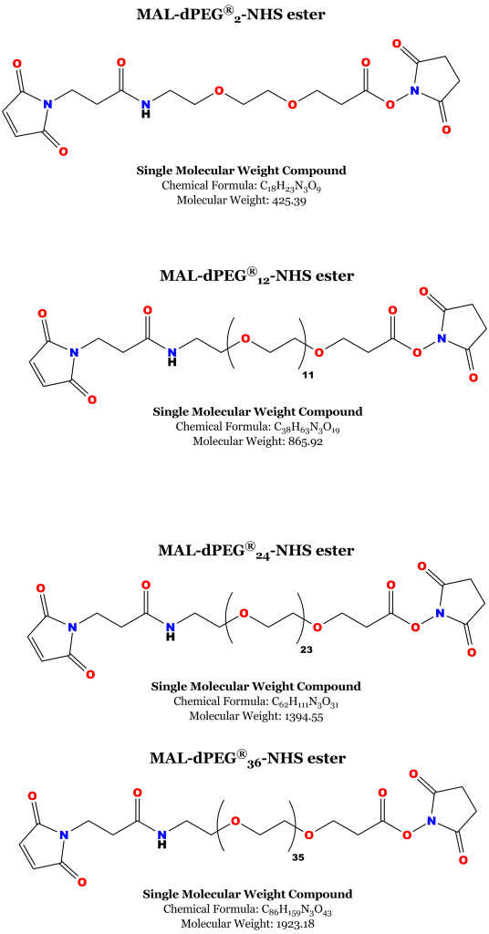
The researchers also investigated the effect of linker length between the mAb and the drug (abbreviated CG1) using Quanta BioDesign’s MAL-dPEG®n-NHS esters. These versatile heterobifunctional linkers come in a variety of specific lengths and are single molecular weight PEG derivatives (i.e., they have no dispersity). The maleimidopropyl group on one end reacts with free sulfhydryl groups forming a thioether linkage, while the NHS ester group on the other end will react with free amines to form a peptide bond. Four lengths of PEG — n = 2, 12, 24, and 36 dPEG® units (27, 56, 105, 144 Angstroms) — were chosen to connect CG1 to the EDC. See Figure 1. Although Figure 1 shows a single CG1-dPEG®n conjugated to the mAb, calculations by Centrose showed that the average EDC had a DAR of four (4). See reference 1, page 5.

Figure 2: MAL-dPEG®n-NHS-ester linkers used to construct the extracellular drug conjugates used in this study.

Extracellular Drug Conjugates Demonstrate In Vitro Efficacy…

The research team examined the efficacy of the extracellular drug conjugates after conjugating CG1 (the cardiac glycoside) to the mAb for dysadherin (a protein marker associated with metastatic cancer). They also measured the efficacy and toxicity of CG1 by itself or conjugated to one of the dPEG® linkers but not conjugated to the EDC.

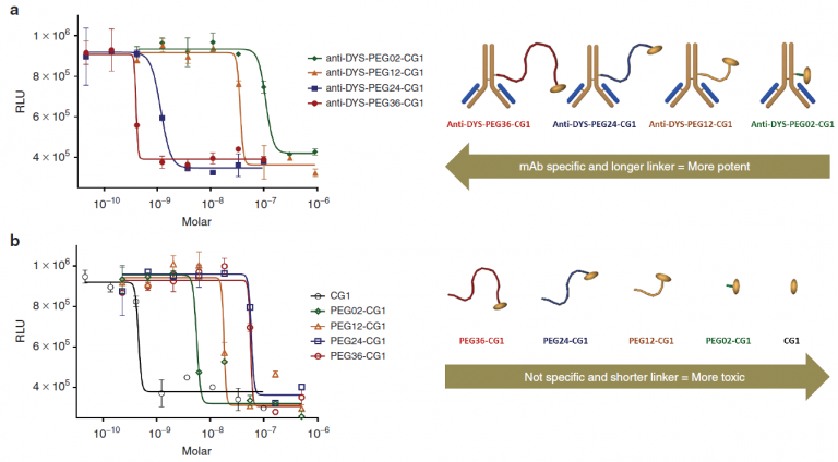
For the EDC-dPEG®n-CG1 conjugates, the Centrose team first measured antibody binding on the surface of the different cell lines. Then, by monitoring cell viability, they tested the cells’ sensitivity to the EDC-dPEG®n-CG1 conjugates at concentrations from 1 to 200,000 pmol/L. The dose-response curve in Figure 3, below, shows that increasing the linker length in EDC-dPEG®n-CG1 conjugates improved target specificity and potency. However, decreasing linker length in dPEG®n-CG1 constructs that were not conjugated to mAb increased toxicity and reduced specificity.

Figure 3: Dose-Response Curve and Potency-Specificity Graph for the Extracellular Drug Conjugates Based on Anti-dysadherin. Note that the potency and specificity increase with linker length for the mAb-dPEG®n-CG1 conjugates, but in the dPEG®n-CG1 constructs not conjugated to mAb, the specificity and toxicity increase as the linker length decreases.

Similar dose-response curves were obtained for some of the other tested cell lines. Cell lines expressing a cell surface antigen closely associated with the NKA were particularly sensitive to the EDC-dPEG®n-CG1 conjugates, but control cell lines (those either not expressing a cell surface antigen or expressing a cell surface antigen not associated with the NKA) were relatively insensitive to the EDC-dPEG®n-CG1 conjugates.

…And They Work In Vivo Also

The in vitro results also translated to in vivo studies in mice. In xenograft studies in mice bearing human pancreatic cancer tumors, EDC-DYS (EDC specific to dysadherin conjugated to dPEG®-CG1, with a DAR of 4) was compared to the standard dosing regimen of gemcitabine. EDC-DYS outperformed gemcitabine in a dose-dependent manner. Similarly, EDC-CD38 (a marker for various lymphomas and multiple myeloma) beat CHOP, a chemotherapy cocktail used as a standard treatment for Ramos B-cell lymphoma. Likewise, EDC-CD20 (another lymphoma marker) exceeded Rituximab’s performance. The control experiments showed that EDC-CONTROL conjugates (mAb targeted to antigens not expressed on the cell surface) did not reduce tumor size in mice.

Extracellular Drug Conjugates Offer New Therapeutic Options

EDCs are a new class of antibody drug conjugate, and they offer new, and potentially superior, therapeutic options for patients. Though similar in design and construction to a standard ADC, an EDC is different. The EDC always resides in the extracellular space, and it targets two cell surface proteins. These two features define the EDC. Neither the mAb nor the cytotoxic drug need to be internalized, released, or broken down in order to act. Many cells evolve to evade chemotherapy by rapidly exporting or neutralizing drugs that are released intracellularly. This unique EDC feature impedes cells in evolving resistance to the EDC.

These results show that EDCs are potentially useful in killing cancers that are resistant to multiple drugs, metastatic, and/or aggressive. Thus, in the future, EDCs may offer new therapeutic options for cancers that are otherwise rather difficult to treat.

Quanta BioDesign’s dPEG® Reagents Were Important to the Success of This Research

Quanta BioDesign’s maleimido-dPEG®n-NHS ester products were important to the success of this research on extracellular drug conjugates. Unlike traditional PEG derivatives, our dPEG® derivatives are single molecular weight compounds. We manufacture all of our products entirely in the USA by a patented, proprietary process. Our dPEG®s have no dispersity. Consequently, standard analytical techniques suffice for analyzing them to determine their purity. With traditional, dispersed PEGs, “purity” becomes a much more elusive term and is more difficult to measure.

References/Endnotes

  1. David J. Marshall, Scott C. Harried, John L. Murphy, et al. Extracellular Antibody Drug Conjugates Exploiting the Proximity of Two Proteins, Molecular Therapy advance online publication 19 July 2016; doi: 10.1038/mt.2016.119
  1. A linker is the component of the antibody drug conjugate that joins a monoclonal antibody (mAb) to a cytotoxic drug. An ideal linker is stable in circulation but should release the cytotoxic drug when the mAb reaches the target. “Linker length” refers to the distance between the mAb and the cytotoxic drug. Distance may be expressed in number of atoms or in units of Angstroms.
  1. For more information on how monoclonal antibodies and antibody drug conjugates work in cancer therapy go here and/or here.
  1. Menger, L, Vacchelli, E, Adjemian, S, Martins, I, Ma, Y, Shen, S et al. (2012). Cardiac glycosides exert anticancer effects by inducing immunogenic cell death. Sci Transl Med 4: 143ra99.

SUPPORT

outstanding technical support

PRODUCT

we offer a full product guarantee

DELIVERY

we offer free delivery to UK universities and non profit organisations