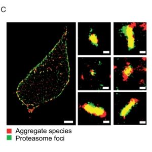
Small protein aggregates (below 450 nm) are highly toxic as they are able to penetrate the cell membrane.
Their small size makes these aggregates especially hard to study.
💡 With Amytracker, however, you can finally see them! 💡
Michael J. Morten and colleagues came up with a new approach that involves using Amytracker 630 and super-resolution microscopy to resolve aggregates involved in neurodegenerative disorders.
In their experiments, Amytracker 630 performed much better than conventional staining methods, such as Thioflavin! 🏅
“We show that Amytracker 630 (AT630), a commercial aggregate-activated fluorophore, has outstanding photophysical properties that enable super-resolution imaging of α-synuclein, tau, and amyloid-β aggregates, achieving ∼4 nm precision”

