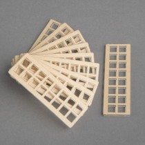
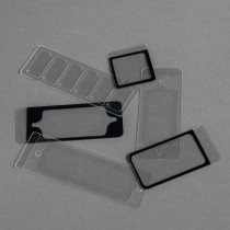
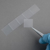
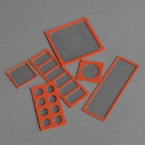
Molecular Surface Engineering – functionalized consumables for LifeScience applications
PolyAn is a nanotechnology company specialized in the functionalization of surfaces using Molecular Surface Engineering (MSE). Since 1996 PolyAn develops and manufactures consumables for multiplex diagnostics and life science research. PolyAn has its production and r&d laboratories at our new site in Berlin which we opened in September 2021.

We are a team with different backgrounds ranging from chemistry, biochemistry, microbiology and engineering with multiple years of experience in surface functionalization. We constantly strive to expand the range of our capabilities and develop new and better performing surfaces. Being based in Berlin, we take advantage of the regional ecosystem of excellent research institutes & universities through co-operations and joint research projects.


Products

Microarray Products

Consumables for microarrays

PolyAn is proud to offer one of the broadest product portfolios of microarray consumables on the market. Our products include:

  • functionalized glass slides
  • functionalized plastic slides
  • functionalized coverslips
  • thin functionalized polymer films
  • functionalized 96-well plates and
  • multipart plates

for DNA-, peptide-, aptamere, carbohydrate (glycans), RPPA or protein microarrays. Applications range from low density microarrays for diagnostic tests to ultra-high density arrays for pharma screening.

PolyAn is able to equip almost any substrate with our reactive matrices and antifouling surfaces. As part of our Molecular Surface Engineering services, we offer functionalized consumable and substrate materials for OEM applications, which are tailored to specified customer requirements.

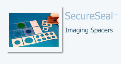
In order to facilitate handling of slides PolyAn also offers a wide range of accessories like the SecureSeal Hybridization Chambers and the ProPlate multi-well chambers. PolyAn is also the European distributor of Grace Bio-Labs’ Nitrocellulose Film slides.

Applications Overview

PolyAn is offering one of the broadest product portfolios of microarray slides on the market. As every biomolecule is a bit different, this enables you to test and choose the optimal surface for your specific application. In order to facilitate the selection of the right surface, PolyAn offers a range of sample packs for standard applications:

  • DNA Microarrays
  • Peptide Microarrays
  • Protein Microarrays
  • Glycan Microarrays

We recommend to start testing the surfaces in the standard 75 mm x 25 mm x 1 mm glass microarray slide format. Once you have selected the optimal surface, it is possible to transfer this surface also onto other support materials, e.g.:

  • Plastic slides
  • Glass slides
  • Coverslips
  • Nitrocellulose film slides
  • Membranes
  • Multi-well plates
  • Metal and metal oxide surfaces

or other more exotic substrates like low autofluorescence films, waveguide materials, microfluidic devices and custom specific formats. We are happy to help you find the best surface for your application. Please contact us.

In order to facilitate handling of glass slides, coverslips and glass plates, PolyAn also offers a range of useful accessories and reagents.

Microarray Slides for DNA applications

All of PolyAn’s reactive surfaces are completely transparent. They are characterized by a low lot-to-lot variation that is specified and monitored by using contact angle measurements as well as qualitative test methods.


ID Title Surface Modifications
104 00 910 Surface test package (1) with 3×5 glass slides for immobilization of oligonucleotides 2D-Epoxy, 3D-Epoxy and 3D-NHS
104 00 915 Surface test package (2) with 3×5 glass slides for directed immobilization of oligonucleotides. 2D-Azide, Streptavidin and 3D-Maleimid
104 00 915 Surface test package (2) with 3×5 glass slides for directed immobilisation of oligonucleotides. Streptavidin, Neutravidin and 3D-Maleimid
104 00 925 Surface test package (4) with 2×5 polymer slides for immobilization of oligonucleotides 3D-Epoxy and 3D-NHS
104 00 926 Surface test package (5) with 3×5 polymer slides for directed immobilization of oligonucleotides Streptavidin, Neutravidin and 3D-Maleimid

More hydrophobic surfaces may result in reduced spot diameter, depending on the spotting buffer composition. Results may vary based on buffers, sample preparation, spotting and scanning instruments.

Please note, that in order to facilitate handling of the glass slides PolyAn also offers a range of useful accessories and reagents.

Microarray Slides for Peptide applications

All of PolyAn’s reactive surfaces are completely transparent. They are characterized by a low lot-to-lot variation that is specified and monitored by using contact angle measurements as well as qualitative test methods.


Surface test package (1) with 3×5 glass slides for immobilization of peptides

Id104 00 930
TitleSurface test package (1) with 3×5 glass slides for immobilization of peptides
SubstrateGlass
FormatPlanar slide
Mean Diameter
Color Labeling
Surface Modifications2D-Epoxy, 3D-Epoxy and 3D-Amino
Solids Content
Product Dimensions25 mm x 75 mm
Packaging
Packaging Volume15 Slides/Pack
Package Weight
Dimensions
Hts Code70 17 9 000
Pads Wells
Pad Size
Well Format
Product Thickness1 mm
DescriptionSelection of surfaces chemistries that are commonly used for immobilization of peptides. The test package is comprised of 5 x 2D-Epoxy glass slides, 5 x 3D-Epoxy glass slides, and 5 x 3D-Amino glass slides. The packages includes standard handling protocols for the different surfaces.

More hydrophobic surfaces may result in reduced spot diameter, depending on the spotting buffer composition. Results may vary based on buffers, sample preparation, spotting and scanning instruments.

Please note, that in order to facilitate handling of the glass slides PolyAn also offers a range of useful accessories and reagents.

Selected Publications

  • Scientific Reports | (2018) 8:4701, “A novel synthetic peptide microarray assay detects Chlamydia species-specific antibodies in animal and human sera” Konrad Sachse, Kh. Shamsur Rahman, Christiane Schnee, Elke Müller, Madlen Peisker, Thomas Schumacher, Evelyn Schubert, Anke Ruettger, Bernhard Kaltenboeck & Ralf Ehricht .

Protein Microarrays

Protein microarrays come in two basic formats, forward and reverse phase. In “forward phase” arrays, captured reagents – whether antibodies, aptamers, or other proteins – are arrayed at defined positions on a glass slide or similar substrate, which is then interrogated with any of a variety of probes, from protein lysate and enzymes to small molecules and nucleic acids.

On the other hand, with a reverse phase array protein lysates are spotted. The arrays are then probed with antibodies, for instance, phosphorylated signaling molecules – one antibody probing multiple samples as opposed to one sample for multiple capture reagents.

Transparent, reactive glass slides for Protein Microarrays

All of PolyAn’s reactive surfaces are completely transparent. They are characterized by a low lot-to-lot variation that is specified and monitored by using contact angle measurements as well as qualitative test methods.

Id Title Surface Modifications
104 00 927 Surface test package (6) with 3×5 polymer slides for immobilization of proteins 3D-Epoxy, 3D-Aldehyde and 3D-NHS
104 00 950 Surface test package (1) with 3×5 glass slides for immobilization of proteins 2D-Epoxy, 2D-Aldehyde and 3D-NHS

More hydrophobic surfaces may result in reduced spot diameter, depending on the spotting buffer composition. Results may vary based on buffers, sample preparation, spotting and scanning instruments.

Please note, that in order to facilitate handling of the glass slides PolyAn also offers a range of useful accessories and reagents.

Nitrocellulose Film Slides

PolyAn is the distributor of the nitrocellulose film slides from Grace Bio-Labs. The following table illustrates the different product families for RPPA and other microarray applications:

AVIDNOVASuperNOVAPATH
Binding Capacity++++++++
Fluorescence background++++++++++
Dynamic range (log scale fluorescence)5-65-67+4-5
Hydrophobicity+++++
ApplicationsBest for any application requiring high binding capacity and colorimetric detection.Reduced fluorescence background with lower binding capacity than AVID. Good signal-to-noise ratio for fluorescence detection.Second generation NOVA, lowest fluorescence background, high binding capacity. Best for fluorescence detection and large dynamic range.Lowest fluorescence background, lower binding capacity, reduced dynamic range. Best signal-to-noise ratio for fluorescence detection.

Microarray Slides for glycan (carbohydrate) applications

Glycan microarrays are presentations of multiple glycans or glycoconjugates printed on a slide for screening with glycan-binding proteins, e.g. lectins, antibodies, bacteria, and viruses. Glycans are usually derivatized with functional groups and can be immobilzed for example on PolyAn’s 2D-Epoxy, 3D-Epoxy and 3D-NHS functionalized glass slides or onto PolyAn’s functionalized 96-well plates.

All of PolyAn’s reactive surfaces are completely transparent. They are characterised by a low lot-to-lot variation that is specified and monitored by using contact angle measurements as well as qualitative test methods.

More hydrophobic surfaces may result in reduced spot diameter, depending on the spotting buffer composition. Results may vary based on buffers, sample preparation, spotting and scanning instruments.

Please note, that in order to facilitate handling of the glass slides PolyAn also offers a range of useful accessories and reagents.

Selected Publications

Shoib S. Siddiqui, Chirag Dhar, Venkatasubramaniam Sundaramurthy, Aniruddha Sasmal, Hai Yu, Esther Bandala-Sanchez, Miaomiao Li, Xiaoxiao Zhang, Xi Chen, Leonard C. Harrison, Ding Xu, Ajit Varki, 2021, Sialoglycan recognition is a common connection linking acidosis, zinc, and HMGB1 in sepsis, PNAS 118 (10), e2018090118. doi: 10.1073/pnas.2018090118.
Yang Ji, Aniruddha Sasmal, Wanqing Li, Lisa Oh, Saurabh Srivastava, Audra A. Hargett, Brian R. Wasik,Hai Yu, Sandra Diaz, Biswa Choudhury, Colin R. Parrish, Darón I. Freedberg, Lee-Ping Wang,Ajit Varki, Xi Chen, 2021, Reversible O Acetyl Migration within the Sialic Acid Side Chain and Its Influence on Protein Recognition, ACS Chem. Biol, doi: 10.1021/acschembio.0c00998.
Chethan D. Shanthamurthy, Shani Leviatan Ben-Arye, Nanjundaswamy Vijendra Kumar, Sharon Yehuda,Ron Amon, Robert J. Woods, Vered Padler-Karavani, Raghavendra Kikkeri, 2021, Heparan Sulfate Mimetics Differentially Affect Homologous Chemokines and Attenuate Cancer Development, J. Med. Chem. 64: 3367-3380. doi: 10.1021/acs.jmedchem.0c01800.
Kevin O. Saunders, Norbert Pardi, Robert Parks, Sampa Santra, Zekun Mu, Laura Sutherland, Richard Scearce, Maggie Barr, Amanda Eaton, Giovanna Hernandez, Derrick Goodman, Michael J. Hogan, Istvan Tombacz, David N. Gordon, R. Wes Rountree, Yunfei Wang, Mark G. Lewis, Theodore C. Pierson, Chris Barbosa, Ying Tam, Xiaoying Shen, Guido Ferrari, Georgia D. Tomaras, David C. Montefiori, Drew Weissman, Barton F. Haynes, 2021, Lipid nanoparticle encapsulated nucleoside-modified mRNA vaccines elicit polyfunctional HIV-1 antibodies comparable to proteins in nonhuman primates. npj Vaccines 6, 50. doi: 10.1038/s41541-021-00307-6.
Tal Noy-Porat, Adva Mechaly, Yinon Levy, Efi Makdasi, Ron Alcalay, David Gur, Moshe Aftalion, Reut Falach, Shani Leviatan Ben-Arye, Shirley Lazar, Ayelet Zauberman, Eyal Epstein, Theodor Chitlaru, Shay Weiss, Hagit Achdout, Jonathan D. Edgeworth, Raghavendra Kikkeri, Hai Yu, Xi Chen, Shmuel Yitzhaki, Shmuel C. Shapira, Vered Padler-Karavani, Ohad Mazor, Ronit Rosenfeld, 2021, Therapeutic antibodies, targeting the SARS-CoV-2spike N-terminal domain, protect lethally infected K18-hACE2 mice, iScience 24 (5), 102479. doi: 10.1016/j.isci.2021.102479.
Sudeshna Saha, Alison Coady, Aniruddha Sasmal, Kunio Kawanishi, Biswa Choudhury, Hai Yu, Ricardo U. Sorensen, , Jaime Inostroza, Ian C. Schoenhofen, Xi Chen, Anja Münster-Kühnel, Chihiro Sato, Ken Kitajima, Sanjay Ram, Victor Nizet, Ajit Varki, 2021, Exploring the Impact of Ketodeoxynonulosonic Acid in Host-Pathogen Interactions Using Uptake and Surface Display by Nontypeable Haemophilus influenza, mBio 12:e03226-20. doi: 10.1128/mBio.03226-20.
Naazneen Khan, Aniruddha Sasmal, Zahra Khedri, Patrick Secrest, Andrea Verhagen, Saurabh Srivastava, Niss, Xi Chen, Hai Yu, Travis Beddoe, Adrienne W. Paton, James C. Paton, Ajit Varki, 2021, Rapid evolution of bacterial AB5 toxin B subunits independent of A subunits: sialic acid binding preferences correlate with host range and toxicity, bioRxiv. doi: 10.1101/2021.05.28.446168.
Fangping Cai, Wei-Hung Chen, Weimin Wu, Julia A. Jones, Misook Choe, Neelakshi Gohain, Xiaoying Shen, Celia LaBranche, Amanda Eaton, Laura Sutherland, Esther M. Lee, Giovanna E. Hernandez, Nelson R. Wu, Richard Scearce, Michael S. Seaman, M. Anthony Moody, Sampa Santra, Kevin Wiehe, Georgia D. Tomaras, Kshitij Wagh, Bette Korber, Mattia Bonsignori, David C. Montefiori, Barton F. Haynes, Nataliade Val, M. Gordon Joyce, Kevin O. Saunders, 2021, Structural and genetic convergence of HIV-1 neutralizing antibodies in vaccinated non-human primates. PLoS Pathog 17(6): e1009624. doi: 10.1371/journal.ppat.1009624.
Aniruddha Sasmal, Naazneen Khan, Zahra Khedri, Benjamin P.Kellman, Saurabh Srivastava, Andrea Verhagen, Hai Yu, Anders Bech Bruntse, Sandra Diaz, Nissi Varki, Travis Beddoe, Adrienne W.Paton, James C.Paton, Xi Chen, Nathan E. Lewis, Ajit Varki, 2021, Sialoglycan microarray encoding reveals differential sialoglycan binding of phylogenetically-related bacterial AB5 toxin B subunits, bioRxiv. doi: 10.1101/2021.05.28.446191.
Ankita Malik, Fridolin Steinbeis, Maria Antonietta Carillo, Peter H. Seeberger, Bernd Lepenies, Daniel Varón Silva, 2020, Immunological Evaluation of Synthetic Glycosylphosphatidylinositol Glycoconjugates as Vaccine Candidates against Malaria, ACS Chem Biol 15, 171-178. doi: 10.1021/acschembio.9b00739.
Jeffrey R. Schneidera, Xiaoying Shen, Chiara Orlandik, Tinashe Nyanheteh, Sheetal Sawantf, Ann M. Cariasb, Archer D. Smith IV, Neil L. Kelleherl, Ronald S. Veazeyo, George K. Lewisk, Georgia D. Tomaras, Thomas J. Hope, 2020, A MUC16 IgG Binding Activity Selects for a Restricted Subset of IgG Enriched for Certain Simian Immunodeficiency Virus Epitope Specificities, Journal of Virology 94 (5). doi: 10.1128/JVI.01246-19.
Salam Bashir, Leopold K. Fezeu, Shani Leviatan Ben-Arye, Sharon Yehuda, Eliran Moshe Reuven, Fabien Szabo de Edelenyi, Imen Fellah-Hebia, Thierry Le Tourneau, Berthe Marie Imbert-Marcille, Emmanuel B. Drouet, Mathilde Touvier, Jean-Christian Roussel, Hai Yu, Xi Chen, Serge Hercberg, Emanuele Cozzi, Jean-Paul Soulillou, Pilar Galan, Vered Padler-Karavani, 2020, Association between Neu5Gc carbohydrate and serum antibodies against it provides the molecular link to cancer: French NutriNet-Santé study, BMC Medicine 18:262. doi: 10.1186/s12916-020-01721-8.
Qifeng Han, Julia A. Jones, Nathan I. Nicely, Rachel K. Reed, Xiaoying Shen, Katayoun Mansouri, Mark Louder, Ashley M. Trama, S. Munir Alam, Robert J. Edwards,Mattia Bonsignori, Georgia D. Tomaras, Bette Korber, David C. Montefiori, John R. Mascola, Michael S. Seaman, Barton F. Haynes, Kevin O. Saunders, 2019, Difficult-to-neutralize global HIV-1 isolates are neutralized by antibodies targeting open envelope conformations, Nat Commun 10, 2898. doi: 10.1038/s41467-019-10899-2.
Colin Ruprechta, Andreas Geissnera, Peter H. Seeberger, Fabian Pfrenglea, 2019, Practical considerations for printing high-density glycan microarrays to study weak carbohydrate-protein interactions, Carbohydrate Research 481, 31-35. doi:10.1016/j.carres.2019.06.006.
Maria Dennis, Joshua Eudailey, Justin Pollara, Arthur S. McMillan, Kenneth D. Cronin, Pooja T. Saha, Alan D. Curtis, , Michael G. Hudgens, Genevieve G. Fouda, Guido Ferrari, Munir Alam, Koen K. A. Van Rompay, Kristina De Paris, Sallie Permar, Xiaoying Shen, 2019, Coadministration of CH31 Broadly Neutralizing Antibody Does Not Affect Development of Vaccine-Induced Anti-HIV-1 Envelope Antibody Responses in Infant Rhesus Macaques, Journal of Virology 93 (5). doi: 10.1128/JVI.01783-18.
Chethan D. Shanthamurthy, Prashant Jain, Sharon Yehuda, João T. Monteiro, Shani Leviatan Ben-Arye, Balamurugan Subramani, Bernd Lepenies, Vered Padler-Karavani, Raghavendra Kikkeri, 2018, Scientific Reports 8:6603, ABO Antigens Active Tri- and Disaccharides Microarray to Evaluate C-type Lectin Receptor Binding Preferences, Scientific Reports 8:6603. doi:10.1038/s41598-018-24333-y.
Torben Schiffner, Jesper Pallesen, Rebecca A. Russell, Jonathan Dodd, Natalia de Val, Celia C. LaBranche, David Montefiori, Georgia D. Tomaras, Xiaoying Shen, Scarlett L. Harris, Amin E. Moghaddam, Oleksandr Kalyuzhniy, Rogier W. Sanders, Laura E. McCoy, John P. Moore, Andrew B. Ward, Quentin J. Sattentau, 2018, Structural and immunologic correlates of chemically stabilized HIV-1 envelope glycoproteins, PLoS Pathog 14(5): e1006986. doi: 10.1371/journal.ppat.1006986.
Madhuri Gade, Catherine Alex, Shani Leviatan Ben-Arye, João T. Monteiro, Sharon Yehuda, Bernd Lepenies, Vered Padler-Karavani, Raghavendra Kikkeri, 2018, Microarray Analysis of Oligosaccharide-Mediated Multivalent Carbohydrate–Protein Interactions and Their Heterogeneity, ChemBioChem 19, 1170. doi: 10.1002/cbic.201800037.
Yingxia Wen, Hung V. Trinh, Christine E. Linton, Chiara Tani, Nathalie Norais, DeeAnn Martinez-Guzman, Priyanka Ramesh, Yide Sun, Frank Situ, Selen Karaca-Griffin, Christopher Hamlin, Sayali Onkar, Sai Tian, Susan Hilt, Padma Malyala, Rushit Lodaya, Ning Li, Gillis Otten, Giuseppe Palladino, Kristian Friedrich, Yukti Aggarwal, Celia LaBranche, Ryan Duffy, Xiaoying Shen, Georgia D. Tomaras, David C. Montefiori, William Fulp, Raphael Gottardo, Brian Burke, Jeffrey B. Ulmer, Susan Zolla-Pazner, Hua-Xin Liao, Barton F. Haynes, Nelson L. Michael, Jerome H. Kim, Mangala Rao, Robert J. O’Connell, Andrea Carfi, Susan W. Barnett, 2018, Generation and characterization of a bivalent protein boost for future clinical trials: HIV-1 subtypes CR01_AE and B gp120 antigens with a potent adjuvant, PLoS ONE 13(4): e0194266. doi: 10.1371/journal.pone.0194266.
Shani Leviatan Ben-Arye, Hai Yu, Xi Chen, Vered Padler-Karavani, 2017, Profiling Anti-Neu5Gc IgG in Human Sera with a Sialoglycan Microarray Assay, J. Vis. Exp. (125), e56094. doi:10.3791/56094.
Felix Broecker, Peter H. Seeberger, 2016, Synthetic Glycan Microarrays, Small Molecule Microarrays, 227–240. doi:10.1007/978-1-4939-6584-7_15.
Sebastian Götze, Anika Reinhardt, Andreas Geissner, Nahid Azzouz, Yu-Hsuan Tsai, Reka Kurucz, Daniel Varón Silva, Peter H Seeberger, 2015, Investigation of the protective properties of glycosylphosphatidylinositol-based vaccine candidates in a Toxoplasma gondii mouse challenge model, Glycobiology 25:9, 984–991, doi: 10.1093/glycob/cwv040.

Surfaces for Microarrays

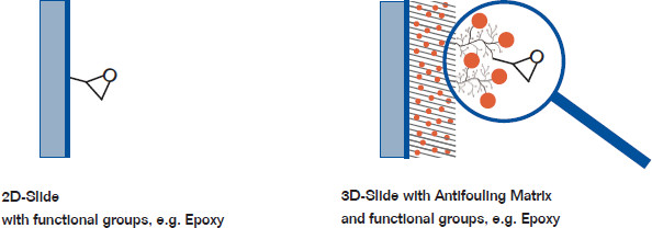
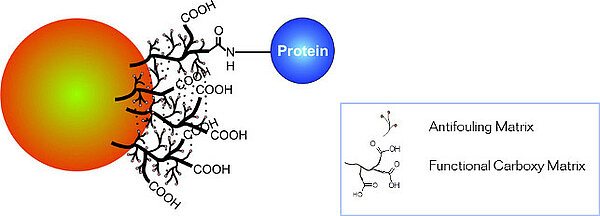
Every biomolecule is different. We are offering one of the broadest range of surfaces for binding of biomolecules on the market, so that you can select the optimal surface for your application. Our product portfolio includes both conventional 2D (2-dimensional)-reactive glass slides as well as PolyAn’s 3D-reactive matrices:

e.g Epoxy 

  • ultra-thin (mono)layer
  • rigid structure
  • cost-effective reactive surface
  • suitable for glass and metal oxides

3D-Slide with Antifoulin and Functional Groups

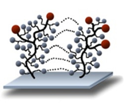
  • tentacular, branched polymer structure, partly crosslinked
  • thickness: 1 – 50 nm, depending on application
  • swellable hydrogel
  • for small molecules fully penetratable
  • variable density / surface concentration of functional groups

Available 2D-reactive matrices

  • 2D-Epoxy
  • 2D-Amino
  • 2D-Aldehyde
  • 2D-Carboxy
  • 2D-Activated Amino (PDITC)
  • 2D-Thiol
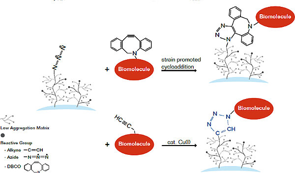
  • 2D-Azide

For cost-sensitive applications PolyAn has developed a range of 2D (2-dimensional)-reactive glass slides that are manufactured from high quality glass with an ultra-flat surface and low inherent fluorescence. The glass is coated with a thin silane layer that will covalently bind most types of bio-molecules. The defect-free surface features uniform silane layers that provide a high covalent coupling efficiency together with a very low background.

Available 3D-reactive matrices

  • 3D-Epoxy matrices
  • 3D-Amino matrices
  • 3D-Aldehyde matrices
  • 3D-Carboxy matrices
  • 3D-NHS matrices
  • 3D-Maleimide
  • Activated Amino (PDITC) matrices
  • Covalently coupled Streptavidin and Neutravidin
  • Other surface chemistries, e.g. 3D-Thiol, Poly-L-Lysine, …

Our 3D-reactive surfaces are functionalized with a three-dimensional (3D)-surface chemistry comprised of a long-chain polymer containing a defined number of reactive groups. The 3D-structure enables a higher loading with reactive groups and incorporates structures that reduce unspecific binding. Optimized loading and low unspecific binding can translate into a higher signal-to-noise ratio.

Dimensions

Our products include plastic and glass slides, coverslips as well as functionalized 96-well plates for DNA-, peptide- or protein microarrays. Applications range from low density microarrays for diagnostic tests to ultra-high density arrays for pharma screening.

The slides are produced with standard dimensions of 25 x 75 x 1 mm. Additionally, we offer customized slides with a surface modification tailored to your specific application. PolyAn also functionalizes metal surfaces with our 3D-reactive matrices.

Customized surfaces

Please do not hesitate to contact us, if you require special surfaces for binding of your biomolecules that are not listed in the products table. We can also functionalize other plate formats and substrates with our surfaces. Additionally, we have access to a wide range of different surface modifications for binding of small molecules, saccharides etc.

3D-Amino Matrix for coupling via electrostatical adsorption of negatively charged biochemical species

An adsorptive immobilization is a non-covalent coupling method on solid supports which is realized by electrostatic, Van der Waals interactions, hydrogen bonds and hydrophobic interactions of the reactants, respectively. An electrostatic interaction is formed by an ion-ion-interaction between the surface and the applied biochemical species. The dissociation energy for typical electrostatic bond is 130 kJ/mol. It is about a third of the strength of an average covalent bond. In order to achieve an optimal adhesion the probe buffer und the adhesion conditions (pH-value) have to be optimized.

Binding of oligonucleotides on the 3D-Amino matrix

The nucleic acid is bound electrostatically on the 3D-Amino surface with its negatively charged backbone or its 5´phosphate group. For immobilization of nulceic acids we recommend an UV- crosslinking after adsorption forming a covalent bond. During the UV irradiation the base Thymine formes radicals which undergoes a H-abstraction in the 3D- Matrix.

Products

PolyAn equips glass slides, coverslips and polymer slides with 3D-Amino surfaces. Additionally, PolyAn has a range of porous materials (membranes, fleece and sinter materials) and microparticles in our product portfolio that are functionalised with 3D-Amino surfaces.

Please do not hesitate to contact us, if you would like to functionalise a different format or substrate with our 3D-Amino surface.

3D-Epoxy Matrix for coupling via the N-terminus of biochemical species

Epoxides are cyclic ethers with a highly strained three member ring. Epoxy rings can be easily reacted with nucleophiles e.g. amines, hydrazines, thiols, hydroxides and carboxyl groups. Compared to NHS-esters or 1,4-Phenylene isothiocyanates (PDITC) the epoxy surface is more stable and has a longer shelf-life. Epoxy-surfaces are stable up temperatures of 40°C and are also more stable against humidity compared to NHS and PDITC-surfaces.

The nucleophilic addition is catalyzed by acid or basic conditions. Under acidic conditions, the oxygen in the ring is positively charged, which facilitates the nucleophilic attack. Under basic conditions the least substituted carbon is attacked by the applied nucleophile in a standard SN2 reaction.

PolyAn equips glass slides, coverslips and polymer slides as well as 96-well plates with 3D-Epoxy surfaces. The 3D-Epoxy 96 well microplates are used mainly if adsorptive binding of peptides or oligonucleotides, for example, to high/medium binding surfaces is ineffective or the binding strength is not sufficient. Areas of application include detection methods such as ELISA, ELI-Spot, protein and peptide arrays and DNA binding.

Please do not hesitate to contact us, if you would like to functionalize a different format or substrate with our 3D-Epoxy surface.

3D-Carboxy Matrix for non-covalent coupling of positively charged biochemical species via electrostatical adsorption

An adsorptive immobilization is a non-covalent coupling method on solid supports which is realized by electrostatic, Van der Waals interactions, hydrogen bonds and hydrophobic interactions of the reactants, respectively. An electrostatic interaction is formed by an ion-ion-interaction between the surface and the applied biochemical species. The dissociation energy for typical electrostatic bond is 130 kJ/mol. It is about a third of the strength of an average covalent bond. In order to achieve an optimal adhesion the probe buffer und the adhesion conditions (pH-value) have to be optimized.

Please note, that the carboxy functionalities can be used for surface activation via 1-Ethyl-3-(3-dimethylaminopropyl) carbo-diimide (EDC), N-hydroxy succinimide (NHS).

PolyAn equips glass slides, coverslips and polymer slides as well as 96-well plates with 3D-Carboxy matrices. Please do not hesitate to contact us, if you would like to functionalize a different format or substrate with our 3D-Carboxy surface.

3D-NHS Matrix for coupling via the N-terminus of biochemical species

The NHS-ester reacts immediately with the NH2- terminus of biochemical species to form a covalent bond with the surface (420 kJ/mol). The reaction of carboxyl functionalities with N- Hydroxy succinimide leads to highly reactive esters, which can be easily reacted with nucleophiles e.g. amines, hydrazines. However, due to its high reactivity the NHS ester is susceptible against hydrolysis and is characterized by a relatively short shelf-life. All NHS-activated surfaces should therefore be processed quickly.

There are a number of different approaches to couple on the NHS-surface:

  1. It is assumed that not all Carboxy groups have reacted to NHS- esters during activation. Thus a negatively charged carboxy surface still remains which in turn supports the physico-chemical adsorption of positively charged probes e.g. NH3+. Hence a protonating media (pH < 5) for the biochemical species getting a positively charge is required.
  2. A nucleophilic attack on the active ester is also catalyzed under basic conditions (pH > 8,5).

After attachment of the biochemical species the surfaces must be blocked with a blocking buffer containing small molecules that can access all reactive groups within the 3D- Matrix.

PolyAn equips glass slides, coverslips and polymer slides as well as 96-well plates with 3D-NHS surfaces. Please do not hesitate to contact us, if you would like to functionalize a different format or substrate with our 3D-NHS surface.

3D-Aldehyde Matrix for coupling via the N-terminus of the biochemical species

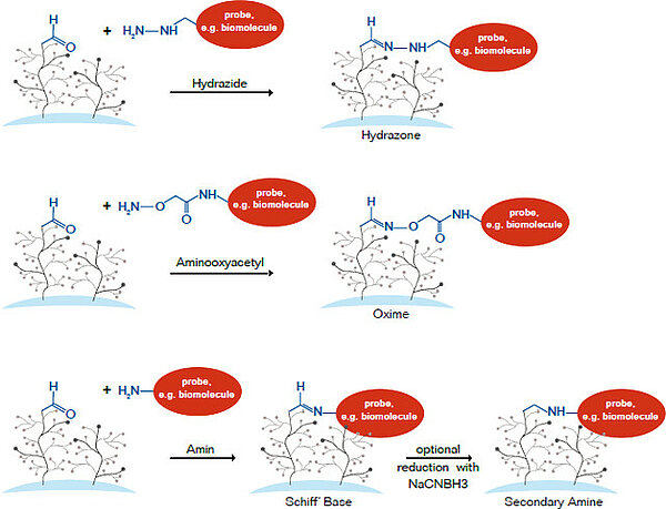
Aldehyde groups react immediately with the NH2-terminus of biochemical species to form a covalent bond with the surface (420 kJ/mol). In an intermediate state the Aldehydes form an instable Imine-group with the Amines (Schiff-base). It is also possible to reduce the Imines with Sodium Borhydride to form stable Amines.

Upon completion the coupling reaction other non-reacted aldehydes must be blocked with small molecules that penetrate the 3D-Matrix and effectively quench all remaining reactive groups.

PolyAn equips glass slides, coverslips and polymer slides with 3D-Aldehyde surfaces. Please do not hesitate to contact us, if you would like to functionalize a different format or substrate with our 3D-Aldehyde surface.

.

Avidin, Streptavidin and Neutravidin Surfaces for non-covalent oriented coupling of Biotin modified biochemical species

Avidin is a glycoprotein comprised of four polypeptides that are connected with carbohydrates via glycosidic bonds. Avidin is a tetrameric protein which forms a highly specific binding site for Biotin. Neutravidin is a deglycosylated form of avidin.

The Avidin (Streptavidin/Neutravidin) -Biotin-bond is one of the strongest known, non-covalent bond in biology/ biochemistry (KD= 10-15 mol/l*). The binding site for Biotin is formed by various amino acids (see adjacent figure: binding site). When using covalently attached Avidin, Streptavidin or Neutravidin the molecules are less susceptible for desorption in the presence of alkaline, acids, in solutions of high ionic strength or at high temperatures. Biotin affine proteins can be distinguished by their isoelectric point, specificity and nonspecific binding as illustrated in the following table.

.

Avidin Streptavidin Neutravidin
Molecular Weight67 kDa53 kDa60 kDa
Biotin Binding Sites444
Isoelectric Point (pl)106.8-7.56.3
SpecificityLowHighHighest
Affinity for Biotin (Kd) 10-15 M10-15 M10-15 M
Nonspecific BindingHighLowLowe

PolyAn equips glass slides, coverslips and polymer slides as well as 96-well plates with Streptavidin and Neutravidin surfaces.

Please do not hesitate to contact us, if you would like to functionalize a different format or substrate with our Streptavidin or Neutravidin surfaces.

3D-Maleimide Matrix for oriented covalent coupling (immobilization) of thiolated biochemical species

Maleimide-esters react immediately with Thiol-groups of biochemical species. The Thiol-groups can be either natively present in the (bio)molecule, e.g. through the amino acid cysteine in proteins, produced via reductive cleavage of disulfide bonds with a reducing agent such as Dithiothreitol (DTT, Cleland´s Reagent), or selectively introduced e.g. with 2-Iminothiolane (Traut’s reagent) for amine-containing molecules.

Similar to NHS-esters, Maleimide surfaces are susceptible to hydrolysis, and thus, should be processed promptly after opening the sealed bags.

PolyAn equips glass slides, coverslips and polymer slides with 3D-Maleimide surfaces. Please do not hesitate to contact us, if you would like to functionalize a different format or substrate with our 3D-Maleimide surface.

Selected Publications

  • Broecker, F. et al., `Synthetic Glycan Microarrays´, Meth Mol Biol, 2017, 1518, 227. DOI: 10.1007/978-1-4939-6584-7_15.
  • Götze, S. et al., `Investigation of the protective properties of glycosylphosphatidylinositol-based vaccine candidates in a Toxoplasma gondii mouse challenge model´, Glycobiology, 2015, 25, 984. DOI: 10.1093/glycob/cwv040.
  • Malik, A. et al., `Immunological Evaluation of Synthetic Glycosylphosphatidylinositol Glycoconjugates as Vaccine Candidates against Malaria´, Chem Biol, 2020, 15, 171. DOI: 10.1021/acschembio.9b00739.

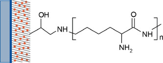
Poly-L-Lysin Matrix

A higher density of surface amines can be achieved with the Poly-L-Lysin which is covalently attached on 3D-surfaces. Slides covalently coated with Poly-L-Lysine can be used as adhesive microscope slides for the electrostatic coupling of biomolecules or biological samples, e.g. DNA, cells, tissues.

PolyAn equips glass slides, coverslips and polymer slides with Poly-L-Lysin. Please do not hesitate to contact us, if you would like to functionalize a different format or substrate with our Poly-L-Lysin surface.

3D-Activated Amino (PDITC) Matrix for coupling via the N-terminus of biochemical species

The PDITC (1,4- Phenylendiisothiocyanate) is a homobifunctional linker, that immediately reacts with nucleophiles e.g. amines, hydrazines, thiols and hydroxides to form stable covalent bonds. After attachment of the biochemical species the surfaces must be blocked with a blocking buffer containing small molecules that can access all reactive groups within the 3D- Matrix.

PolyAn equips glass slides, coverslips and polymer slides with 3D-PDITC surfaces. Please do not hesitate to contact us, if you would like to functionalize a different format or substrate with our 3D-PDITC surface.

Functionalized Glass Slides

Microarray Glass Slides

PolyAn is proud to offer one of the broadest product portfolios of microarray substrates on the market. Our products include plastic and glass slides as well as functionalized 96-well plates for DNA-, peptide-, glycan- or protein microarrays. Applications range from low density microarrays for diagnostic tests to ultra-high density arrays for pharma screening.

The slides are produced with standard dimensions of 25 x 75 x 1 mm. Additionally, we offer customized slides and alternative glass formats with surface modifications tailored to your specific application. Please also have a look at our ProPlate, HybriWell and SecureSeal products that facilitate handling of slides.

For cost-sensitive applications PolyAn has developed a range of 2D (2-dimensional)-reactive glass slides that are manufactured from high quality glass with an ultra-flat surface and low inherent fluorescence. The glass is coated with a thin silane layer that will covalently bind most types of bio-molecules. The defect-free surface features uniform silane layers that provide a high covalent coupling efficiency together with a very low background. The slides are easy to use, and are fully compatible with all commercially available arraying and scanning instruments.


ID Surface Modifications Product Dimensions Packaging Volume
104 00 321 2D-Aldehyde 25.0 mm x 75.6 mm 25 Slides/Box
104 00 021 2D-Amino 25.0 mm x 75.6 mm 25 Slides/Box
104 00 121 2D-Carboxy 25.0 mm x 75.6 mm 25 Slides/Box
104 00 221 2D-Epoxy 25.0 mm x 75.6 mm 25 Slides/Box
104 00 411 2D-NHS 25.0 mm x 75.6 mm 25 Slides/Box
104 00 521 2D-Thiol 25.0 mm x 75.6 mm 25 Slides/Box
104 00 421 2D-Activated Amino (PDITC) 25.0 mm x 75.6 mm 25 Slides/Box
104 00 621 2D-Azide 25.0 mm x 75.6 mm 25 Slides/Box
ID Surface Modifications Product Dimensions Packaging Volume
104 00 001 3D-Amino 25.0 mm x 75.6 mm 25 Slides/Box
104 00 101 3D-Carboxy 25.0 mm x 75.6 mm 25 Slides/Box
104 00 201 3D-Epoxy 25.0 mm x 75.6 mm 25 Slides/Box
104 00 301 3D-Aldehyde 25.0 mm x 75.6 mm 25 Slides/Box
104 00 401 3D-NHS 25.0 mm x 75.6 mm 25 Slides/Box
104 00 402 3D-NHS (hydrophilic) 25.0 mm x 75.6 mm 25 Slides/Box
104 00 431 3D-Activated Amino (PDITC) 25.0 mm x 75.6 mm 25 Slides/Box
104 00 441 3D-Maleimide 25.0 mm x 75.6 mm 25 Slides/Box
104 00 501 3D-Thiol 25.0 mm x 75.6 mm 25 Slides/Box
104 01 201 Covalently coated Poly-L-Lysin 25.0 mm x 75.6 mm 25 Slides/Box
104 02 205 Covalently coated Streptavidin 25.0 mm x 75.6 mm 25 Slides/Box
104 03 205 Covalently coated Neutravidin 25.0 mm x 75.6 mm 25 Slides/Box
104 04 205 Covalently coated Avidin 25.0 mm x 75.6 mm 25 Slides/Box

PolyAn’s 3D-functionalised microarray Slides are functionalized with a three-dimensional (3D)-surface chemistry comprised of a long-chain polymer containing a defined number of reactive groups. This polymer is covalently linked to the surface of the slide.

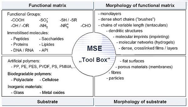
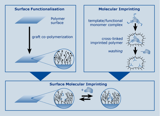
Our MSE-technology gently binds the functional layer onto the surface without damaging the base substrate. The morphology of the functional surface and thus the number of the reactive groups can be fine-tuned within a narrow range. This yields a number of advantages:

Low fluorescence background Covalent binding of functional layer on the substrate without changing the initial autofluorescence.
Low unspecific binding Combination of reactive functional groups with PolyAn antifouling matrix.
Topography Tuneable surface
hydrophilicity / hydrophobicity (contact angle).
Uniform spot morphology Narrow variation of surface properties and homogeneous distribution of reactive groups.
Optimal density and high accessibility of reactive groups Morphology and thickness of functional layer tailored to the desired application.

Packaging:

The slides are packaged in inert atmospheric boxes, with a capacity of 25 or 5 slides per box.

Storage

PolyAn’s slides are characterised by a long shelf-life when stored dry, at 4-8°C and protected from sunlight. All slides are packaged in boxes under Argon atmosphere to avoid contamination with particles. The Argon atmosphere also minimises degradation of the reactive surface through contact with air or humidity.

Functionalized Polymer Slides

Especially for diagnostic applications PolyAn is offering a broad range of functionalized microarray polymer slides. For reasons of reliability and bio-safety, patient-centric biomarker testing is always performed in single-use cartridges with a one-step process (sample in – result out). These cartridges should be disposable after use, so the materials of the cartridge should be non-hazardous and recyclable.

PolyAn’s Polymer Slides are comprised of a low auto fluorescence plastic. The material is highly robust against breakage making it the material of choice when working with hazardous materials. It is suitable for injection molding and hot embossing processes.

The slides are produced with standard dimensions of 25 x 75 x 1 mm. Additionally, we offer the functionalization of your plastic including microfluidics with a surface modification that is tailored to your specific application. Please also have a look at our ProPlate, HybriWell and SecureSeal products that facilitate handling of slides.

ID Surface Modifications Product Dimensions Packaging Volume
104 00 051 3D-Amino 25 mm x 75 mm 25 Slides/Box
104 00 151 3D-Carboxy 25 mm x 75 mm 25 Slides/Box
104 00 251 3D-Epoxy 25 mm x 75 mm 25 Slides/Box
104 00 351 3D-Aldehyde 25 mm x 75 mm 25 Slides/Box
104 00 451 3D-NHS 25 mm x 75 mm 25 Slides/Box
104 00 481 3D-Activated Amino (PDITC) 25 mm x 75 mm 25 Slides/Box
104 00 551 3D-Thiol 25 mm x 75 mm 25 Slides/Box
104 01 251 Covalently coated Poly-L-Lysin 25 mm x 75 mm 25 Slides/Box
104 01 441 3D-Maleimide 25 mm x 75 mm 25 Slides/Box
104 02 255 Covalently coated Streptavidin 25 mm x 75 mm 25 Slides/Box
104 03 255 Covalently coated Neutravidin 25 mm x 75 mm 25 Slides/Box
104 04 255 Covalently coated Avidin 25 mm x 75 mm 25 Slides/Box

PolyAn’s 3D-functionalised microarray Slides are functionalized with a three-dimensional (3D)-surface chemistry comprised of a long-chain polymer containing a defined number of reactive groups. This polymer is covalently linked to the surface of the slide.

Our MSE-technology gently binds the functional layer onto the surface without damaging the base substrate. The morphology of the functional surface and thus also the number of the reactive groups can be fine-tuned within a narrow range. This yields a number of advantages:

Low fluorescence background Covalent binding of functional layer on the substrate without changing the initial autofluorescence.
Low unspecific binding Combination of reactive functional groups with PolyAn antifouling matrix.
Topography Tuneable surface
hydrophilicity / hydrophobicity (contact angle).
Uniform spot morphology Narrow variation of surface properties and homogeneous distribution of reactive groups.
Optimal density and high accessibility of reactive groups Morphology and thickness of functional layer tailored to the desired application.

Packaging

The slides are packaged in inert atmospheric boxes, with a capacity of 25 or 5 slides per box.

Storage

PolyAn’s slides are characterised by a long shelf-life when stored dry, at 4-8°C and protected from sunlight. All slides are packaged in boxes under Argon atmosphere to avoid contamination with particles. The Argon atmosphere also minimises degradation of the reactive surface through contact with air or humidity.

Functionalized Coverslips

For high-resolution microscopic imaging or scanning applications PolyAn offers functionalized coverslips. All of PolyAn’s standard functional coatings can also be applied to coverslip glass.

All coverslips have the standard dimension of 25 x 60mm. Type #1.5 is used as a standard thickness. All coverslips are packed in boxes of 5. Other functionalities, thicknesses and sizes are available upon request. PolyAn also offers functionalised substrates that are coated with gold or other noble metals as well as waveguide materials.

ID Surface Modifications Product Dimensions Packaging Volume
104 00 026 2D-Amino 25 mm x 60 mm 5 coverslips/ Box
104 00 126 2D-Carboxy 25 mm x 60 mm 5 coverslips/ Box
104 00 226 2D-Epoxy 25 mm x 60 mm 5 coverslips/ Box
104 00 326 2D-Aldehyde 25 mm x 60 mm 5 coverslips/ Box
104 00 526 2D-Thiol 25 mm x 60 mm 5 coverslips/ Box
104 00 626 2D-Azide 25 mm x 60 mm 5 coverslips/ Box

For cost-sensitive applications PolyAn has developed a range of 2D (2-dimensional)-reactive coverslips that are manufactured from high quality glass with an ultra-flat surface and low inherent fluorescence. The glass is coated with a thin silane layer that will covalently bind most types of bio-molecules. The defect-free surface features uniform silane layers that provide a high covalent coupling efficiency together with a very low background.

ID Surface Modifications Product Dimensions Packaging Volume
104 00 006 3D-Amino 25 mm x 60 mm 5 coverslips/ Box
104 00 106 3D-Carboxy 25 mm x 60 mm 5 coverslips/ Box
104 00 206 3D-Epoxy 25 mm x 60 mm 5 coverslips/ Box
104 00 306 3D-Aldehyde 25 mm x 60 mm 5 coverslips/ Box
104 00 407 3D-NHS 25 mm x 60 mm 5 coverslips/ Box
104 00 436 3D-Activated Amino (PDITC) 25 mm x 60 mm 5 coverslips/ Box
104 00 446 3D-Maleimide 25 mm x 60 mm 5 coverslips/ Box
104 00 507 3D-Thiol 25 mm x 60 mm 5 coverslips/ Box
104 01 206 Covalently coated Poly-L-Lysin 25 mm x 60 mm 5 coverslips/ Box
104 02 206 Covalently coated Streptavidin 25 mm x 60 mm 5 coverslips/ Box
104 03 206 Covalently coated Neutravidin 25 mm x 60 mm 5 coverslips/ Box
104 00 446 3D-Maleimide 25 mm x 60 mm 5 coverslips/ Box

PolyAn’s 3D-functionalised microarray Slides are functionalized with a three-dimensional (3D)-surface chemistry comprised of a long-chain polymer containing a defined number of reactive groups. This polymer is covalently linked to the surface of the slide.

Our MSE-technology gently binds the functional layer onto the surface without damaging the base substrate. The morphology of the functional surface and thus also the number of the reactive groups can be fine-tuned within a narrow range. This yields a number of advantages:

Low fluorescence background Covalent binding of functional layer on the substrate without changing the initial autofluorescence.
Low unspecific binding Combination of reactive functional groups with PolyAn antifouling matrix.
Topography Tuneable surface
hydrophilicity / hydrophobicity (contact angle).
Uniform spot morphology Narrow variation of surface properties and homogeneous distribution of reactive groups.
Optimal density and high accessibility of reactive groups Morphology and thickness of functional layer tailored to the desired application.

PolyAn’s 3D-functionalised microarray Slides are functionalized with a three-dimensional (3D)-surface chemistry comprised of a long-chain polymer containing a defined number of reactive groups. This polymer is covalently linked to the surface of the coverslip.

Our MSE-technology gently binds the functional layer onto the surface without damaging the base substrate. The morphology of the functional surface and thus the number of the reactive groups can be fine-tuned within a narrow range. This yields a number of advantages:


Low fluorescence background Covalent binding of functional layer on the substrate without changing the initial autofluorescence.
Low unspecific binding Combination of reactive functional groups with PolyAn antifouling matrix.
Topography Tuneable surface
hydrophilicity / hydrophobicity (contact angle).
Uniform spot morphology Narrow variation of surface properties and homogeneous distribution of reactive groups.
Optimal density and high accessibility of reactive groups Morphology and thickness of functional layer tailored to the desired application.

Packaging:

The coverslips are packaged in inert atmospheric boxes, with a capacity of 5 coverslips per box.

Storage

PolyAn’s coverslips are characterised by a long shelf-life when stored dry, at 4-8°C and protected from sunlight. All coverslips are packaged in boxes under Argon atmosphere to avoid contamination with particles. The Argon atmosphere also minimises degradation of the reactive surface through contact with air or humidity.

Functionalized Polymer Films

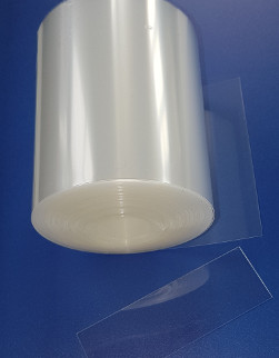
Functionalized Cyclo Olefin Polymer (COP) films with low autofluorescence that can be easily cut into suitable formats post printing. COP is characterized by a low autofluorescence. The finished film-chip can be easily integrated into cartridges or microfluidic devices.

COP-films with the following matrices are available:

The sheets are available in slide format (75 mm x 25 mm) as well as larger sheets (75 mm x 110 mm). Customized pre-scored sheets are available upon request.

Please download the Application Note “Assembling of spotted, pre-cut COP sensor foil with DNA-microarray on a microfluidic hybridization cartridge” here.

ID Title
104 00 056 3D-Amino COP film, 25x75mm, 188µm thickness, 5 sheets/box
104 00 252 3D-Epoxy COP film, 25x75mm, 188µm thickness, 25 sheets/box
104 00 256 3D-Epoxy COP film, 25x75mm, 188µm thickness, 5 sheets/box
104 00 456 3D-NHS COP film, 25x75mm, 188µm thickness, 5 sheets/box

Please do not hesitate to contact us, if you require a special surface for binding of your biomolecules that is not listed in the products table. We can also functionalize microplates, slides, beads and other substrates with our surfaces. We are looking forward to your inquiry.

Functionalized multi-well Plates

Functionalized 96-well plates for microarrays

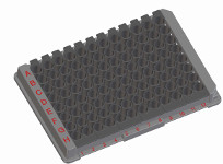
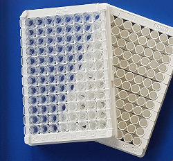
In order to take advantage of the existing liquid handling and automation solutions for 96-well SBS formats, PolyAn is offering a range of functionalized multiwell products which are equipped with the same reactive surfaces as our glass and plastic slides.

Id00 680 251
Title3D-Epoxy 96 well plate, white, 12 x 8-strip, flat bottom
SubstratePS plate
Format96 well plate, white, 12 x 8-strip, flat bottom
Mean Diameter
Color Labeling
Surface Modifications3D-Epoxy
Solids Content
Product Dimensions85 x 128 x 14 mm
Packaging
Packaging Volume4 plates/Box
Package Weight158g
Dimensions128 x 86 x 45mm
Hts Code39 26 90 97
Pads Wells96
Pad Size
Well Formatflat bottom
Product Thickness
DescriptionPolyAn’s 3D-Epoxy functionalized 96-well microplates are used for covalent immobilization of biomolecules. Epoxides are cyclic ethers with a highly strained three member ring. Epoxy rings can be easily reacted with nucleophiles e.g. amines, hydrazines, thiols, hydroxides and carboxyl groups.

Functionalized HTA 96 well plates

The HTA96-well plates of Greiner Bio-One have been specifically designed for microarrays. They are comprised of a low autofluorescence injection moulded plastic which PolyAn equips with our 3D-reactive surfaces. Plates with the following matrices are available:

Please do not hesitate to contact us, if you would like to learn more about this novel design and/or if you would like to test some samples.

Key features:

  • Fast printing on planar surface
  • No electrostatic effects
  • Standard SBS format
  • All of PolyAn’s 3D-reactive matrices available for coupling
  • Mechanically robust, low autofluorescence injection moulded plastic
Id00 680 251
Title3D-Epoxy 96 well plate, white, 12 x 8-strip, flat bottom
SubstratePS plate
Format96 well plate, white, 12 x 8-strip, flat bottom
Mean Diameter
Color Labeling
Surface Modifications3D-Epoxy
Solids Content
Product Dimensions85 x 128 x 14 mm
Packaging
Packaging Volume4 plates/Box
Package Weight158g
Dimensions128 x 86 x 45mm
Hts Code39 26 90 97
Pads Wells96
Pad Size
Well Formatflat bottom
Product Thickness
DescriptionPolyAn’s 3D-Epoxy functionalized 96-well microplates are used for covalent immobilization of biomolecules. Epoxides are cyclic ethers with a highly strained three member ring. Epoxy rings can be easily reacted with nucleophiles e.g. amines, hydrazines, thiols, hydroxides and carboxyl groups.

Standard polystyrene 96-well plates in different designs which are equipped with PolyAn’s 3D-Epoxy and 3D-NHS reactive matrices for covalent coupling of biomolecules.

Key features:

  • Compatible with all liquid handling systems for ELISA
  • Covalent binding of nucleophiles to the plate surface without complex coupling chemistry
  • Integrated antifouling matrix reduces unspecific binding
  • No cross-contamination between wells possible
  • Most cost-efficient solution

Multipart plates

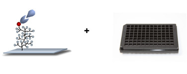
PolyAn’s multipart plates are comprised of a functionalized glass plate (75 x 110 mm, 1mm thickness) which is combined with the ProPlate superstructure after the printing process.

ProPlate® MTP features a biocompatible, pressure sensitive, adhesive bonding system applied to a bottomless microtiter plate to form a leak-proof seal between wells to isolate microarrays printed on glass or plastic during processing. Designs are available in black polystyrene for the 96 well (round) and 384 well (square) plates, and in black polycarbonate for the 96 well (square) plate, which maximizes the array printing area. Application of the substrate is as simple as peeling off a liner to expose the adhesive and pressing the content to the surface. Alignment of arrays within the wells may be facilitated by means of a fixture (sold separately). This product is often customized for content providers seeking a go-to-market solution for their arrays printed on difficult to bond substrates. Lids and adhesive plate seals are also available both as a packaging solution to protect content and as a means to prevent evaporation during reagent incubation steps. ProPlate MTP products are compatible with several available microarray imagers and most robotic liquid handling platforms.

Id00 680 251
Title3D-Epoxy 96 well plate, white, 12 x 8-strip, flat bottom
SubstratePS plate
Format96 well plate, white, 12 x 8-strip, flat bottom
Mean Diameter
Color Labeling
Surface Modifications3D-Epoxy
Solids Content
Product Dimensions85 x 128 x 14 mm
Packaging
Packaging Volume4 plates/Box
Package Weight158g
Dimensions128 x 86 x 45mm
Hts Code39 26 90 97
Pads Wells96
Pad Size
Well Formatflat bottom
Product Thickness
DescriptionPolyAn’s 3D-Epoxy functionalized 96-well microplates are used for covalent immobilization of biomolecules. Epoxides are cyclic ethers with a highly strained three member ring. Epoxy rings can be easily reacted with nucleophiles e.g. amines, hydrazines, thiols, hydroxides and carboxyl groups.

Key features:

  • Fast printing on planar surface
  • No electrostatic effects
  • All of PolyAn’s reactive matrices available for coupling
  • Functionalized bottom plate can be comprised of glass, PMMA, COP and other plastics

Functionalized glass sheets

Functionalized glass sheets without superstructure in 1 mm thickness. The size of the glass sheets is optimized for use with standard 96-well and 384-well superstructures. Other surfaces are available upon request.

Please do not hesitate to contact us, if you require a special surface for binding of your biomolecules that is not listed in the products table. We can also functionalise other plate formats and substrates with our surfaces. Additionally, we have access to a wide range of different surface modifications for binding of small molecules, saccharides etc.

Nitrocellulose Film Slides

ONCYTE® AVID Nitrocellulose Film Slides

Nitrocellulose-coated microarray substrates used for protein, glycan and cell lysate arrays, tissue and cell printing and other applications where binding affinity, binding capacity and preservation of native bioactivities are important.
The nitrocellulose Film-slide and Film-plate technology, trademarked ONCYTE®, was invented by Grace Bio-Labs scientists. The ONCYTE® product is a three dimensional microporous film cast on a variety of solid surfaces (vis. Glass, plastic) comprised of a combination of nitrocellulose polymer and proprietary chemistry that provides an ideal surface for marker discovery and validation as well as study of protein function.

IdTitle
305002ONCYTE® AVID 2- 20mm x 20mm NC pads per slide, Plain Glass 25 x 75 x 1mm - 20 PACK
305003ONCYTE® AVID 3- 18mm x 16mm NC pads per slide, Plain Glass 25 x 75 x 1mm - 20 PACK
305004ONCYTE® AVID 4- 15mm x 15mm NC pads per slide, Plain Glass 25 x 75 x 1mm - 20 PACK
305008ONCYTE® AVID 8- 6mm x 6mm NC pads per slide, Plain Glass 25 x 75 x 1mm - 20 PACK
305012ONCYTE® AVID 12- 6mm x 6mm NC pads per slide, Plain Glass 25 x 75 x 1mm - 20 PACK
305016ONCYTE® AVID 16- 6mm x 6mm NC pads per slide, Plain Glass 25 x 75 x 1mm - 20 PACK
305024ONCYTE® AVID 24- 5mm x 6mm NC pads per slide, Plain Glass 25 x 75 x 1mm - 20 PACK
305064ONCYTE® AVID 64- 2.5mm x 2.5mm NC pads per slide, Plain Glass 25 x 75 x 1mm - 20 PACK
305096ONCYTE® AVID 96- 6mm Diameter, NC Round Pads for Microtiter Plate Glass Substrate, 74 x 110 x 1mm - EACH
305101ONCYTE® AVID 1- 15mm x 69mm NC pad per slide, Plain Glass 25 x 75 x 1mm - 20 PACK
305102ONCYTE® AVID 2- 15mm x 32mm NC pads per slide, Plain Glass 25 x 75 x 1mm - 20 PACK
305103ONCYTE® AVID 3- 20mm x 20mm NC Pads per Slide, Plain Glass 25 x 75 x 1mm - 20 PACK
305108ONCYTE® AVID 8- 6mm x 15mm NC pads per slide , Plain Glass 25 x 75 x 1mm - 20 PACK
305116ONCYTE® AVID 16- 6.5mm x 6.5mm NC pads per slide, Plain Glass 25 x 75 x 1mm - 20 PACK
305118ONCYTE® AVID 8- 6mm x 20mm NC pads per slide, Plain Glass 25 x 75 x 1mm - 20 PACK
305170ONCYTE® AVID 1- 20mm x 51mm NC pad per slide, Plain Glass 25 x 75 x 1mm - 20 PACK
305177ONCYTE® AVID 1- 20mm x 60mm NC pad per slide, Plain Glass 25 x 75 x 1mm - 20 PACK
305196ONCYTE® AVID 96- 6mm x 6mm NC Square Pads for Microtiter Plate Glass Substrate, 74 x 110 x 1mm - EACH
305270ONCYTE® AVID 1- 20mm x 51mm NC pad per slide, Plain Glass 25 x 75 x 1mm - 20 PACK
305278ONCYTE® AVID 1- 21mm x 71mm NC pad per slide, Plain Glass 25 x 75 x 1mm - 20 PACK
305384ONCYTE® AVID 384- 2.5mm x 2.5mm NC Square Pads for Microtiter Plate Glass Substrate, 74 x 110 x 1mm - EACH
315002ONCYTE® AVID 2-20mm x 20mm NC Pad Per Slide, Plain Glass 25 x 75 x 1mm, 20/PK, 20mm X 2.5mm Code 128 Barcoded
315003ONCYTE® AVID 3-18mm x 16mm NC Pad Per Slide, Plain Glass 25 x 75 x 1mm, 20/PK, 20mm X 2.5mm Code 128 Barcoded
315004ONCYTE® AVID 4-15mm x 15mm NC Pad Per Slide, Plain Glass 25 x 75 x 1mm, 20/PK, 20mm X 2.5mm Code 128 Barcoded
315008ONCYTE® AVID 8-6mm x 6mm NC Pad Per Slide, Plain Glass 25 x 75 x 1mm, 20/PK, 20mm X 2.5mm Code 128 Barcoded
315012ONCYTE® AVID 12-6mm x 6mm NC Pad Per Slide, Plain Glass 25 x 75 x 1mm, 20/PK, 20mm X 2.5mm Code 128 Barcoded
315016ONCYTE® AVID 16-6mm x 6mm NC Pad Per Slide, Plain Glass 25 x 75 x 1mm, 20/PK, 20mm X 2.5mm Code 128 Barcoded
315024ONCYTE® AVID 24-5mm x 6mm NC Pad Per Slide, Plain Glass 25 x 75 x 1mm, 20/PK, 20mm X 2.5mm Code 128 Barcoded
315101ONCYTE® AVID 1-15mm x 69mm NC Pad Per Slide, Plain Glass 25 x 75 x 1mm, 20/PK, 20mm X 2.5mm Code 128 Barcoded
315102ONCYTE® AVID 2-15mm x 32mm NC Pad Per Slide, Plain Glass 25 x 75 x 1mm, 20/PK, 20mm X 2.5mm Code 128 Barcoded
315103ONCYTE® AVID 3-20mm x 20mm NC Pad Per Slide, Plain Glass 25 x 75 x 1mm, 20/PK, 20mm X 2.5mm Code 128 Barcoded
315108ONCYTE® AVID 8-6mm x 15mm NC Pad Per Slide, Plain Glass 25 x 75 x 1mm, 20/PK, 20mm X 2.5mm Code 128 Barcoded
315116ONCYTE® AVID 16-6.5mm x 6.5mm NC Pad Per Slide, Plain Glass 25 x 75 x 1mm, 20/PK, 20mm X 2.5mm Code 128 Barcoded
315118ONCYTE® AVID 8-6mm x 20mm NC Pad Per Slide, Plain Glass 25 x 75 x 1mm, 20/PK, 20mm X 2.5mm Code 128 Barcoded
315170ONCYTE® AVID 1-20mm x 51mm NC Pad Per Slide, Plain Glass 25 x 75 x 1mm, 20/PK, 20mm X 2.5mm Code 128 Barcoded
315177ONCYTE® AVID 1-20mm x 60mm NC Pad Per Slide, Plain Glass 25 x 75 x 1mm, 20/PK, 20mm X 2.5mm Code 128 Barcoded
325002ONCYTE® AVID 2-20mm x 20mm NC Pad Per Slide, Plain Glass 25 x 75 x 1mm, 20/PK, 20mm X 8.5mm Code 128 Barcoded
325008ONCYTE® AVID 8-6mm x 6mm NC Pad Per Slide, Plain Glass 25 x 75 x 1mm, 20/PK, 20mm X 8.5mm Code 128 Barcoded
325170ONCYTE® AVID 1-20mm x 51mm NC Pad Per Slide, Plain Glass 25 x 75 x 1mm, 20/PK, 20mm X 8.5mm Code 128 Barcoded
325177ONCYTE® AVID 1-20mm x 60mm NC Pad Per Slide, Plain Glass 25 x 75 x 1mm, 20/PK, 20mm X 8.5mm Code 128 Barcoded
335002ONCYTE® AVID 2-20mm x 20mm NC Pad Per Slide, Plain Glass 25 x 75 x 1mm, 20/PK, 22mm X 14mm - CODABAR Barcoded
335008ONCYTE® AVID 8-6mm x 6mm NC Pad Per Slide, Plain Glass 25 x 75 x 1mm, 20/PK, 22mm X 14mm - CODABAR Barcoded
335012ONCYTE® AVID 12-6mm x 6mm NC Pad Per Slide, Plain Glass 25 x 75 x 1mm, 20/PK, 22mm X 14mm - CODABAR Barcoded
335170ONCYTE® AVID 1-20mm x 51mm NC Pad Per Slide, Plain Glass 25 x 75 x 1mm, 20/PK, 22mm X 14mm - CODABAR Barcoded

ONCYTE® AVID -Nitrocellulose film slides with highest binding capacity, large dynamic range. Original formula from Grace Bio-Labs. Slides are 25 x 75 x 1 mm and have 20 slides in a Pack

The ONCYTE® NOVA product is a three dimensional microporous film cast on Glass comprised of a combination of nitrocellulose polymer and proprietary chemistry that provides an ideal surface for marker discovery and validation as well as study of protein function.

ONCYTE® NOVA Nitrocellulose porous film slides are the first generation of porous nitrocellulose with reduced fluorescence background compared to AVID slides.

IdTitle
505002ONCYTE® NOVA 2- 20mm x 20mm NC pads per slide, Plain Glass 25 x 75 x 1mm - 20 PACK
505003ONCYTE® NOVA 3- 18mm x 16mm NC pads per slide, Plain Glass 25 x 75 x 1mm - 20 PACK
505004ONCYTE® NOVA 4- 15mm x 15mm NC pads per slide, Plain Glass 25 x 75 x 1mm - 20 PACK
505008ONCYTE® NOVA 8- 6mm x 6mm NC pads per slide, Plain Glass 25 x 75 x 1mm - 20 PACK
505012ONCYTE® NOVA 12- 6mm x 6mm NC pads per slide, Plain Glass 25 x 75 x 1mm - 20 PACK
505016ONCYTE® NOVA 16- 6mm x 6mm NC pads per slide, Plain Glass 25 x 75 x 1mm - 20 PACK
505024ONCYTE® NOVA 24- 5mm x 6mm NC Pads per slide, Plain Glass 25 x 75 x 1mm - 20 PACK
505064ONCTYE® NOVA 64- 2.5mm x 2.5mm NC pads per slide, Plain Glass 25 x 75 x 1mm - 20 PACK
505096ONCYTE® NOVA 96- 6mm Diameter, NC Round Pads for Microtiter Plate Glass Substrate, 74 x 110 x 1mm - EACH
505101ONCYTE® NOVA 1- 15mm x 69mm NC pad per slide, Plain Glass 25 x 75 x 1mm - 20 PACK
505102ONCYTE® NOVA 2- 15mm x 32mm NC pads per slide, Plain Glass 25 x 75 x 1mm - 20 PACK
505103ONCYTE® NOVA 3- 20mm x 20mm NC pads per slide, Plain Glass 25 x 75 x 1mm - 20 PACK
505108ONCYTE® NOVA 8- 6mm x 15mm NC pads per slide, Plain Glass 25 x 75 x 1mm - 20 PACK
505116ONCYTE® NOVA 16- 6.5mm x 6.5mm NC pads per slide, Plain Glass 25 x 75 x 1mm - 20 PACK
505118ONCYTE® NOVA 8- 6mm x 20mm NC pads per slide, Plain Glass 25 x 75 x 1mm - 20 PACK
505170ONCYTE® NOVA 1- 20mm x 51mm NC pad per slide, Plain Glass 25 x 75 x 1mm - 20 PACK
505177ONCYTE® NOVA 1- 20mm x 60mm NC pad per slide, Plain Glass 25 x 75 x 1mm - 20 PACK
505196ONCYTE® NOVA 96- 6mm x 6mm NC Square Pads for Microtiter Plate Glass Substrate, 74 x 110 x 1mm - EACH
505278ONCYTE® NOVA 1- 21mm x 71mm NC pad per slide, Plain Glass 25 x 75 x 1mm - 20 PACK
505384ONCYTE® NOVA 384- 2.5mm X 2.5mm NC Square Pads for Microtiter Plate Glass Substrate, 74 x 110 x 1mm - EACH

We can easily customize pad size and placement for your application. Please do not hesitate to contact us for a tailor-made solution.

SuperNOVA: Porous Nitrocellulose Surface for Fluorescence Detection

Fluorescence detection is a preferred method for many quantitative protein microarray assays in research and diagnostic applications due to its excellent sensitivity and potential for multiplexing. Use of multiplexed fluorescence-based assays on porous nitrocellulose though has been hampered by high backgrounds at shorter excitation wavelengths (such as the 532 nm fluorescence channel).

Grace Bio-Labs has therefore developed ONCYTE® SuperNOVA microporous nitrocellulose film slides to increase protein microarray assay sensitivity and dynamic range using fluorescent endpoints at multiple wavelengths. Use of SuperNOVA slides can lower the limit of detection with fluorescent endpoints by 3 – 4 orders of magnitude over competing nitrocellulose film slides.

IdTitle
705002ONCYTE® SuperNOVA 2- 20mm x 20mm NC pads per slide, Plain Glass 25 x 75 x 1mm - 20 PACK
705003ONCYTE® SuperNOVA 3- 18mm x 16mm NC pads per slide, Plain Glass 25 x 75 x 1mm - 20 PACK
705004ONCYTE® SuperNOVA 4- 15mm x 15mm NC pads per slide, Plain Glass 25 x 75 x 1mm - 20 PACK
705008ONCYTE® SuperNOVA 8- 6mm x 6mm NC pads per slide, Plain Glass 25 x 75 x 1mm - 20 PACK
705012ONCYTE® SuperNOVA 12- 6mm x 6mm NC pads per slide, Plain Glass 25 x 75 x 1mm - 20 PACK
705016ONCYTE® SuperNOVA 16- 6mm x 6mm NC pads per slide, Plain Glass 25 x 75 x 1mm - 20 PACK
705024ONCYTE® SuperNOVA 24- 5mm x 6mm NC pads per slide, Plain Glass 25 x 75 x 1mm - 20 PACK
705064ONCYTE® SuperNOVA 64- 2.5mm x 2.5mm NC pads per slide, Plain Glass 25 x 75 x 1mm - 20 PACK
705096ONCYTE® SuperNOVA 96- 6mm Diameter NC Round Pads for Microtiter Plate Glass Substrate, 74 x 110 x 1mm - EACH
705101ONCYTE® SuperNOVA 1- 15mm x 69mm NC pad per slide, Plain Glass 25 x 75 x 1mm - 20 PACK
705102ONCYTE® SuperNOVA 2- 15mm x 32mm NC pads per slide, Plain Glass 25 x 75 x 1mm - 20 PACK
705103ONCYTE® SuperNOVA 3- 20mm x 20mm NC pads per slide, Plain Glass 25 x 75 x 1mm - 20 PACK
705108ONCYTE® SuperNOVA 8- 6mm x 15mm NC pads per slide, Plain Glass 25 x 75 x 1mm - 20 PACK
705116ONCYTE® SuperNOVA 16- 6.5mm x 6.5mm NC pads per slide, Plain Glass 25 x 75 x 1mm - 20 PACK
705118ONCYTE® SuperNOVA 8- 6mm x 20mm NC pads per slide, Plain Glass 25 x 75 x 1mm - 20 PACK
705124ONCYTE® SuperNOVA 24- 5mm x 6mm NC pads per slide, Plain Glass 25 x 75 x 1mm - 20 PACK
705170ONCYTE® SuperNOVA 1- 20mm x 51mm NC pad per slide, Plain Glass 25 x 75 x 1mm - 20 PACK
705177ONCYTE® SuperNOVA 1- 20mm x 60mm NC pad per slide, Plain Glass 25 x 75 x 1mm - 20 PACK
705196ONCYTE® SuperNOVA 96- 6mm X 6mm NC Square Pads for Microtiter Plate Glass Substrate, 74 x 110 x 1mm - EACH
705278ONCYTE® SuperNOVA 1- 21mm x 71mm NC pad per slide, Plain Glass 25 x 75 x 1mm - 20 PACK
705384ONCYTE® SuperNOVA 384- 2.5mm x 2.5mm NC Square Pads for Microtiter Plate Glass Substrate, 74 x 110 x 1mm - EACH

ONCYTE(R) SuperNOVA-Nitrocellulose film slides optimized for low background fluorescence without sacrificing dynamic range. Glass slides are 25 x 75 x 1 mm and have 20 slides in a Pack.

ONCYTE® AVID Nitrocellulose Film Slides

Nitrocellulose-coated microarray substrates used for protein, glycan and cell lysate arrays, tissue and cell printing and other applications where binding affinity, binding capacity and preservation of native bioactivities are important.

The nitrocellulose Film-slide and Film-plate technology, trademarked ONCYTE®, was invented by Grace Bio-Labs scientists. The ONCYTE® product is a three dimensional microporous film cast on a variety of solid surfaces (vis. Glass, plastic) comprised of a combination of nitrocellulose polymer and proprietary chemistry that provides an ideal surface for marker discovery and validation as well as study of protein function.

IdTitle
305002ONCYTE® AVID 2- 20mm x 20mm NC pads per slide, Plain Glass 25 x 75 x 1mm - 20 PACK
305003ONCYTE® AVID 3- 18mm x 16mm NC pads per slide, Plain Glass 25 x 75 x 1mm - 20 PACK
305004ONCYTE® AVID 4- 15mm x 15mm NC pads per slide, Plain Glass 25 x 75 x 1mm - 20 PACK
305008ONCYTE® AVID 8- 6mm x 6mm NC pads per slide, Plain Glass 25 x 75 x 1mm - 20 PACK
305012ONCYTE® AVID 12- 6mm x 6mm NC pads per slide, Plain Glass 25 x 75 x 1mm - 20 PACK
305016ONCYTE® AVID 16- 6mm x 6mm NC pads per slide, Plain Glass 25 x 75 x 1mm - 20 PACK
305024ONCYTE® AVID 24- 5mm x 6mm NC pads per slide, Plain Glass 25 x 75 x 1mm - 20 PACK
305064ONCYTE® AVID 64- 2.5mm x 2.5mm NC pads per slide, Plain Glass 25 x 75 x 1mm - 20 PACK
305096ONCYTE® AVID 96- 6mm Diameter, NC Round Pads for Microtiter Plate Glass Substrate, 74 x 110 x 1mm - EACH
305101ONCYTE® AVID 1- 15mm x 69mm NC pad per slide, Plain Glass 25 x 75 x 1mm - 20 PACK
305102ONCYTE® AVID 2- 15mm x 32mm NC pads per slide, Plain Glass 25 x 75 x 1mm - 20 PACK
305103ONCYTE® AVID 3- 20mm x 20mm NC Pads per Slide, Plain Glass 25 x 75 x 1mm - 20 PACK
305108ONCYTE® AVID 8- 6mm x 15mm NC pads per slide , Plain Glass 25 x 75 x 1mm - 20 PACK
305116ONCYTE® AVID 16- 6.5mm x 6.5mm NC pads per slide, Plain Glass 25 x 75 x 1mm - 20 PACK
305118ONCYTE® AVID 8- 6mm x 20mm NC pads per slide, Plain Glass 25 x 75 x 1mm - 20 PACK
305170ONCYTE® AVID 1- 20mm x 51mm NC pad per slide, Plain Glass 25 x 75 x 1mm - 20 PACK
305177ONCYTE® AVID 1- 20mm x 60mm NC pad per slide, Plain Glass 25 x 75 x 1mm - 20 PACK
305196ONCYTE® AVID 96- 6mm x 6mm NC Square Pads for Microtiter Plate Glass Substrate, 74 x 110 x 1mm - EACH
305270ONCYTE® AVID 1- 20mm x 51mm NC pad per slide, Plain Glass 25 x 75 x 1mm - 20 PACK
305278ONCYTE® AVID 1- 21mm x 71mm NC pad per slide, Plain Glass 25 x 75 x 1mm - 20 PACK
305384ONCYTE® AVID 384- 2.5mm x 2.5mm NC Square Pads for Microtiter Plate Glass Substrate, 74 x 110 x 1mm - EACH
315002ONCYTE® AVID 2-20mm x 20mm NC Pad Per Slide, Plain Glass 25 x 75 x 1mm, 20/PK, 20mm X 2.5mm Code 128 Barcoded
315003ONCYTE® AVID 3-18mm x 16mm NC Pad Per Slide, Plain Glass 25 x 75 x 1mm, 20/PK, 20mm X 2.5mm Code 128 Barcoded
315004ONCYTE® AVID 4-15mm x 15mm NC Pad Per Slide, Plain Glass 25 x 75 x 1mm, 20/PK, 20mm X 2.5mm Code 128 Barcoded
315008ONCYTE® AVID 8-6mm x 6mm NC Pad Per Slide, Plain Glass 25 x 75 x 1mm, 20/PK, 20mm X 2.5mm Code 128 Barcoded
315012ONCYTE® AVID 12-6mm x 6mm NC Pad Per Slide, Plain Glass 25 x 75 x 1mm, 20/PK, 20mm X 2.5mm Code 128 Barcoded
315016ONCYTE® AVID 16-6mm x 6mm NC Pad Per Slide, Plain Glass 25 x 75 x 1mm, 20/PK, 20mm X 2.5mm Code 128 Barcoded
315024ONCYTE® AVID 24-5mm x 6mm NC Pad Per Slide, Plain Glass 25 x 75 x 1mm, 20/PK, 20mm X 2.5mm Code 128 Barcoded
315101ONCYTE® AVID 1-15mm x 69mm NC Pad Per Slide, Plain Glass 25 x 75 x 1mm, 20/PK, 20mm X 2.5mm Code 128 Barcoded
315102ONCYTE® AVID 2-15mm x 32mm NC Pad Per Slide, Plain Glass 25 x 75 x 1mm, 20/PK, 20mm X 2.5mm Code 128 Barcoded
315103ONCYTE® AVID 3-20mm x 20mm NC Pad Per Slide, Plain Glass 25 x 75 x 1mm, 20/PK, 20mm X 2.5mm Code 128 Barcoded
315108ONCYTE® AVID 8-6mm x 15mm NC Pad Per Slide, Plain Glass 25 x 75 x 1mm, 20/PK, 20mm X 2.5mm Code 128 Barcoded
315116ONCYTE® AVID 16-6.5mm x 6.5mm NC Pad Per Slide, Plain Glass 25 x 75 x 1mm, 20/PK, 20mm X 2.5mm Code 128 Barcoded
315118ONCYTE® AVID 8-6mm x 20mm NC Pad Per Slide, Plain Glass 25 x 75 x 1mm, 20/PK, 20mm X 2.5mm Code 128 Barcoded
315170ONCYTE® AVID 1-20mm x 51mm NC Pad Per Slide, Plain Glass 25 x 75 x 1mm, 20/PK, 20mm X 2.5mm Code 128 Barcoded
315177ONCYTE® AVID 1-20mm x 60mm NC Pad Per Slide, Plain Glass 25 x 75 x 1mm, 20/PK, 20mm X 2.5mm Code 128 Barcoded
325002ONCYTE® AVID 2-20mm x 20mm NC Pad Per Slide, Plain Glass 25 x 75 x 1mm, 20/PK, 20mm X 8.5mm Code 128 Barcoded
325008ONCYTE® AVID 8-6mm x 6mm NC Pad Per Slide, Plain Glass 25 x 75 x 1mm, 20/PK, 20mm X 8.5mm Code 128 Barcoded
325012ONCYTE® AVID 12-6mm x 6mm NC Pad Per Slide, Plain Glass 25 x 75 x 1mm, 20/PK, 20mm X 8.5mm Code 128 Barcoded
325170ONCYTE® AVID 1-20mm x 51mm NC Pad Per Slide, Plain Glass 25 x 75 x 1mm, 20/PK, 20mm X 8.5mm Code 128 Barcoded
325177ONCYTE® AVID 1-20mm x 60mm NC Pad Per Slide, Plain Glass 25 x 75 x 1mm, 20/PK, 20mm X 8.5mm Code 128 Barcoded
335002ONCYTE® AVID 2-20mm x 20mm NC Pad Per Slide, Plain Glass 25 x 75 x 1mm, 20/PK, 22mm X 14mm - CODABAR Barcoded
335008ONCYTE® AVID 8-6mm x 6mm NC Pad Per Slide, Plain Glass 25 x 75 x 1mm, 20/PK, 22mm X 14mm - CODABAR Barcoded
335012ONCYTE® AVID 12-6mm x 6mm NC Pad Per Slide, Plain Glass 25 x 75 x 1mm, 20/PK, 22mm X 14mm - CODABAR Barcoded
335170ONCYTE® AVID 1-20mm x 51mm NC Pad Per Slide, Plain Glass 25 x 75 x 1mm, 20/PK, 22mm X 14mm - CODABAR Barcoded

ONCYTE® AVID -Nitrocellulose film slides with highest binding capacity, large dynamic range. Original formula from Grace Bio-Labs. Slides are 25 x 75 x 1 mm and have 20 slides in a Pack

Accessories for Microarrays

Microarray Reagents Reagents for reactive surfaces

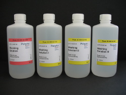
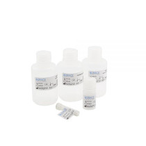
PolyAn provides washing and blocking buffers that are optimized for PolyAn’s 2D- and 3D-reactive surfaces. The PolyAn-buffers promotes highly efficient coupling of biomolecules and increases the signal/noise ratio by minimizing unspecific binding.

Id Title
000 02 501 PolyAn Blocking Buffer 100 + PolyAn Washing Buffer Set 100
000 02 505 PolyAn Blocking Buffer 250 + PolyAn Washing Buffer Set 250
000 02 510 PolyAn Blocking Buffer 500 + PolyAn Washing Buffer Set 500

Reagents for Nitrocellulose Film Slides

Grace Bio-Labs microarray regents have been specifically formulated to achieve the full potential of porous nitrocellulose, accelerating experimental design and data collection and validation. They provide reliable tools for the development of reproducible and sensitive microarray-based assays, from printing to the long-term preservation of the printed nitrocellulose film slide. Retention of protein function and structure are guaranteed during downstream microarray applications.

The Protein Array Assay System and the Reverse Phase Array System provide each a set of reagents necessary to obtain optimal results for forward phase microarrays applications (antibody and antigen capture array) and reverse phase protein array (protein expression profiling, biomarker discovery etc.).

IdTitle
105100Super G Blocking Buffer, 100mL
105101Super G Blocking Buffer, 500mL
105102Super G Plus Protein Preservative - 100mL
105103Super G Plus Protein Preservative - 500mL
105106Q-Block Protein Microarray Blocking Buffer - 100mL
105107Q-Block Protein Microarray Blocking Buffer - 500mL
105111(2X) PATH® Blocking Buffer
10520110X PBST Wash Buffer - 100mL
10520510X PBST Wash Buffer - 500mL
105207(2X) PATH® Rinse Buffer
105208(4X) PATH® Sample Diluent
105209(4X) PATH® Wash Buffer
105407(4X) PATH® Print Buffer
105501GBL Protein Array Buffer, 10mL
105502PATH® Protein Microarray Kit
1055051 Bottle of Spot Tuning Solution - 1.25mL
105901Protein Array Assay System

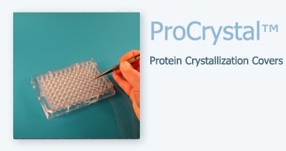
ProPlate™ multi-well chambers

ProPlates were specifically designed to enable automated robotic liquid handling. Two main configurations are available:

  • The ProPlate Microtiter Plate is comprised of an SBS compliant microtiter plate superstructure and feature a SesureSeal™ adhesive bonding system applied to the bottom.
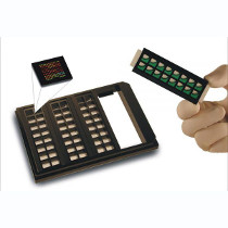
  • The ProPlate Multi-Array Slide System integrates microscope array technology with automated microtiter plate processing. Individual modules fit into a tray with a standard microtiter plate footprint.

The multi-well chambers can be used with PolyAn’s functionalized glass slides and the ONCYTE® nitrocellulose-coated slides. They integrate microscope slide based microarray technology with high-throughput microtiter plate processing. Select a well configuration from our repertoire or contact us if you need a customized version.

ProPlate™ Microtiter Plates

IdTitle
204960ProPlate 96-well microtiter plate - Square wells - EACH
204969ProPlate 96-well microtiter plate - Round wells - EACH
206384ProPlate 384-well microtiter plate - EACH

ProPlate Slide Chamber System

IdTitle
204830ProPlate Delrin Non-Numbered Snap Clips - 20 PACK
204838ProPlate Stainless Steel, Spring Clips -NON-NUMBERED - 20 PACK
204874Seal Strips - 50 pack
205005ProPlate 16-well gasket only - Square wells- 10 PACK
246870ProPlate tray and cover set - for Spring clips, with seal strips and applicator - Each
246871ProPlate 1-well gasket only- 10 PACK
246872ProPlate 2-well gasket only- 10 PACK
246873ProPlate 3-well gasket only- 10 PACK
246874ProPlate 4-well gasket only- 10 PACK
246875ProPlate 64-well gasket only - 10 PACK
246877ProPlate 16-well gasket only - Round wells- 10 PACK
246878ProPlate 8-well gasket only- 10 PACK

ProPlate Slide Modules

IdTitle
204862ProPlate 16 Well Slide Module - 7mm X 7mm, Delrin Snap Clips - 2 PACK
206862ProPlate 16-well slide module - Square wells, No clips- 10 PACK
244862ProPlate 16-well slide module - Square wells, Steel Spring Clips - 2 PACK
246850ProPlate 16-well slide module - Round Wells, Steel Spring Clips- 10 PACK
246851ProPlate 1-well slide module - No clips- 10 PACK
246852ProPlate 2-well slide module - No clips- 10 PACK
246853ProPlate 3-well slide module - No clips- 10 PACK
246854ProPlate 4-well slide module - No clips- 10 PACK
246855ProPlate 64-well slide module - No clips- 10 PACK
246858ProPlate 8-well slide module - No clips- 10 PACK
246860ProPlate 16-well slide module - Round wells, Delrin clips - 2 PACK
246861ProPlate 1-well slide module - Delrin clips - 2 PACK
246862ProPlate 2-well slide module - Delrin clips - 2 PACK
246863ProPlate 3-well slide module - Delrin clips - 2 PACK
246864ProPlate 4-well slide module - Delrin clips - 2 PACK
246865ProPlate 64-well slide module - Delrin clips - 2 PACK
246868ProPlate 8-well slide module - Delrin clips - 2 PACK
248860ProPlate 16-well slide module - Round Wells, Steel Spring Clips - 2 PACK
248861ProPlate 1-well slide module - Steel Spring Clips - 2 PACK
248862ProPlate 2-well slide module - Steel Spring Clips - 2 PACK
248863ProPlate 3-well slide module - Steel Spring Clips - 2 PACK
248864ProPlate 4-well slide module - Steel Spring Clips - 2 PACK
248865ProPlate 64-well slide module - Steel Spring Clips - 2 PACK
248868ProPlate 8-well slide module - Steel Spring Clips - 2 PACK

ProPlate SBS Compliant Tray Sets

IdTitle
204860ProPlate 16-well tray set - square wells, delrin clips - Each
246880ProPlate 16-well tray set - Round Wells, Steel Spring Clips - EACH
246881ProPlate 1-well tray set - Steel Spring Clips - EACH
246882ProPlate 2-well tray set - Steel Spring Clips - EACH
246883Proplate 3-well tray set - Steel Spring Clips - EACH
246884Proplate 4-well tray set - Steel Spring Clips - EACH
246888ProPlate 8-well tray set - Steel Spring Clips - EACH
246890ProPlate 16-well tray set - Square wells, steel spring clips - EACH
247865ProPlate 64-well tray set - square wells, delrin clips - EACH
247880ProPlate 16-well tray set - Round wells, delrin clips - EACH
247881ProPlate 1-well tray set - Delrin clips - EACH
247882ProPlate 2-well tray set - Delrin clips - EACH
247883ProPlate 3-well tray set - Delrin clips - EACH
247884ProPlate 4-well tray set - Delrin clips - EACH
247888ProPlate 8-well tray set - Delrin clips - EACH
249865ProPlate 64-well tray set - Steel Spring Clips - EACH

ProPlate Accessories & Replacement Gaskets

IdTitle
204870ProPlate Tray and Cover Set for Delrin Snap Clip Modules. Holds 4 Slide Modules - Includes 10 Seal Strips and 1 Applicator
204871ProPlate tray and cover - for Delrin Clips- EACH
246879ProPlate Tray and Cover - for Spring clips - EACH

ProPlate™ multi-well chambers

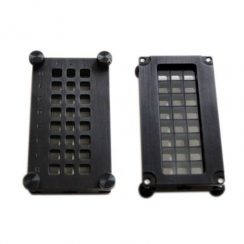
ProChambers™ MultiWell Chambers are made from a durable, flat, precision machined aluminum to ensure proper slide alignment and leak-free arrays. The chambers have a footprint compatible with multi-channel pipettes or automated loading. The top chamber face is embossed with a letter and number panel for easy identification of each subarray.

The ArraySlide 16 chamber fits one microarray slide and sections it into 16 individual surfaces for multiple simultaneous protein, gene expression, or screening analysis studies. The unit includes a lower carrier tray, upper structure plate, and gasket (patent pending). The 7 mm x 7 mm surfaces are positioned 9 mm apart in a 2 x 8 format for easy loading by multi-channel pipettes or automated robots.

ProChambers™ MultiWell Chambers

IdTitle
64550816 Well ProChamber Microarray System, 2 x 8 Well, 7.5 mm x 7.5 mm, 9 mm Center- EACH

FlexWell™ removable incubation chambers for microarrays using the slide format.

Features:

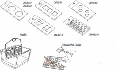
  • Silicone FlexWell™ forms wells on slides using a clean release adhesive to isolate up to 16 specimens per slide
  • Slides and FlexWell™ fit into a reusable slide tray organizing wells into the footprint of a SBS compliant microtiter plate
  • Gaskets may be sealed for mixing and to prevent evaporation
  • Available in eight-well and 16-well (shown) formats

FlexWell™ incubation chambers form wells on slides using a clean release adhesive to isolate up to 16 specimens per slide using multiwall chambers. The gaskets are coated with SecureSeal™ PLUS adhesive layer with removable liner, while the top surface is sealed with FlexWell™ Adhesive Seal Strips for mixing and to prevent evaporation. Multiple slides and FlexWells™ may be inserted into a reusable FlexWell™ tray, organizing wells into the footprint of a SBS compliant microtiter plate to facilitate handling.

Please note, that we can customize chamber shape, size and depth for your application. Ask for technical support if your experimental conditions involve solvents or extreme temperature or pH, and for more information on FlexWell incubation chambers.

ProChambers™ MultiWell Chambers

IdTitle
204064FlexWell 64-3.5 X 3.5mm Wells, 1.8mm Depth 25 X 75mm OD, Black Silicone - Adhesive One Side - 5 PACK
204908FlexWell 8-6.5 X 6.5mm Wells, 3.2mm Depth 25 X 40mm OD Clear Silicone - Adhesive One Side - 10 PACK
204916FlexWell 16-6.5 X 6.5mm Wells, 3.2mm Depth 25 X 75mm OD Clear Silicone - Adhesive One Side - 10 PACK
204970Flexwell 4 Slide Tray Holds Up to 4 Slides , Tray Measures 75.9mm X 127.6mm OD - EACH
204974Flexwell Seal Strips 24mm X 77mm Seal Area - 50 PACK
204996FlexWell 96 Square Well 7.25 X 7.25mm, 9mm Center to Center Well Spacing, 4.5mm Depth, 110 X 75mm OD. Clear Silicone - Adhesive One Side, GASKET ONLY - 5 PACK
204997FlexPlate 96 Square Well 7.25 X 7.25mm, 9mm Center to Center Well Spacing, 4.5mm Depth, 110 X 75mm OD. Clear Silicone - Adhesive One Side, Black Frame - EACH

HybriWell™ Sealing System seals securely to a microscope slide surface in seconds.

  • Multiple formats
  • Dual loading ports, seal tabs included
  • Clean release
  • Small volume, minimize reagent use

HybriWell™ Sealing System bonds securely to a microscope slide surface in seconds to confine small reagent volumes with samples and eliminate evaporation. Each package includes illustrated instructions for use, applicator and 200 adhesive port seals. Any of the designs listed may be easily customized to increase or decrease chamber depth. Hybriwells™ are recommended for most protein and nucleic acid assays. If you are using Cy5 or ® Alexa Fluor 647 direct-labeled DNA probes please refer to our new “fluorescent friendly” chambers. All current designs may be fabricated using our “fluorescent friendly” adhesive material.

ProChambers™ MultiWell Chambers

IdTitle
440904HBW HybriWell Sealing System 21mm X 60mm X 0.14mm Depth, SecureSeal Adhesive Chamber, 3.2mm Dia. Ports - 200 Port Seals Included (Optimized for NAPPA Arrays) - 100 PACK
611101HBW13 / 1-13mm Dia. X 0.15mm Depth / 18UL Approx. Vol. per Chamber, 25mm X 28mm OD / SecureSeal Adhesive Chamber / 1.5mm Dia. Ports - 200 Port Seals Included - 100 PACK
611102HBW20-HybriWell / 20mm Dia. X 0.15mm Depth / 30UL Approx. Vol., 25mm X 30mm OD / SecureSeal Adhesive Chamber / 1.5mm Dia. Ports - 200 Port Seals Included - 100 PACK
611103HBW1932-HybriWell, 19mm X 32mm X 0.15mm Depth / 30-50UL Approx. Vol., 26mm X 43mm OD / SecureSeal Adhesive Chamber / 1.5mm Dia. Ports - 200 Port Seals Included - 100 PACK
611104HBW2222-HybriWell / 21.6mm X 21.6mm X 0.15mm Depth / 30-50UL Approx. Vol., 25.5mm X 30mm OD / SecureSeal Adhesive Chamber / 1.5mm Dia. Ports - 200 Port Seals Included - 100 PACK
611105HBW75-HybriWell / 21mm X 40mm X 0.15mm Depth / 50-100UL Approx. Vol., 25mm X 50mm OD / SecureSeal Adhesive Chamber / 3.2mm Dia. Ports - 200 Port Seals Included - 100 PACK
611106HBW4545-HybriWell, 45mm X 45mm X 0.15mm Depth / 150-300UL Approx. Vol. / 51mm X 56mm OD / SecureSeal Adhesive Chamber / 1.5mm Dia. Ports - 200 Port Seals Included - 50 PACK
611115HBW2230-HybriWell, 22mm X 30mm X 0.15mm Depth / 40-70UL Approx. Vol. per Chamber, 25.5mm X 50mm OD / SecureSeal Adhesive Chamber 1.5mm Dia. Ports - 200 Port Seals Included - 100 PACK
611201"HBW2240FL-HybriWell FL, 22mm X 40mm X 0.12mm Depth / 90 - 100UL Approx. Vol., 25.5mm X 50mm OD / Fluor "Friendly" Adhesive Chamber / 1.5mm Dia. Ports - 200 Port Seals Included - 100 PACK",,,,,,,"25.5 x 56
611202"HBW2260FL-HybriWell FL, 22mm X 60mm X 0.12mm Depth / 140 - 165UL Approx. Vol., 25mm X 69mm OD / Fluor "Friendly" Adhesive Chamber / 1.5mm Dia. Ports - 200 Port Seals Included - 100 PACK",,,,,,,"25 x 64
611204"HBW2222FL-1L-HybriWell FL, 22mm X 22mm X 0.12mm Depth / 15 - 25UL Approx. Vol., 25.5mm X 30mm OD / Fluor "Friendly" Adhesive Chamber / 1.5mm Dia. Ports - 200 Port Seals Included - 100 PACK",,,,,,,"25.5 x 30
611205"HBW75FL-1L-HybriWell FL / 21mm X 40mm X 0.12mm Depth / 50 - 100UL Approx. Vol., 25mm X 50mm OD / Fluor "Friendly" Adhesive Chamber / 3.2mm Dia. Ports - 200 Port Seals Included - 100 PACK",,,,,,,"25 x 56
612101HBW2240-HybriWell 22mm X 40mm X 0.25mm Depth / 180-200UL Approx. Vol., 25.5mm X 50mm OD / SecureSeal Adhesive Chamber / 1.5mm Dia. Ports - 200 Port Seals Included - 100 PACK
612102HBW2260-HybriWell 22mm X 60mm X 0.25mm Depth / 280-330UL Approx. Vol., 25mm X 69mm OD / SecureSeal Adhesive Chamber / 1.5mm Dia. Ports - 200 Port Seals Included - 100 PACK
612103HBW2265-HybriWell 22mm X 65mm X 0.25mm Depth / 310-425UL Approx. Vol., 25mm X 75mm OD / SecureSeal Adhesive Chamber / 3.2mm Dia. Ports - 200 Port Seals Included - 100 PACK
612105HBW6021-HybriWell 21mm X 60mm X 0.15mm Depth /275-320UL Approx. Vol., 25mm X 75mm OD / SecureSeal Adhesive Chamber, End Tab, 3.2mm Dia. Ports - 200 Port Seals Included - 100 PACK
612106HBW3S-HybriWell 3-20mm X 21mm X 0.25mm Depth / 100UL Approx. Vol. PER CHMBR, 25mm X 75mm OD / SecureSeal Adhesive Chamber / 1.5mm Dia. Ports / 200 Port Seals Included - 100 PACK
612107HBW6L-2L-HybriWell / 6-9.8mm X 20mm X 0.25mm Depth / 50UL Approx. Vol. PER CHMBR, 25mm X 75mm OD / SecureSeal Adhesive Chamber / 1.5mm Dia. Ports - 200 Port Seals Included - 100 PACK
612301HBW2160-HybriWell 1-21mm X 60mm X 0.25mm Depth / 275-320UL Approx. Vol. per Chamber, 25mm X 70mm OD / SecureSeal Adhesive Chamberwith Corner Tab, 3mm Dia. Ports - 200 Port Seals Included - 100 PACK
615101HybriWell Assortment - 8 PACK

Product description

Disposable seals comprised of a plastic cover and chamber walls fabricated from double-sided adhesive to create the desired depth. Access ports in the chamber surface allow for the addition or removal of reactants. Ports are easily sealed using Adhesive Seal Tabs. Sealed chambers are watertight and ideally suited for submerged or overnight incubations. RNase free, hydrophobic surfaces will not trap or bind probes. Disposable chamber removes cleanly and easily even after heating. Two standard adhesive types (SecureSeal™ and Fluorescent Friendly) are available in a variety of chamber sizes, and depths. This product is easily customized for applications that employ the use of “exotic” surface chemistries by selecting from our portfolio of adhesive options.

SecureSeal™ Adhesive vs Fluorescent Friendly Adhesive:

Standard versions of the HybriWell™ are manufactured using double-sided, clear SecureSeal™ Adhesive. The Fluorescent Friendly Adhesive was developed in response to reports from few laboratories that they were experiencing signal quenching using CY5 and AF647 using SecureSeal™ Adhesive. We contacted multiple HybriWell™ customers using these fluors for hybridization and results were mixed with many of the labs having no issues whatsoever. After creating a matrix of variables, the information we know to date is:

  • The phenomenon only occurs during hybridization assays above 55C
  • Only occurs if the fluor is directly conjugated to the probe (no problem in a sandwich assay).
  • CY3 and other fluors are not affected.
  • Some labs do not have the quenching issue at all under any of the above conditions.

As far as we know, some component of the hybridization buffer is reacting with some component of the adhesive used to make standard HybriWell™ and SecureSeal™ products resulting in signal quenching. We are not able to identify which component may be the culprit as many of the customers having the problem were using pre-made hybe buffers , the manufacturers of which will not share the formulation with us. We feel that it is appropriate to alert customers to the potential risk of this quenching phenomenon if they are using SecureSeal™ adhesive under any of the conditions we’ve described. We identified the Fluorescent Friendly adhesive as a material which does not quench signal under any conditions and it was tested by the labs which reported the problem and all reported favorable results.

HybriWells™ are sold with a standard compliment of Adhesive Seal Tabs (QTY 200) which may be applied to the filling ports to prevent evaporation. Many customers do not find it necessary or desirable to use the tabs, therefore to reduce cost we do not include a full complement of port seals with all versions of the product. For example, if you purchased HybriWell 6L, a seal with 6 chambers X 2 ports each, you would require 1200 port seals to cover all of the filling ports, therefore 800 additional port seals would need to be purchased separately.

The HybriWell™ product group represents the one most often modified for a customer’s particular application, OEM component for private label kits and research and development projects. Applications include commercial microarray kits, flowcells and chambers for genome sequencing, tissue incubation and content packaging.

HybriSlip™ Hybridization Covers

HybriSlips™ are rigid, light-weight, thin plastic coverslips that minimize friction and facilitate uniform reagent distribution during incubation steps which require small reagent volume.

HybriSlips™ are RNase and DNase free, hydrophobic covers that will not trap or bind probes to their surfaces like coverglass. Working surfaces are protected from nuclease contamination by clean liners and are ready to use without pretreatment. HybriSlips™ remain flat and unlike Parafilm, will not curl, even at high temperatures. HybriSlips™ are clear, will not chip or break, and are lighter weight than glass, minimizing friction and facilitating uniform reagent distribution

HybriSlips™ are easily customized providing users with options tailored to suit their application. Changes in size, shape and quantity per package are the most common requests. HybriSlip™ plastic material is also stocked in thickness 0.18 mm.

ProChambers™ MultiWell Chambers

IdTitle
702430HS2430-HybriSlip Hybridization Cover, 24mm X 30mm X 0.25mm Thickness - 100 PACK
702525HS2525-HybriSlip Hybridization Cover, 25mm X 25mm X 0.25mm Thickness - 1,000 PACK
712222HS22-HybriSlip Hybridization Cover, 22- 22mmx22mm X 0.25mm Thickness - 100 PACK
712525HS2525-HybriSlip Hybridization Cover, 25mm X 25mm X 0.25mm Thickness - 100 PACK
714022HS40-HybriSlip Hybridization Cover, 22mm X 40mm X 0.25mm Thickness - 100 PACK
714024HS4024-HybriSlip Hybridization Cover, 24mm X 40mm X 0.25mm Thickness - 100 PACK
714550HS4550-HybriSlip Hybridization Cover, 45mm X 50mm X 0.25mm Thickness - 50 PACK
716022HS60-HybriSlip Hybridization Cover, 22mm X 60mm X 0.25mm Thickness - 100 PACK
716024HS6024-HybriSlip Hybridization Cover, 24mm X 60mm X 0.25mm Thickness - 100 PACK
722222HS22-HybriSlip Hybridization Cover, 22mm X 22mm X 0.25mm Thickness - 1000 PACK
722430HS2430CS-HybriSlip Hybridization Cover, 24mm X 30mm X 0.25mm Thickness - 1000 PACK
724022HS40-CS-HybriSlip Hybridization Cover, 22mm X 40mm X 0.25mm Thickness - 1000 PACK
724024HybriSlip Hybridization Cover, 24mm X 40mm X 0.25mm Thickness - 1000 PACK
724550HS4550-CS-HybriSlip Hybridization Cover, 45mm X 50mm X 0.25mm Thickness - 250 PACK
726022HS60-CS-HybriSlip Hybridization Cover, 22mm X 60mm X 0.25mm Thickness - 1000 PACK
726024HS6024-CS-HybriSlip Hybridization Cover, 24mm X 60mm X 0.25mm Thickness - 1000 PACK
730018HS18R-HybriSlip Hybridization Cover - Round -18mm Dia., 0.25mm Thickness - 100 PACK
730022HS22R-HybriSlip Hybridization Cover - Round -22mm Dia., 0.25mm Thickness - 100 PACK

SecureSeal™

SecureSeal™ hybridization chambers provide optimum surface to volume fluid dynamics

SecureSeal™ Hybridization Chambers are thin, silicone-gasketed chambers providing optimal surface-to-volume fluid dynamics for hybridization assays on large or multiple specimens and microarrays on glass or film coated slides. These adhesive-backed chambers are designed for single-use, very-low volume incubations and are ideal for autoradiographic, fluorescent or chemiluminescent end-point applications requiring small, enclosed fluid volumes.

Features:

  • Design minimizes friction, promotes reagent mixing, and facilitates uniform hybridization.
  • Sealable access ports in the chamber surface allow for the addition and removal of reactants.
  • Adhesive seal tabs create leak-proof chambers that are temperature resistant.
  • SecureSeal™ adhesive bonds chambers to glass in seconds and removes cleanly and easily even after heating.
  • SecureSeals™ are ideally suited for protocols utilizing autoradiographic, fluorescent or chemiluminescent end points.

SecureSeal Hybridization Chambers

IdTitle
621502SA50-SecureSeal 1-13mm Dia. X 0.8mm Depth, 22mm X 25mm OD, 1.5mm Dia. Ports - 120 Port Seals - 50 PACK
621506SA2260-SecureSeal 1-22mm X 60mm X 0.8mm Depth, 25mm X 65mm OD, 1.5mm Dia. Ports - 120 Port Seals - 25 PACK
621507SA2171-SecureSeal 1-21.5mm X 71.5mm X 0.8mm Depth ID, 25.5mm X 75.5mm OD, 1.5mm Dia. Ports - 120 Port Seals - 25 PACK
622506SA2657-SecureSeal 1-22mm X 53mm X 0.8mm Depth ID, 25mm X 57mm OD, 1.5mm Dia. Ports - 120 Port Seals - 25 PACK
622507SA4545-SecureSeal 1-45mm X 45mm X 0.8mm Depth ID / 51mm X 51mm OD, 1.5mm Dia. Ports - 120 Port Seals - 20 PACK
622508SA3S-0.5-SecureSeal 3-20mm X 21mm X 0.8mm Depth ID, 25mm X 75mm OD, 1.5mm Dia. Ports - 120 Port Seals - 25 PACK
622510SA16S-SecureSeal 16-7mm X 7mm X 1.0mm Depth ID, 26mm X 43mm OD, 1.5mm Dia. Ports - 120 Port Seals - 25 PACK
622511SA2X2121-SecureSeal 2-21mm X 21mm X 0.8mm Depth ID, 25mm X 51mm OD, 1.5mm Dia. Ports - 120 Port Seals - 25 PACK
622512SA2x2134-SecureSeal 2-21.5mm X 34.5mm X 0.6mm Depth ID, 25mm X 75mm OD, 1.5mm Dia. Ports - 120 Port Seals - 25 PACK
622513SA2160-SecureSeal 1- 21mm X 60mm X 0.8mm Depth / 25mm X 65mm OD, 1.5mm Dia. Ports - 120 Port Seals - 25 PACK
623503SA200-SecureSeal 1-22mm X 22mm X 0.8mm Depth ID, 25mm X 25mm OD, 1.5mm Dia. Ports - 120 Port Seals - 50 PACK
623504SA500-SecureSeal 1-22mm X 40mm X 0.8mm Depth ID, 25mm X 44mm OD, 1.5mm Dia. Ports - 120 Port Seals - 50 PACK
623507SA600-SecureSeal 1-22mm X 60mm X 0.8mm Depth ID, 25mm X 65mm OD, 1.5mm Dia. Ports - 120 Port Seals - 50 PACK
629200ST200 Adhesive Seal Tabs, 7.62mm Dia. - 200 PACK
3181209SS1X13 SecureSeal Imaging Spacer 4118Y-A - 100 PACK

Micro- and Nanoparticles (beads)

Micro- and Nanoparticles

PolyAn is offering a portfolio of monodisperse PMMA (poly methyl methacrylate) particles for multiplex bead assays, calibration of flow cytometers and calibration of fluorescence imaging systems as well as a wide range of other applications in LifeScience research. PolyAn’s non-porous particles are comprised of a PMMA core with a 3D surface modification:

  • Transparent PMMA beads (microparticles)
  • Functionalized, transparent PMMA beads (microparticles)
  • Fluorescence encoded beads for standardisation and calibration
  • Functionalized fluorescence encoded beads
  • Fluorescence encoded beads for multiplex bead assays
  • Calibration slide for fluorescence imaging systems
  • Fluorescence lifetime encoded beads
  • Fluorescence encoded PMMA-Nanobeads (carboxylated, aldehyde modified)

PolyAn’s microparticles can be colour encoded with up to six fluorescent dyes. The fluorophores are directly incorporated into the core of the bead during the microparticle formation. This ensures a much more homogeneous distribution of the dyes within the beads when compared to conventional diffusion controlled dyeing processes. Additionally, the fluorophores are caged within the polymeric PMMA matrix and thus less likely to leak-out. The homogenous distribution of fluorophores is illustrated in the adjacent CLSM image.

* Image courtesy of Bundesanstalt für Materialforschung und -prüfung

PolyAn’s microparticles are functionalized using our proprietary Molecular Surface Engineering (MSE) Technology. PolyAn’s reactive matrices are suitable as a platform for a wide range of coupling methods. Our microparticles are characterized by low non-specific adsorption and low aggregation behaviour.

While we publish an overview of our products, it is by no means comprehensive. If the particle you require is not listed, we urge you to contact us to check our complete inventory. Please note, that we also offer a customization service for your specific application.

Microparticle Characteristics Base material

Our polymer microparticles are based on a PMMA (poly methyl methacrylate) core with a covalently bound reactive 3-dimensional surface.

Using PMMA ensures an excellent optical brilliance and a low autofluorescence compared to other microparticle materials. The refractive index of 1.48 is close to the refractive index of cells (ca. 1.38). Our microparticles have a density of 1.18 g/cm³ and a glass transition temperature (Tg) of about 110°C. PolyAn’s PMMA is biocompatible.

Size and size distribution

PolyAn produces microparticles in the range between 2 – 20 µm. Each bead population is monodisperse with a maximum Coefficient of Variation (CV) of less than 5%.

For applications that require smaller bead sizes, PolyAn also offers PMMA submicron particles in the size range between 100 nm – 500 nm.

Imagesources: *Agricultural Research Service, Wikimedia Commons, **Medipan GmbH, ***Universität Potsdam

Applications Overview

PolyAn’s beads can be used as valuable tools for achieving accurate and consistent results within a flow cytometry study, and generating comparable data between instruments and laboratories. The bead populations are valuable tools for assay development.

PolyAn’s PMMA beads with narrow fluorescence coefficients of variation (CVs) are used for Q&B (alignment, non-fluorescent and dimed population in addition to the bead fluorescence population of choice for background and sensitivity). They can also be used as compensation controls including antibody binding and help estimate the cell size.

  • Spectrum Calibration Beads with up to 8 fluorescence intensities
  • Fluorescent PMMA microparticles
  • Functionalized, fluorescent PMMA microparticles
  • Functionalized, fluorescent submicon particles

Bead based flow cytometric assays for multiplex applications both in in-vitro diagnostics and LifeScience screening applications. PolyAn’s multiplex beads for flow cytometers feature up to 25 populations of microspheres coated with different ligands to interrogate multiple targets within a single sample.

  • 25-plex PolyAn Red4 beads
  • 8-plex PolyAn Blue beads
  • 6-plex PolyAn Red5 beads

By combining the PolyAn Red4 and PolyAn Blue Plex beads it is possible to further increase the number of populations to 33.

Addressable bead populations with different intensities of fluorescence and/or size can be used for the development of multiplexed bead assays. PolyAn’s multiplex beads optimized for use with fluorescence microscope based imaging systems (Scanning Cytometry, SCM).

  • 18-plex dual colour encoded PMMA microparticles
  • 6-plex PolyAn Red5 Multiplex Bead Set

PolyAn offers calibration slides for microscope based imaging systems that are used to standardize measurements of fluorescence intensity based on immobilized fluorescent calibration beads.

  • Fluorescence Calibration Slides

PolyAn’s bio-compatible PMMA bead suspensions can be used in conjunction with living cells to help process cell assays more quickly by indication of the correct focus level in fluorescence microscopy. They can also be used for calibration in 3D-printing or single cell spotting.

  • Focus beads
  • TechNote single bead spotting
  • Fluorescent submicron particles

In the classic latex agglutination tests, beads are coated with antigen for detection of antibody in serum or blood samples. If present, the antibody bridges the antigen-coated microparticles, causing agglutination or aggregation. Positive results are visually apparent as the homogeneous, milky white suspension takes on a grainy or sandy appearance.

  • 1-2 µm transparent 3D-Carboxy LA Beads

Differences in fluorescence-lifetime are for example used in fluorescence lifetime imaging microscopy (FLIM). FLIM is an imaging technique for producing an image based on the differences in the exponential decay rate of the fluorescence from a fluorescent sample. PolyAn offers beads which are encoded with different fluorescent lifetimes.

  • FLT Beads

Fluorescent microspheres are useful for fluid tracing, cell tracking, or phagocytosis studies. PolyAn offers a broad portfolio of beads at different sizes and a wide selection of fluorophores.

  • Fluorescence encoded PMMA beads

Chemically and mechanically robust Polyethylene microparticles that are functionalised with a reactive surface matrix to efficiently immobilise catalysts, enzymes and other molecules.

  • Functionalized PP microparticles

Surfaces for PMMA Microparticles

Surface functionalized Microparticles

PolyAn’s high-performance polymer microparticles are functionalized with a 3D surface chemistry comprised of a long-chain polymer with a defined number of reactive groups. In contrast to conventional coating procedures, the reactive polymer is covalently linked to the surface.

PolyAn offers the following surfaces for immobilization of DNA, peptides and proteins:

  • 3D-Carboxy for EDC/EDAC mediated coupling
  • 3D-Aldehyde for one-step binding of biomolecules
  • 3D-Alkyne and 3D-Azide for “click chemistry”
  • Streptavidin and Neutravidin for coupling of biotinylated biomolecules
  • Protein A/G for binding of IgG

Unspecific binding and aggregation of biomolecules is reduced by our antifouling functionality. Our rigorous quality control procedures according to ISO 9001 ensure the constant loading and low batch-to-batch variation necessary for molecular diagnostics and pharma screening.

By using MSE-technology, a thin polymer shell of a few nanometers, consisting of reactive groups, is formed on the surface. This occurs without changing the excellent physical and optical properties of the PMMA-core.

3D-Carboxy functionalized microparticles

PolyAn´s carboxy-microparticles with antifouling behaviour are suitable for coupling proteins and antibodies. Depending on the bead-size the loading with proteins, antibodies and other high molecular weight molecules to the surface is in the range of 0.2 µg/mg.

PolyAn can functionalize all the beads from our portfolio with the Carboxy surfaces:

  • Transparent beads
  • Fluorescence encoded microparticles
  • Multiplex bead populations with 8-25 peaks for flow cytometry and fluorescence imaging systems
  • Fluorescence encoded submicon beads (nanoparticles)

Please do not hesitate to contact us (mail@poly-an.de), if you have any questions regarding our products. We are looking forward to your inquiry.

3D-Aldehyde functionalized beads for direct coupling of biomolecules

Aldehyde groups react immediately with the NH2-terminus or other suitable functional groups of the probe to form a (covalent) bond with the surface. Thus, no activation of the bead surface is necessary prior to binding of the probe. The 3D-Aldehyde matrix has an integrated spacer structure to ensure optimal binding conditions. If necessary, the loading with Aldehyde groups can be adapted to the specific application.

Key features

  • Aldehyde groups bind to Amines, Hydrazines and Aminoalkoxyacetyl modified biomolecules
  • Suitable for one-step coupling of biomolecules, no activation of surface with Glutaraldehyde necessary
  • Different Aldehyde loadings on beads are possible
  • Wide range of sizes to choose, i.e. 100 – 500 nm submicron beads as well as 2 µm – 20 µm microspheres
  • Both transparent and fluorescence encoded beads as well as multiplex beads are available

PolyAn can functionalize all beads from our portfolio with an Aldehyde surface:

  • Transparent beads
  • Fluorescence encoded microparticles
  • Multiplex bead populations with up to 25 peaks for flow cytometry and fluorescence imaging systems
  • Fluorescence encoded submicron beads (nanoparticles)

As part of our functionalization services PolyAn is now also offering the custom modification of beads with probes. Please do not hesitate to contact us, if you have any questions regarding our products. We are looking forward to your inquiry.

Selected publications

For a detailed description of PolyAn’s 3D-Aldehyde bead surfaces please read:

Roloff, A.; Nirmalananthan-Budau, N.; Rühle, B.; Borcherding, H.; Thiele, T.; Schedler, U.; Resch-Genger, U.; Quantification of Aldehydes on Polymeric Microbead Surfaces via Catch and Release of Reporter Chromophores, Analytical Chemistry 2019, 91, 14, 8827-8834.

Streptavidin and Neutravidin surfaces

PolyAn´s streptavidin or neutravidin functionalized microparticles are suitable for coupling biotin-modified biomolecules, e.g. oligonucleotides, peptides, proteins etc..

PolyAn offers to functionalize the surface with Avidin, Streptavidin and Neutravidin, respectively. The table below illustrates the differences between the variants (data of molecules in solution, not immobilized on bead surface):

AvadinStreptavidinNeutravidin
Molecular Weight67 kDa53 kDa60 kDa
Biotin-binding Sites444
Iscoelectric Point (pl)106.8-7.56.3
SpecificityLowHighHighest
Affinity for Biotin (Kd)10-15 M10-15 M10-15 M
Nonspecific BindingHighLowLowest

PolyAn can functionalize all the beads from our portfolio with the Streptavidin Avidin and Neutravidin surfaces, respectively:

  • Transparent beads
  • Fluorescence encoded beads
  • Multiplex bead populations with 8-16 peaks for flow cytometry and fluorescence imaging systems

Please do not hesitate to contact us , if you have any questions regarding our products. We are looking forward to your inquiry.

.

3D-Alkyne and 3D-Azide surface for “click chemistry”

Click chemistry is a chemical paradigm introduced by K. Barry Sharpless in 2001. Click chemistry describes chemical reactions for orthogonal, stereo-specific coupling while working in easily removable or benign solvents. The technology is inspired by the fact that chemical reactions in nature also generate substances by joining small modular units. One prominent example of click-chemistry is the reaction of Alkynes with Azide groups.

PolyAn has developed new 3D-Alkyne and 3D-Azide surfaces for directed and bio-orthogonal coupling of biomolecules:

  • Alternative to conventional Streptavidin-Biotin chemistry
  • Less unspecific interactions compared to Streptavidin
  • No reactions of alkyne with regular biomolecule functionalities, e.g. hydroxy, amino and carboxy

PolyAn can functionalize all the beads from our portfolio with the new 3D-Alkyne and 3D-Azide surfaces, respectively:

  • Transparent beads
  • Fluorescence encoded beads
  • Multiplex bead populations with up to 25 peaks for flow cytometry and fluorescence imaging systems

As part of our functionalization services PolyAn is now also offering the custom modification of beads with oligonucleotides or peptides as well as 3D-MTZ (Methyltetrazine) and 3D-DBCO (Dibenzylcyclooctyne) surfaces, respectively. Please do not hesitate to contact us , if you have any questions regarding our products. We are looking forward to your inquiry.

Transparent PolyAn microparticles with 3D-Alkyne surface coupled to fluorescence labeled azido peptide via ”click chemistry”.
[Fluorescence microscope image: Olympus IX71, 60x: visualisation of positive ligand binding]

Multiplex Bead Assays

PolyAn’s Multiplex Bead Assay Particles are designed for the development of multiplex assays that can be analyzed using a flow cytometer or fluorescence imaging based platforms, e.g. fluorescence microscope based systems:

  • Flow Cytometry Systems: PolyAn Red4 – up to 25 populations of single color encoded beads
  • Flow Cytometry Systems: PolyAn Blue – up to 8 populations of single color encoded beads, that can be combined with the 25-plex PolyAn Red4 Multiplex Bead Set
  • Flow Cytometry and fluorescence microscope based systems: PolyAn Red5 – up to 6 populations of single color encoded beads
  • Fluorescence microscope systems: Dual color encoded Multiplex Beads – up to 18 populations of dual color encoded beads
  • Customized development of multiplex sets for OEM-applications

PolyAn’s multiplex assay technology utilizes multiple bead populations differentiated by size and different levels of fluorescence intensity and/or fluorophores. With multiple sizes of beads and multiple levels of fluorescence intensity in each bead size, the PolyAn Plex technology can measure up to 25 analytes simultaneously in a single reaction.

Each bead set is conjugated with a specific biomolecule (such as an oligonucleotide, peptide or antibody) on the surface and serves as the capture bead for that particular analyte. When a selected panel of capture beads are mixed together and incubated with an unknown sample containing target analytes, each analyte will be bound by its specific capture bead.

After washing, detection antibodies are added and each detection antibody will bind to its specific analyte bound on the capture beads, thus forming capture bead-analyte-detection antibody sandwiches.

PolyAn offers the design of multiplex beads for proprietary analytics systems. Please do not hesitate to contact us, if you are interested in the OEM-production of multiplex beads for your specific systems.

PolyAn Red4 encoded Multiplex Beads

PolyAn Plex bead kits provide a platform for the design of multiplexed suspension arrays that can be run on standard flow cytometers. PolyAn offers a 25-plex (peaks) set of beads that can be distinguished by both different fluorescence intensities of our PolyAn Red4 dye (Excitation: 590–680 nm/Emission: 660–780 nm) as well as three different sizes (3.5 μm, 5 μm and 9 μm).

Id Title
106 50 003 3.5µm, 3D-Carboxy PolyAn Red4 Multiplex Beads, 8 populations (peaks)
106 50 005 5µm, 3D-Carboxy PolyAn Red4 Multiplex Beads, 10 populations (peaks)
106 50 009 8.5µm, 3D-Carboxy PolyAn Red4 Multiplex Beads, 7 populations (peaks)
106 51 003 3.5µm, 3D-Alkyne PolyAn Red4 Multiplex Beads, 8 populations (peaks)
106 51 005 5µm, 3D-Alkyne PolyAn Red4 Multiplex Beads, 10 populations (peaks)
106 51 009 8.5µm, 3D-Alkyne PolyAn Red4 Multiplex Beads, 7 populations (peaks)
106 52 003 3.5µm, Streptavidin PolyAn Red4 Multiplex Beads, 8 populations (peaks)
106 52 005 5µm, Streptavidin PolyAn Red4 Multiplex Beads, 10 populations (peaks)
106 52 009 8.5µm, Streptavidin PolyAn Red4 Multiplex Beads, 7 populations (peaks)
106 53 003 3.5µm, Neutravidin PolyAn Red4 Multiplex Beads, 8 populations (peaks)
106 53 005 5µm, Neutravidin PolyAn Red4 Multiplex Beads, 10 populations (peaks)
106 53 009 8.5µm, Neutravidin PolyAn Red4 Multiplex Beads, 7 populations (peaks)
106 54 003 3.5µm, 3D-Aldehyde PolyAn Red4 Multiplex Beads, 8 populations (peaks)
106 54 005 5µm, 3D-Aldehyde PolyAn Red4 Multiplex Beads, 10 populations (peaks)
106 54 009 8.5µm, 3D-Aldehyde PolyAn Red4 Multiplex Beads, 7 populations (peaks)
106 55 003 3.5µm, 3D-Azide PolyAn Red4 Multiplex Beads, 8 populations (peaks)
106 55 005 5µm, 3D-Azide PolyAn Red4 Multiplex Beads, 10 populations (peaks)
106 55 009 8.5µm, 3D-Azide PolyAn Red4 Multiplex Beads, 7 populations (peaks)
106 56 003 3.5µm, Protein A/G PolyAn Red4 Multiplex Beads, 8 populations (peaks)
106 56 005 5µm, Protein A/G PolyAn Red4 Multiplex Beads, 10 populations (peaks)
106 56 009 8.5µm, Protein A/G PolyAn Red4 Multiplex Beads, 7 populations (peaks)

The standard packaging volume is 1.5 mL/population with a solids content of 0.5%. A custom modification with antibodies, peptides or oligonucleotides is available upon request.

The image below illustrates the read-out of all 25 populations of the PolyAn Plex Red4 beads at one setting using different flow cytometers.

Images courtesy of Helmholtz Zentrum München (Monoclonal Antibody Core Facility & Research Group) and **Humboldt Universität Berlin (Systems Immunology Lab), respectively.

Selected Publications and References

  • Egia-Mendikute, L., Bosch, A., Prieto-Fernández, E. et al. Sensitive detection of SARS-CoV-2 seroconversion by flow cytometry reveals the presence of nucleoprotein-reactive antibodies in unexposed individuals, 2021, Commun Biol 4, 485.

PolyAn Red5 encoded Multiplex Beads

PolyAn Plex bead kits provide a platform for the design of multiplexed suspension arrays that can be run on standard flow cytometers as well as fluorescence microscope based systems. PolyAn offers a 6-plex (peaks) set of beads that can be distinguished by different fluorescence intensities of our PolyAn Red5 dye (Excitation: 490–680 nm/Emission: 660–730 nm).

Products

Id Title
107 11 009 8.5µm, 3D-Carboxy PolyAn Red5 Multiplex Beads, 6 populations (peaks)
107 12 009 8.5µm, 3D-Alkyne PolyAn Red5 Multiplex Beads, 6 populations (peaks)
107 13 009 8.5µm, Streptavidin PolyAn Red5 Multiplex Beads, 6 populations (peaks)
107 14 009 8.5µm, Neutravidin PolyAn Red5 Multiplex Beads, 6 populations (peaks)
107 15 009 8.5µm, 3D-Aldehyde PolyAn Red5 Multiplex Beads, 6 populations (peaks)
107 16 009 8.5µm, 3D-Azide PolyAn Red5 Multiplex Beads, 6 populations (peaks)
107 17 009 8.5µm, Protein A/G PolyAn Red5 Multiplex Beads, 6 populations (peaks)

The standard packaging volume is 1.5 mL/population with a solids content of 0.5%. A custom modification with antibodies, peptides or oligonucleotides is available upon request.

PolyAn Blue encoded Multiplex Beads

PolyAn Plex bead kits provide a platform for the design of multiplexed suspension arrays that can be run on standard flow cytometers. PolyAn offers a 8-plex (peaks) set of beads that can be distinguished by different fluorescence intensities of our PolyAn Blue dye (Excitation: 350–400 nm/Emission: 400–480 nm).

Products

Id Title
107 50 005 5µm, 3D-Carboxy PolyAn Blue Multiplex Beads, 8 populations (peaks)
107 51 005 5µm, 3D-Alkyne PolyAn Blue Multiplex Beads, 8 populations (peaks)
107 52 005 5µm, Streptavidin PolyAn Blue Multiplex Beads, 8 populations (peaks)
107 53 005 5µm, Neutravidin PolyAn Blue Multiplex Beads, 8 populations (peaks)
107 54 005 5µm, 3D-Aldehyde PolyAn Blue Multiplex Beads, 8 populations (peaks)
107 55 005 5µm, 3D-Azide PolyAn Blue Multiplex Beads, 8 populations (peaks)
107 56 005 5µm, Protein A/G PolyAn Blue Multiplex Beads, 8 populations (peaks)

The standard packaging volume is 1.5 mL/population with a solids content of 0.5%. A custom modification with antibodies, peptides or oligonucleotides is available upon request.

Dual color encoded Multiplex Beads

The spectral characteristics of the dyes used for the dual colour encoded system are:

Color 1: Excitation 420 – 480 nm    Color 2: Excitation 515 – 540 nm
Emission 485 – 540 nm Emission 535 – 570 nm

PolyAn’s dual color encoded have been developed specifically for read-out using a fluorescence microscope in combination with a suitable pattern recognition software.

Products

Id Mean Diameter Colour Labeling Surface Modifications
105 12 011 11 µm dual colour 3D-Carboxy AF
105 12 015 15 µm dual colour 3D-Carboxy AF
105 31 011 11 µm dual colour Streptavidin
105 31 015 15 µm dual colour Streptavidin
105 32 011 11 µm dual colour Neutravidin
105 32 015 15 µm dual colour Neutravidin
105 33 011 11 µm dual colour 3D-Alkyne
105 33 015 15 µm dual colour 3D-Alkyne
105 34 011 11 µm dual colour 3D-Azide
105 34 015 15 µm dual colour 3D-Azide
105 35 011 11 µm dual colour Protein A/G
105 35 015 15 µm dual colour Protein A/G
105 36 011 11 µm dual colour 3D-Aldehyde
105 36 015 15 µm dual colour 3D-Aldehyde


The standard packaging volume is 1.5 mL/population with a solids content of 0.5%. Please do not hesitate to contact us, if you are interested in a set of multiplex bead populations that is tailored to your specific application and/or read-out system.

Technology for dual-color bead encoding

The definition and differentiation between bead populations used in multiplex bead assays can be achieved using a combination of fluorescence emission wavelength (colour), fluorescence intensity and bead size. This encoding principle is illustrated in the image above.

PolyAn offers both dual-colour encoding and single-dye-encoding. Dual-colour encoded beads allow a narrower definition of the bead population, but have higher requirements regarding the read-out software.

By combining fluorescence encoding and different particle size populations up to 100 populations can be defined. Using size as well as fluorescence makes it easier to differentiate between populations. The overall system becomes more robust and the requirements with regards to the read-out system can be reduced.

Selected publications

  • C. Liebsch, S. Roediger, A. Boehm, J. Nitschke, J. Weinreich, A. Fruth, D. Roggenbuck, W. Lehmann, U. Schedler, T. Juretzek, P. Schierack, Solid-phase microbead array for the multiplex O-serotyping of Escherichia coli, Microchim Acta 2017 184:1405–1415.
  • Scholz, J., Grossmann, K., Knütter, I., Hiemann, R., Sowa, M., Röber, N., Rödiger, S., Schierack, P., Reinhold, D., Bogdanos, D., Meroni, P.L., Radice, A., Conrad, K., Roggenbuck, D. (2015). Second generation analysis of antinuclear antibody (ANA) by combination of screening and confirmatory testing. Clin Chem Lab Med. May 15.
  • Spiess, A.N., Deutschmann, C., Burdukiewicz, M., Himmelreich, R., Klat, K., Schierack, P., Rodiger, S. (2015). Impact of Smoothing on Parameter Estimation in Quantitative DNA Amplification Experiments. Clinical Chemistry. Feb;61(2):379-88.
  • Sowa, M., Großmann, K., Scholz, J., Röber, N., Rödiger, S., Schierack, P., Conrad, K., Roggenbuck, D., Hiemann, R. (2014). Der CytoBead-Assay – Eine neue Möglichkeit der multiparametrischen Autoantikörperanalytik bei systemischen Autoimmunerkrankungen. J Lab Med 2014; 38(6): 309–317.
  • Rödiger, S., Liebsch, C., Schmidt, C, Lehman, W., Resch-Genger, U., Schedler, U., Schierack, P. (2014). Nucleic acid detection based on the use of microbeads: a review. Microchimica Acta. Volume 181, Issue 11-12, pp 1151-1168.
  • Rödiger, S., Lehmann, W., Schierack, P., Schröder, C. (2013) Mikropartikelsysteme für die Nukleinsäurediagnostik. BioSpectrum, 02/2013, 153-154
  • Schierack, P., Rödiger, S., Kuhl, C., Hiemann, R., Roggenbuck, D., Li, G., Weinreich, J., Berger, E., Nolan, L.K., Nicholson, B., Römer, A., Frömmel, U., Wieler, L.H., Schröder, C. (2013). Porcine E. coli: Virulence-Associated Genes, Resistance Genes and Adhesion and Probiotic Activity Tested by a New Screening Method. PLoS One. 2013 Apr 26;8(4):e59242.
  • Rödiger, S., Schierack, P., Böhm, A., Nitschke, J., Berger, I., Frömmel, U., Schmidt, C., Ruhland, M., Schimke, I., Roggenbuck, D., Lehmann, W., Schröder, C. (2013). A highly versatile microscope imaging technology platform for the multiplex real-time detection of biomolecules and autoimmune antibodies. Adv Biochem Eng Biotechnol. 133:35-74.
  • Grossmann, K., Roggenbuck, D., Schröder, C., Conrad, K., Schierack, P., Sack, U. (2011). Multiplex assessment of non-organ-specific autoantibodies with a novel microbead-based immunoassay. Cytometry A. Feb;79(2):118-25.
  • Frömmel, U., Berger, I., Rödiger, S., Schierack, P., Schröder, C., Multiplex-PCR Mikropartikel-Assay zum Nachweis bakterieller Gene, 01/2011; Pabst Publishers, ISBN: 978-3-89967-703-4

.

Calibration & Imaging Tools

Calibration tools for fluorescence imaging systems

PolyAn has developed new standard reference materials to help correct and validate the performance of analytical instruments that detect, measure and identify substances based on fluorescence:

  • Fluorescence Calibration Slide
  • Focus Beads

Fluorescence analytical procedures are increasingly being used in areas such as biotechnology, clinical diagnostics, drug development and environmental monitoring, where standards for instrument qualification and method validation are important considerations.

Calibration slide for fluorescence imaging systems

  • for the routine calibration of fluorescence microscopes
  • for automated fluorescence imaging systems, e.g. scanning cytometry

PolyAn’s calibration slides are designed for the routine calibration of confocal fluorescence microscopes and other fluorescence imaging systems. They are prepared by mounting statistically distributed monodisperse PMMA beads that contain ultra-stable fluorophores onto standard 75 x 25 x 1 mm glass slides. The beads are protected from mechanical stress with a coverglass.

Products

Id Title
104 200 05 PolyAn DAPI Calibration Slide
104 200 10 PolyAn FITC Calibration Slide
104 200 20 PolyAn APC Channel Calibration Slide

We can also mount other fluorescent and non-fluorescent particles in any size between 5-15 µm onto our slides. Please do not hesitate to contact us (mail@poly-an.de), if you are interested in a set of calibration slides that is tailored to your specific application and/or read-out system.

Optical key features:

  • Ultra thin monolayer on glass slides (alternative formats available upon request) ensures that there are no out of focus particles
  • Homogeneous particle size and fluorescence intensity
  • Single particles, no particle aggregates and homogeneous, statistical particle distribution
  • Suitable for all automated fluorescence imaging systems

The calibration slides are used to determine the sensitivity and system performance.

Stability of fluorophores

For calibration applications PolyAn uses fluorophores that meet the highest quality requirements for fluorescence stability even after very long exposure times. This ensures that the calibration slides can be used up to 100-200 times with minimal loss of fluorescence intensity. The calibration slides have a good longterm stability, e.g. less than 0.5 % decrease in fluorescence intensity after storage for 1 month at 37°C.

Selected publications & downloads

  • Roggenbuck D, Hiemann R, Bogdanos D, Reinhold D, Conrad K., Standardization of automated interpretation of immunofluorescence tests. Clin Chim Acta. 2013 Jun 5;421:168-9. doi: 10.1016/j.cca.2013.03.019. Epub 2013 Mar 26.
  • Roggenbuck D, Hiemann R,  Schierack P,  Reinhold D,  Conrad K., Digital immunofluorescence enables automated detection of antinuclear antibody endpoint titers avoiding serial dilution . In: Clinical chemistry and laboratory medicine. – Berlin [u.a.] : De Gruyter; Bd. 52.2014, 2, S. e9-e11

PolyAn product flyer Fluorescence Calibration Slide

Spectrum Calibration Beads for flow cytometers

PolyAn’s Spectrum Calibration Beads are designed for calibration of flow cytometers and other fluorescence imaging systems. Each color encoded PMMA bead population (peak) contains a mixture of fluorophores that allows performance validation at all wavelengths.

8-peak Spectrum Calibration Beads with increasing fluorophore content for all channels. One transparent population (only in FL3 detectable) and 7 fluorescence encoded populations. Measurement with QA Quantum P flow cytometer. Excitation laser line at 488 nm.

Key features

  • Contains a mixture of fluorophores that enable the Spectrum Calibration Beads to be excited at any wavelength from 365 nm to 650 nm.
  • Fluorophores are homogeneously encapsulated in the PMMA matrix. A special shell prevents the leaching of fluorophores.
  • Allows the calibration of the FITC, PE, PE-TR, PE-Cy5, and APC channels (or analogue fluorophores) with the same set of particles.
  • Set of up to 8 similar size microparticle populations (peaks) with different fluorescence intensities.

Products

Id Title Solids Content
107 00 006 6µm, Spectrum Flow Cytometer Calibration Beads, 8 populations (peaks) 0.15%
107 01 006 6 µm, Spectrum Flow Cytometer Calibration Beads, 5 populations (peaks) 0.15%
107 02 006 6 µm, Spectrum Flow Cytometer Calibration Beads, 1 population (peak) 0.15%
107 02 010 10 µm, Spectrum Flow Cytometer Calibration Beads, 1 population (peak) 0.15%
107 03 006 6 µm, Spectrum Flow Cytometer Calibration Beads – SCB 0.2.4, 1 population (peak) 107 03 006

Individual packaging, other sizes and alternative fluorescence intensities (peaks) are available upon request. Please contact our customer service for a custom development.

Selected Publications and References

  • P. Rosendalh, K. Plak, A. Jacobi, M. Kraeter, N. Toepfner, O. Otto, C. Herold, M. Winzi, M. Herbig, Y. Ge, S. Girardo, K. Wagner, B. Baum, J. Guck: Real-time fluorescence and deformability cytometry, 2018, Nature Methods 15, 355-358.
  • Volkmann, K.v. et al., “Comparison study of flow cytometers combining novel ultra-bright calibration particles with state-of-the-art characterization methods” (download poster here)

Use of focus beads for imaging of cell-based assays

Quickly finding the correct focus level is one of the key challenges for automated, microscope based fluorescence maging systems. In most applications DAPI-focussing on cell nuclei is used to determine the bottom of slides, plates or other carrier materials. However, this method can lead to errors when the cells detach from the bottom surface or are insufficiently stained.

PolyAn addresses this problem with our new 2µm blue Polymethylmethacrylate (PMMA) beads:

  • 2 µm beads have a comparable size to cell nuclei.
  • PMMA-grade is not cell-toxic and does not interfere with the cell behavior.
  • Color encoded in the DAPI channel, transparent in all other channels.
  • Well-defined fuorescence intensity. Problems caused by insuffcient staining of the cell nuclei can thus be avoided.
  • Easy handling: the beads can be easily added to the cell suspension. They quickly sink to the bottom and thus indicate the correct focus level.
  • Application: standards in a range of cell assays, e.g. bioflim assays, adhesion assays or detection of bacteria.

The beads have a well-defined fluorescence intensity. Problems caused by insufficient staining of the cell nuclei can thus be avoided. PolyAn‘s focus beads can be used as standards in a range of cell assays, e.g. biofilm assays, adhesion assays, detection of bacteria.

Id Title Mean Diameter Colour Labeling Excitation Emission Solids Content
105 89 002 PolyAn Focus Beads (PolyAn Blue) 2 µm PolyAn Blue 350-400 nm / 400-480 nm 1%

In the examples illustrated below the read-out was done using the VideoScan HCU*, a fluorescence imaging system based on a fluorescence microscope**. The microscope focusses quickly on the focus beads independent whether bacteria adhere or not:

Focus beads (blue, DAPI channel) in combination with EPEC-bacteria on a GP2-coated plate. Only the surface of the bacteria has been dyed with O26 E.coli-antibody sera and FITC-conjugated secondary antibody.

Focus beads (blue, DAPI channel) in combination with E.Coli-bacteria on a GP2-coated plate. The bacteria have been coloured using PI (DNA).

Publications & downloads

* Rödiger, S. et.al. Adv. Biochem. Eng. Biotechnol. 2013, 133, 35–74

** Schierack, P. et.al. Gut (BMJ Group), 2014

PolyAn product flyer Focus Beads

Transparent PMMA Microparticles

PolyAn Blue encoded Multiplex Beads

PolyAn Plex bead kits provide a platform for the design of multiplexed suspension arrays that can be run on standard flow cytometers. PolyAn offers a 8-plex (peaks) set of beads that can be distinguished by different fluorescence intensities of our PolyAn Blue dye (Excitation: 350–400 nm/Emission: 400–480 nm).

Products

Id Title Packaging Volume
105 00 002 Transparent PMMA Beads, 2µm diameter 10 mL
105 00 005 Transparent PMMA Beads, 5µm diameter 10 mL
105 00 009 Transparent PMMA Beads, 9µm diameter 10 mL
105 00 012 Transparent PMMA Beads, 12µm diameter 10 mL
105 00 016 Transparent PMMA Beads, 16µm diameter 10 mL
105 00 020 Transparent PMMA Beads, 20µm diameter 10 mL

Other particle sizes are available upon request. All beads can be equipped with a wide range of functional surfaces. Please do not hesitate to contact us.

Functionalized PMMA Microparticles

PolyAn offers functionalized, transparent polymer beads with a very narrow particle size distribution for particle analysis using flow cytometry or other screening applications.

PolyAn can custom modify beads with antibodies, oligonucleotides and peptides, respectively. Please contact us, if you are interested in our modification services.

Transparent, functionalized PMMA Microparticles

Id Mean Diameter Surface Modifications Packaging Volume
105 01 002 2 µm 3D-Carboxy 4 mL
105 01 005 5 µm 3D-Carboxy 4 mL
105 01 009 9 µm 3D-Carboxy 4 mL
105 01 012 12 µm 3D-Carboxy 4 mL
105 01 016 16 µm 3D-Carboxy 4 mL
105 01 020 20 µm 3D-Carboxy 4 mL
105 01 037 37 µm 3D-Carboxy 4 mL
105 10 002 2 µm Low Aggregation 4 mL
105 10 005 5 µm Low Aggregation 4 mL
105 10 009 9 µm Low Aggregation 4 mL
105 10 012 12 µm Low Aggregation 4 mL
105 10 016 16 µm Low Aggregation 4 mL
105 10 020 20 µm Low Aggregation 4 mL
105 21 002 2 µm Streptavidin 1.5 mL
105 21 005 5 µm Streptavidin 1.5 mL
105 21 009 9 µm Streptavidin 1.5 mL
105 21 012 12 µm Streptavidin 1.5 mL
105 21 016 16 µm Streptavidin 1.5 mL
105 21 020 20 µm Streptavidin 1.5 mL
105 21 037 37 µm Streptavidin 1.5 mL
105 22 002 2 µm Neutravidin 1.5 mL
105 22 005 5 µm Neutravidin 1.5 mL
105 22 009 9 µm Neutravidin 1.5 mL
105 22 012 12 µm Neutravidin 1.5 mL
105 22 016 16 µm Neutravidin 1.5 mL
105 22 020 20 µm Neutravidin 1.5 mL
105 22 037 37 µm Neutravidin 1.5 mL
105 24 002 2 µm 3D-Alkyne 1.5 mL
105 24 005 5 µm 3D-Alkyne 1.5 mL
105 24 009 9 µm 3D-Alkyne 1.5 mL
105 24 012 12 µm 3D-Alkyne 1.5 mL
105 24 016 16 µm 3D-Alkyne 1.5 mL
105 24 020 20 µm 3D-Alkyne 1.5 mL
105 28 002 2 µm Protein A/G 1.5 mL
105 28 005 5 µm Protein A/G 1.5 mL
105 28 009 9 µm Protein A/G 1.5 mL
105 28 012 12 µm Protein A/G 1.5 mL
105 28 016 16 µm Protein A/G 1.5 mL
105 28 020 20 µm Protein A/G 1.5 mL
108 35 002 2 µm 3D-Azide 1.5 mL
108 35 005 5 µm 3D-Azide 1.5 mL
108 35 009 9 µm 3D-Azide 1.5 mL
108 35 012 12 µm 3D-Azide 1.5 mL
108 35 016 16 µm 3D-Azide 1.5 mL
108 35 020 20 µm 3D-Azide 1.5 mL
108 35 037 37 µm 3D-Azide 1.5 mL
108 36 002 2 µm 3D-Aldehyde 1.5 mL
108 36 005 5 µm 3D-Aldehyde 1.5 mL
108 36 009 9 µm 3D-Aldehyde 1.5 mL
108 36 012 12 µm 3D-Aldehyde 1.5 mL
108 36 016 16 µm 3D-Aldehyde 1.5 mL
108 36 020 20 µm 3D-Aldehyde 1.5 mL
108 36 037 37 µm 3D-Aldehyde 1.5 mL

Other particle sizes are available upon request. All beads can be equipped with a wide range of functional surfaces. Please do not hesitate to contact us.

PolyAn can custom modify beads with antibodies, oligonucleotides and peptides, respectively. In order to optimize the performance for your specific application, other particle sizes and functional groups with different loading capacities are available upon request. Please do not hesitate to contact us to discuss your application.

Selected Publications and References

  • E. Hemmig, Y. Temiz, O. Gökçe, R. D. Lovchik, E. Delarmarche: Transposing lateral flow immunoassays to capillary-driven microfluidics using self-coalescence modules and capillary-assembled receptor carriers, 2020, Analytical Chemistry 92(1), 940-946.
  • A. L. Wooster, T. S. Anderson, D. B. Lowe: Expression and characterization of solubleepitope-defined major histocompatibility complex (MHC) from stable eukaryotic cell lines, 2019, Journal of Immunological Methods 464, 22-30.
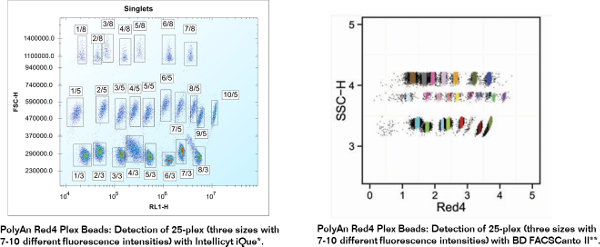
Fluorescent PMMA Microparticles

Fluorescent PMMA Microparticles


Fluorescent PolyAn’s fluorescent microparticles are available in various sizes, emission spectra and fluorescence intensities. The fluorescent PMMA microparticles are suitable for use in flow cytometry, fluorescence microscopy, phagocytosis studies and cell labelling. They can be used in image based systems as well as in other screACening applications. Typical applications include calibration of flow cytometers, calibration of fluorescence microscopes and multiplex bead assays.
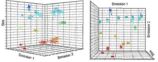
With PolyAn’s production process up to six fluorophores can be incorporated into the beads during the bead polymerisation process. This ensures a much more homogeneous distribution of the dyes within the beads when compared to conventional diffusion controlled dyeing processes. The fluorophores are also caged within the polymeric PMMA matrix and are less likely to leak-out.

Products

Id Mean Diameter Color Labeling Excitation Emission Solids Content
105 40 002 2 µm PolyAn Red 510–580 nm / 570–630 nm 1.0 %
105 40 005 5 µm PolyAn Red 510–580 nm / 570–630 nm 1.0 %
105 40 009 9 µm PolyAn Red 510–580 nm / 570–630 nm 1.0 %
105 40 012 12 µm PolyAn Red 510–580 nm / 570–630 nm 1.0 %
105 40 016 16 µm PolyAn Red 510–580 nm / 570–630 nm 1.0 %
105 40 020 20 µm PolyAn Red 510–580 nm / 570–630 nm 1.0 %
105 60 002 2 µm PolyAn Green 415–480 nm / 470–550 nm 1.0 %
105 60 005 5 µm PolyAn Green 415–480 nm / 470–550 nm 1.0 %
105 60 009 9 µm PolyAn Green 415–480 nm / 470–550 nm 1.0 %
105 60 012 12 µm PolyAn Green 415–480 nm / 470–550 nm 1.0 %
105 60 016 16 µm PolyAn Green 415–480 nm / 470–550 nm 1.0 %
105 60 020 20 µm PolyAn Green 415–480 nm / 470–550 nm 1.0 %
105 70 002 2 µm PolyAn Pink 450-565 nm / 540-620 nm 1.0 %
105 70 005 5 µm PolyAn Pink 450-565 nm / 540-620 nm 1.0 %
105 70 009 9 µm PolyAn Pink 450-565 nm / 540-620 nm 1.0 %
105 70 012 12 µm PolyAn Pink 450-565 nm / 540-620 nm 1.0 %
105 70 016 16 µm PolyAn Pink 450-565 nm / 540-620 nm 1.0 %
105 70 020 20 µm PolyAn Pink 450-565 nm / 540-620 nm 1.0 %
105 89 005 5 µm PolyAn Blue 350–400 nm / 400–480 nm 1.0 %
105 89 009 9 µm PolyAn Blue 350–400 nm / 400–480 nm 1.0 %
105 89 012 12 µm PolyAn Blue 350–400 nm / 400–480 nm 1.0 %
105 89 016 16 µm PolyAn Blue 350–400 nm / 400–480 nm 1.0 %
105 89 020 20 µm PolyAn Blue 350–400 nm / 400–480 nm 1.0 %
106 00 002 2 µm PolyAn Red4 590–680 nm / 660–780 nm 1.0 %
106 00 005 5 µm PolyAn Red4 590–680 nm / 660–780 nm 1.0 %
106 00 009 9 µm PolyAn Red4 590–680 nm / 660–780 nm 1.0 %
106 00 012 12 µm PolyAn Red4 590–680 nm / 660–780 nm 1.0 %
106 00 016 16 µm PolyAn Red4 590–680 nm / 660–780 nm 1.0 %
106 00 020 20 µm PolyAn Red4 590–680 nm / 660–780 nm 1.0 %
106 10 002 2 µm PolyAn Red5 490–680 nm / 660–730 nm 1.0 %
106 10 005 5 µm PolyAn Red5 490–680 nm / 660–730 nm 1.0 %
106 10 009 9 µm PolyAn Red5 490–680 nm / 660–730 nm 1.0 %
106 10 012 12 µm PolyAn Red5 490–680 nm / 660–730 nm 1.0 %
106 10 016 16 µm PolyAn Red5 490–680 nm / 660–730 nm 1.0 %
106 10 020 20 µm PolyAn Red5 490–680 nm / 660–730 nm 1.0 %

The standard packaging volume for our fluorescent, unmodified bead is 1.5 mL. The beads are available in any size between 2 – 20 µm. Both the fluorescence intensity and the spectral characteristics can be tailored to your specific requirements as part of our Molecular Surface Engineering Service.

Our fluorescence encoded beads can also be functionalized with 3D-Carboxy, Streptavidin, Neutravidin, Protein A/G, 3D-Azide and 3D-Alkyne surfaces. A custom modification with antibodies or oligonucleotides is available upon request. Please do not hesitate to contact us!

Selected Publications and References

  • A. Rodríguez-Pena, J Uranga-Solchaga, C. Ortiz-de-Solórzano, I. Cortés-Domínguez: Spheroscope: A custom-made miniaturized microscope for tracking tumour spheroids in microfluidic devices, 2020, Scientific Reports 10,

Functionalized Fluorescent Microparticles

PolyAn offers a wide variety of functionalized, fluorescent particles. They can be used in flow cytometry, image based systems as well as in other imaging systems. PolyAn also offers different multiplex bead sets for flow cytometry and fluorescence microscopes.

The fluorescence intensity of the beads can be tailored to specific applications and read-out systems. We are happy to help you select the right fluorescence intensity for your application. Customized coupling of biomolecules is available upon request. Please do not hesitate to contact us if you have a specific request!

Low Aggregation fluorescent microparticles

Id Color Labeling Excitation Emission Solids Content Mean Diameter
105 41 002 PolyAn Red 510–580 nm / 570–630 nm 0.5 % 2 µm
105 41 005 PolyAn Red 510–580 nm / 570–630 nm 0.5 % 5 µm
105 41 009 PolyAn Red 510–580 nm / 570–630 nm 0.5 % 9 µm
105 41 012 PolyAn Red 510–580 nm / 570–630 nm 0.5 % 12 µm
105 41 016 PolyAn Red 510–580 nm / 570–630 nm 0.5 % 16 µm
105 41 020 PolyAn Red 510–580 nm / 570–630 nm 0.5 % 20 µm
105 67 002 PolyAn Green 415–480 nm / 470–550 nm 0.5 % 2 µm
105 67 005 PolyAn Green 415–480 nm / 470–550 nm 0.5 % 5 µm
105 67 009 PolyAn Green 415–480 nm / 470–550 nm 0.5 % 9 µm
105 67 012 PolyAn Green 415–480 nm / 470–550 nm 0.5 % 12 µm
105 67 016 PolyAn Green 415–480 nm / 470–550 nm 0.5 % 16 µm
105 67 020 PolyAn Green 415–480 nm / 470–550 nm 0.5 % 20 µm
105 90 002 PolyAn Blue 350-400 nm / 400-480 nm 0.5 % 2 µm
105 90 005 PolyAn Blue 350-400 nm / 400-480 nm 0.5 % 5 µm
105 90 009 PolyAn Blue 350-400 nm / 400-480 nm 0.5 % 9 µm
105 90 012 PolyAn Blue 350-400 nm / 400-480 nm 0.5 % 12 µm
105 90 016 PolyAn Blue 350-400 nm / 400-480 nm 0.5 % 16 µm
105 90 020 PolyAn Blue 350-400 nm / 400-480 nm 0.5 % 20 µm
106 01 002 PolyAn Red4 590–680 nm / 660–780 nm 0.5 % 2 µm
106 01 005 PolyAn Red4 590–680 nm / 660–780 nm 0.5 % 5 µm
106 01 009 PolyAn Red4 590–680 nm / 660–780 nm 0.5 % 9 µm
106 01 012 PolyAn Red4 590–680 nm / 660–780 nm 0.5 % 12 µm
106 01 016 PolyAn Red4 590–680 nm / 660–780 nm 0.5 % 16 µm
106 01 020 PolyAn Red4 590–680 nm / 660–780 nm 0.5 % 20 µm
106 18 002 PolyAn Red5 490–680 nm / 660–730 nm 0.5 % 2 µm
106 18 005 PolyAn Red5 490–680 nm / 660–730 nm 0.5 % 5 µm
106 18 009 PolyAn Red5 490–680 nm / 660–730 nm 0.5 % 9 µm
106 18 012 PolyAn Red5 490–680 nm / 660–730 nm 0.5 % 12 µm
106 18 016 PolyAn Red5 490–680 nm / 660–730 nm 0.5 % 16 µm
106 18 020 PolyAn Red5 490–680 nm / 660–730 nm 0.5 % 20 µm
106 71 002 PolyAn Pink 450-565 nm / 540-620 nm 0.5 % 2 µm
106 71 005 PolyAn Pink 450-565 nm / 540-620 nm 0.5 % 5 µm
106 71 009 PolyAn Pink 450-565 nm / 540-620 nm 0.5 % 9 µm
106 71 012 PolyAn Pink 450-565 nm / 540-620 nm 0.5 % 12 µm
106 71 016 PolyAn Pink 450-565 nm / 540-620 nm 0.5 % 16 µm
106 71 020 PolyAn Pink 450-565 nm / 540-620 nm 0.5 % 20 µm

3D-Carboxy fluorescent microparticles

Id Color Labeling Excitation Emission Solids Content Mean Diameter
105 42 002 PolyAn Red 510–580 nm / 570–630 nm 0.5 % 2 µm
105 42 005 PolyAn Red 510–580 nm / 570–630 nm 0.5 % 5 µm
105 42 009 PolyAn Red 510–580 nm / 570–630 nm 0.5 % 9 µm
105 42 012 PolyAn Red 510–580 nm / 570–630 nm 0.5 % 12 µm
105 42 016 PolyAn Red 510–580 nm / 570–630 nm 0.5 % 16 µm
105 42 020 PolyAn Red 510–580 nm / 570–630 nm 0.5 % 20 µm
105 52 002 PolyAn Orange 470-540 nm / 520-580 nm 0.5 % 2 µm
105 52 005 PolyAn Orange 470-540 nm / 520-580 nm 0.5 % 5 µm
105 52 009 PolyAn Orange 470-540 nm / 520-580 nm 0.5 % 9 µm
105 52 012 PolyAn Orange 470-540 nm / 520-580 nm 0.5 % 12 µm
105 52 016 PolyAn Orange 470-540 nm / 520-580 nm 0.5 % 16 µm
105 52 020 PolyAn Orange 470-540 nm / 520-580 nm 0.5 % 20 µm
105 61 002 PolyAn Green 415–480 nm / 470–550 nm 0.5 % 2 µm
105 61 005 PolyAn Green 415–480 nm / 470–550 nm 0.5 % 5 µm
105 61 009 PolyAn Green 415–480 nm / 470–550 nm 0.5 % 9 µm
105 61 012 PolyAn Green 415–480 nm / 470–550 nm 0.5 % 12 µm
105 61 016 PolyAn Green 415–480 nm / 470–550 nm 0.5 % 16 µm
105 61 020 PolyAn Green 415–480 nm / 470–550 nm 0.5 % 20 µm
105 91 002 PolyAn Blue 350-400 nm / 400-480 nm 0.5 % 2 µm
105 91 005 PolyAn Blue 350-400 nm / 400-480 nm 0.5 % 5 µm
105 91 009 PolyAn Blue 350-400 nm / 400-480 nm 0.5 % 9 µm
105 91 012 PolyAn Blue 350-400 nm / 400-480 nm 0.5 % 12 µm
105 91 016 PolyAn Blue 350-400 nm / 400-480 nm 0.5 % 16 µm
105 91 020 PolyAn Blue 350-400 nm / 400-480 nm 0.5 % 20 µm
106 02 002 PolyAn Red4 590–680 nm / 660–780 nm 0.5 % 2 µm
106 02 005 PolyAn Red4 590–680 nm / 660–780 nm 0.5 % 5 µm
106 02 009 PolyAn Red4 590–680 nm / 660–780 nm 0.5 % 9 µm
106 02 012 PolyAn Red4 590–680 nm / 660–780 nm 0.5 % 12 µm
106 02 016 PolyAn Red4 590–680 nm / 660–780 nm 0.5 % 16 µm
106 02 020 PolyAn Red4 590–680 nm / 660–780 nm 0.5 % 20 µm
106 11 002 PolyAn Red5 490–680 nm / 660–730 nm 0.5 % 2 µm
106 11 005 PolyAn Red5 490–680 nm / 660–730 nm 0.5 % 5 µm
106 11 009 PolyAn Red5 490–680 nm / 660–730 nm 0.5 % 9 µm
106 11 012 PolyAn Red5 490–680 nm / 660–730 nm 0.5 % 12 µm
106 11 016 PolyAn Red5 490–680 nm / 660–730 nm 0.5 % 16 µm
106 11 020 PolyAn Red5 490–680 nm / 660–730 nm 0.5 % 20 µm
106 72 002 PolyAn Pink 450-565 nm / 540-620 nm 0.5 % 2 µm
106 72 005 PolyAn Pink 450-565 nm / 540-620 nm 0.5 % 5 µm
106 72 009 PolyAn Pink 450-565 nm / 540-620 nm 0.5 % 9 µm
106 72 012 PolyAn Pink 450-565 nm / 540-620 nm 0.5 % 12 µm
106 72 016 PolyAn Pink 450-565 nm / 540-620 nm 0.5 % 16 µm
106 72 020 PolyAn Pink 450-565 nm / 540-620 nm 0.5 % 20 µm

3D-Aldehyde coated fluorescent microparticles


Id Color Labeling Excitation Emission Solids Content Mean Diameter
105 49 002 PolyAn Red 510–580 nm / 570–630 nm 0.5 % 2 µm
105 49 005 PolyAn Red 510–580 nm / 570–630 nm 0.5 % 5 µm
105 49 009 PolyAn Red 510–580 nm / 570–630 nm 0.5 % 9 µm
105 49 012 PolyAn Red 510–580 nm / 570–630 nm 0.5 % 12 µm
105 49 016 PolyAn Red 510–580 nm / 570–630 nm 0.5 % 16 µm
105 49 020 PolyAn Red 510–580 nm / 570–630 nm 0.5 % 20 µm
105 57 002 PolyAn Orange 470-540 nm / 520-580 nm 0.5 % 2 µm
105 57 005 PolyAn Orange 470-540 nm / 520-580 nm 0.5 % 5 µm
105 57 009 PolyAn Orange 470-540 nm / 520-580 nm 0.5 % 9 µm
105 57 012 PolyAn Orange 470-540 nm / 520-580 nm 0.5 % 12 µm
105 57 016 PolyAn Orange 470-540 nm / 520-580 nm 0.5 % 16 µm
105 57 020 PolyAn Orange 470-540 nm / 520-580 nm 0.5 % 20 µm
105 66 002 PolyAn Green 415–480 nm / 470–550 nm 0.5 % 2 µm
105 66 005 PolyAn Green 415–480 nm / 470–550 nm 0.5 % 5 µm
105 66 009 PolyAn Green 415–480 nm / 470–550 nm 0.5 % 9 µm
105 66 012 PolyAn Green 415–480 nm / 470–550 nm 0.5 % 12 µm
105 66 016 PolyAn Green 415–480 nm / 470–550 nm 0.5 % 16 µm
105 66 020 PolyAn Green 415–480 nm / 470–550 nm 0.5 % 20 µm
105 96 002 PolyAn Blue 350–400 nm / 400–480 nm 0.5 % 2 µm
105 96 005 PolyAn Blue 350–400 nm / 400–480 nm 0.5 % 5 µm
105 96 009 PolyAn Blue 350–400 nm / 400–480 nm 0.5 % 9 µm
105 96 012 PolyAn Blue 350–400 nm / 400–480 nm 0.5 % 12 µm
105 96 016 PolyAn Blue 350–400 nm / 400–480 nm 0.5 % 16 µm
105 96 020 PolyAn Blue 350–400 nm / 400–480 nm 0.5 % 20 µm
106 08 002 PolyAn Red4 590–680 nm / 660–780 nm 0.5 % 2 µm
106 08 005 PolyAn Red4 590–680 nm / 660–780 nm 0.5 % 5 µm
106 08 009 PolyAn Red4 590–680 nm / 660–780 nm 0.5 % 9 µm
106 08 012 PolyAn Red4 590–680 nm / 660–780 nm 0.5 % 12 µm
106 08 016 PolyAn Red4 590–680 nm / 660–780 nm 0.5 % 16 µm
106 08 020 PolyAn Red4 590–680 nm / 660–780 nm 0.5 % 20 µm
106 17 002 PolyAn Red5 490–680 nm / 660–730 nm 0.5 % 2 µm
106 17 005 PolyAn Red5 490–680 nm / 660–730 nm 0.5 % 5 µm
106 17 009 PolyAn Red5 490–680 nm / 660–730 nm 0.5 % 9 µm
106 17 012 PolyAn Red5 490–680 nm / 660–730 nm 0.5 % 12 µm
106 17 016 PolyAn Red5 490–680 nm / 660–730 nm 0.5 % 16 µm
106 17 020 PolyAn Red5 490–680 nm / 660–730 nm 0.5 % 20 µm
106 80 002 PolyAn Pink 450-565 nm / 540-620 nm 0.5 % 2 µm
106 80 005 PolyAn Pink 450-565 nm / 540-620 nm 0.5 % 5 µm
106 80 009 PolyAn Pink 450-565 nm / 540-620 nm 0.5 % 9 µm
106 80 012 PolyAn Pink 450-565 nm / 540-620 nm 0.5 % 12 µm
106 80 016 PolyAn Pink 450-565 nm / 540-620 nm 0.5 % 16 µm
106 80 020 PolyAn Pink 450-565 nm / 540-620 nm 0.5 % 20 µm

Streptavidin coated fluorescent microparticles

Id Color Labeling Excitation Emission Solids Content Mean Diameter
105 43 002 PolyAn Red 510–580 nm / 570–630 nm 0.5 % 2 µm
105 43 005 PolyAn Red 510–580 nm / 570–630 nm 0.5 % 5 µm
105 43 009 PolyAn Red 510–580 nm / 570–630 nm 0.5 % 9 µm
105 43 012 PolyAn Red 510–580 nm / 570–630 nm 0.5 % 12 µm
105 43 016 PolyAn Red 510–580 nm / 570–630 nm 0.5 % 16 µm
105 43 020 PolyAn Red 510–580 nm / 570–630 nm 0.5 % 20 µm
105 53 002 PolyAn Orange 470-540 nm / 520-580 nm 0.5 % 2 µm
105 53 005 PolyAn Orange 470-540 nm / 520-580 nm 0.5 % 5 µm
105 53 009 PolyAn Orange 470-540 nm / 520-580 nm 0.5 % 9 µm
105 53 012 PolyAn Orange 470-540 nm / 520-580 nm 0.5 % 12 µm
105 53 016 PolyAn Orange 470-540 nm / 520-580 nm 0.5 % 16 µm
105 53 020 PolyAn Orange 470-540 nm / 520-580 nm 0.5 % 20 µm
105 62 002 PolyAn Green 415–480 nm / 470–550 nm 0.5 % 2 µm
105 62 005 PolyAn Green 415–480 nm / 470–550 nm 0.5 % 5 µm
105 62 009 PolyAn Green 415–480 nm / 470–550 nm 0.5 % 9 µm
105 62 012 PolyAn Green 415–480 nm / 470–550 nm 0.5 % 12 µm
105 62 016 PolyAn Green 415–480 nm / 470–550 nm 0.5 % 16 µm
105 62 020 PolyAn Green 415–480 nm / 470–550 nm 0.5 % 20 µm
105 92 002 PolyAn Blue 350-400 nm / 400-480 nm 0.5 % 2 µm
105 92 005 PolyAn Blue 350-400 nm / 400-480 nm 0.5 % 9 µm
105 92 009 PolyAn Blue 350-400 nm / 400-480 nm 0.5 % 5 µm
105 92 012 PolyAn Blue 350-400 nm / 400-480 nm 0.5 % 12 µm
105 92 016 PolyAn Blue 350-400 nm / 400-480 nm 0.5 % 16 µm
105 92 020 PolyAn Blue 350-400 nm / 400-480 nm 0.5 % 20 µm
106 03 002 PolyAn Red4 590–680 nm / 660–780 nm 0.5 % 2 µm
106 03 005 PolyAn Red4 590–680 nm / 660–780 nm 0.5 % 5 µm
106 03 009 PolyAn Red4 590–680 nm / 660–780 nm 0.5 % 9 µm
106 03 012 PolyAn Red4 590–680 nm / 660–780 nm 0.5 % 12 µm
106 03 016 PolyAn Red4 590–680 nm / 660–780 nm 0.5 % 16 µm
106 03 020 PolyAn Red4 590–680 nm / 660–780 nm 0.5 % 20 µm
106 14 002 PolyAn Red5 490–680 nm / 660–730 nm 0.5 % 2 µm
106 14 005 PolyAn Red5 490–680 nm / 660–730 nm 0.5 % 5 µm
106 14 009 PolyAn Red5 490–680 nm / 660–730 nm 0.5 % 9 µm
106 14 012 PolyAn Red5 490–680 nm / 660–730 nm 0.5 % 12 µm
106 14 016 PolyAn Red5 490–680 nm / 660–730 nm 0.5 % 16 µm
106 14 020 PolyAn Red5 490–680 nm / 660–730 nm 0.5 % 20 µm
106 73 002 PolyAn Pink 450-565 nm / 540-620 nm 0.5 % 2 µm
106 73 005 PolyAn Pink 450-565 nm / 540-620 nm 0.5 % 5 µm
106 73 009 PolyAn Pink 450-565 nm / 540-620 nm 0.5 % 9 µm
106 73 012 PolyAn Pink 450-565 nm / 540-620 nm 0.5 % 12 µm
106 73 016 PolyAn Pink 450-565 nm / 540-620 nm 0.5 % 16 µm
106 73 020 PolyAn Pink 450-565 nm / 540-620 nm 0.5 % 20 µm

Neutravidin coated fluorescent microparticles


Id Color Labeling Excitation Emission Solids Content Mean Diameter
105 54 002 PolyAn Orange 470-540 nm / 520-580 nm 0.5 % 2 µm
105 54 005 PolyAn Orange 470-540 nm / 520-580 nm 0.5 % 5 µm
105 54 009 PolyAn Orange 470-540 nm / 520-580 nm 0.5 % 9 µm
105 54 012 PolyAn Orange 470-540 nm / 520-580 nm 0.5 % 12 µm
105 54 016 PolyAn Orange 470-540 nm / 520-580 nm 0.5 % 16 µm
105 54 020 PolyAn Orange 470-540 nm / 520-580 nm 0.5 % 20 µm
105 68 002 PolyAn Green 415–480 nm / 470–550 nm 0.5 % 2 µm
105 68 005 PolyAn Green 415–480 nm / 470–550 nm 0.5 % 5 µm
105 68 009 PolyAn Green 415–480 nm / 470–550 nm 0.5 % 9 µm
105 68 012 PolyAn Green 415–480 nm / 470–550 nm 0.5 % 12 µm
105 68 016 PolyAn Green 415–480 nm / 470–550 nm 0.5 % 16 µm
105 68 020 PolyAn Green 415–480 nm / 470–550 nm 0.5 % 20 µm
105 97 002 PolyAn Blue 350–400 nm / 400–480 nm 0.5 % 2 µm
105 97 005 PolyAn Blue 350–400 nm / 400–480 nm 0.5 % 5 µm
105 97 009 PolyAn Blue 350–400 nm / 400–480 nm 0.5 % 9 µm
105 97 012 PolyAn Blue 350–400 nm / 400–480 nm 0.5 % 12 µm
105 97 016 PolyAn Blue 350–400 nm / 400–480 nm 0.5 % 16 µm
105 97 020 PolyAn Blue 350–400 nm / 400–480 nm 0.5 % 20 µm
106 09 002 PolyAn Red4 590–680 nm / 660–780 nm 0.5 % 2 µm
106 09 005 PolyAn Red4 590–680 nm / 660–780 nm 0.5 % 5 µm
106 09 009 PolyAn Red4 590–680 nm / 660–780 nm 0.5 % 9 µm
106 09 012 PolyAn Red4 590–680 nm / 660–780 nm 0.5 % 12 µm
106 09 016 PolyAn Red4 590–680 nm / 660–780 nm 0.5 % 16 µm
106 09 020 PolyAn Red4 590–680 nm / 660–780 nm 0.5 % 20 µm
106 19 002 PolyAn Red5 490–680 nm / 660–730 nm 0.5 % 2 µm
106 19 005 PolyAn Red5 490–680 nm / 660–730 nm 0.5 % 5 µm
106 19 009 PolyAn Red5 490–680 nm / 660–730 nm 0.5 % 9 µm
106 19 012 PolyAn Red5 490–680 nm / 660–730 nm 0.5 % 12 µm
106 19 016 PolyAn Red5 490–680 nm / 660–730 nm 0.5 % 16 µm
106 19 020 PolyAn Red5 490–680 nm / 660–730 nm 0.5 % 20 µm
106 46 002 PolyAn Red 510–580 nm / 570–630 nm 0.5 % 2 µm
106 46 005 PolyAn Red 510–580 nm / 570–630 nm 0.5 % 5 µm
106 46 009 PolyAn Red 510–580 nm / 570–630 nm 0.5 % 9 µm
106 46 012 PolyAn Red 510–580 nm / 570–630 nm 0.5 % 12 µm
106 46 016 PolyAn Red 510–580 nm / 570–630 nm 0.5 % 16 µm
106 46 020 PolyAn Red 510–580 nm / 570–630 nm 0.5 % 20 µm
106 74 002 PolyAn Pink 450-565 nm / 540-620 nm 0.5 % 2 µm
106 74 005 PolyAn Pink 450-565 nm / 540-620 nm 0.5 % 5 µm
106 74 009 PolyAn Pink 450-565 nm / 540-620 nm 0.5 % 9 µm
106 74 012 PolyAn Pink 450-565 nm / 540-620 nm 0.5 % 12 µm
106 74 016 PolyAn Pink 450-565 nm / 540-620 nm 0.5 % 16 µm
106 74 020 PolyAn Pink 450-565 nm / 540-620 nm 0.5 % 20 µm

3D-Alkyne fluorescent microparticles


Id Color Labeling Excitation Emission Solids Content Mean Diameter
105 48 002 PolyAn Red 510–580 nm / 570–630 nm 0.5 % 2 µm
105 48 005 PolyAn Red 510–580 nm / 570–630 nm 0.5 % 5 µm
105 48 009 PolyAn Red 510–580 nm / 570–630 nm 0.5 % 9 µm
105 48 012 PolyAn Red 510–580 nm / 570–630 nm 0.5 % 12 µm
105 48 016 PolyAn Red 510–580 nm / 570–630 nm 0.5 % 16 µm
105 48 020 PolyAn Red 510–580 nm / 570–630 nm 0.5 % 20 µm
105 56 002 PolyAn Orange 470-540 nm / 520-580 nm 0.5 % 2 µm
105 56 005 PolyAn Orange 470-540 nm / 520-580 nm 0.5 % 5 µm
105 56 009 PolyAn Orange 470-540 nm / 520-580 nm 0.5 % 9 µm
105 56 012 PolyAn Orange 470-540 nm / 520-580 nm 0.5 % 12 µm
105 56 016 PolyAn Orange 470-540 nm / 520-580 nm 0.5 % 16 µm
105 56 020 PolyAn Orange 470-540 nm / 520-580 nm 0.5 % 20 µm
105 64 002 PolyAn Green 415–480 nm / 470–550 nm 0.5 % 2 µm
105 64 005 PolyAn Green 415–480 nm / 470–550 nm 0.5 % 5 µm
105 64 009 PolyAn Green 415–480 nm / 470–550 nm 0.5 % 9 µm
105 64 012 PolyAn Green 415–480 nm / 470–550 nm 0.5 % 12 µm
105 64 016 PolyAn Green 415–480 nm / 470–550 nm 0.5 % 16 µm
105 64 020 PolyAn Green 415–480 nm / 470–550 nm 0.5 % 20 µm
105 94 002 PolyAn Blue 350–400 nm / 400–480 nm 0.5 % 2 µm
105 94 005 PolyAn Blue 350–400 nm / 400–480 nm 0.5 % 5 µm
105 94 009 PolyAn Blue 350–400 nm / 400–480 nm 0.5 % 9 µm
105 94 012 PolyAn Blue 350–400 nm / 400–480 nm 0.5 % 12 µm
105 94 016 PolyAn Blue 350–400 nm / 400–480 nm 0.5 % 16 µm
105 94 020 PolyAn Blue 350–400 nm / 400–480 nm 0.5 % 20 µm
106 05 002 PolyAn Red4 590–680 nm / 660–780 nm 0.5 % 2 µm
106 05 005 PolyAn Red4 590–680 nm / 660–780 nm 0.5 % 5 µm
106 05 009 PolyAn Red4 590–680 nm / 660–780 nm 0.5 % 9 µm
106 05 012 PolyAn Red4 590–680 nm / 660–780 nm 0.5 % 12 µm
106 05 016 PolyAn Red4 590–680 nm / 660–780 nm 0.5 % 16 µm
106 05 020 PolyAn Red4 590–680 nm / 660–780 nm 0.5 % 20 µm
106 13 002 PolyAn Red5 490–680 nm / 660–730 nm 0.5 % 2 µm
106 13 005 PolyAn Red5 490–680 nm / 660–730 nm 0.5 % 5 µm
106 13 009 PolyAn Red5 490–680 nm / 660–730 nm 0.5 % 9 µm
106 13 012 PolyAn Red5 490–680 nm / 660–730 nm 0.5 % 12 µm
106 13 016 PolyAn Red5 490–680 nm / 660–730 nm 0.5 % 16 µm
106 13 020 PolyAn Red5 490–680 nm / 660–730 nm 0.5 % 20 µm
106 76 002 PolyAn Pink 450-565 nm / 540-620 nm 0.5 % 2 µm
106 76 005 PolyAn Pink 450-565 nm / 540-620 nm 0.5 % 5 µm
106 76 009 PolyAn Pink 450-565 nm / 540-620 nm 0.5 % 9 µm
106 76 012 PolyAn Pink 450-565 nm / 540-620 nm 0.5 % 12 µm
106 76 016 PolyAn Pink 450-565 nm / 540-620 nm 0.5 % 16 µm
106 76 020 PolyAn Pink 450-565 nm / 540-620 nm 0.5 % 20 µm

3D-Azide fluorescent microparticles


Id Color Labeling Excitation Emission Solids Content Mean Diameter
105 58 002 PolyAn Orange 470-540 nm / 520-580 nm 0.5 % 2 µm
105 58 005 PolyAn Orange 470-540 nm / 520-580 nm 0.5 % 5 µm
105 58 009 PolyAn Orange 470-540 nm / 520-580 nm 0.5 % 9 µm
105 58 012 PolyAn Orange 470-540 nm / 520-580 nm 0.5 % 12 µm
105 58 016 PolyAn Orange 470-540 nm / 520-580 nm 0.5 % 16 µm
105 58 020 PolyAn Orange 470-540 nm / 520-580 nm 0.5 % 20 µm
105 65 002 PolyAn Green 415–480 nm / 470–550 nm 0.5 % 2 µm
105 65 005 PolyAn Green 415–480 nm / 470–550 nm 0.5 % 5 µm
105 65 009 PolyAn Green 415–480 nm / 470–550 nm 0.5 % 9 µm
105 65 012 PolyAn Green 415–480 nm / 470–550 nm 0.5 % 12 µm
105 65 016 PolyAn Green 415–480 nm / 470–550 nm 0.5 % 16 µm
105 65 020 PolyAn Green 415–480 nm / 470–550 nm 0.5 % 20 µm
105 95 002 PolyAn Blue 350–400 nm / 400–480 nm 0.5 % 2 µm
105 95 005 PolyAn Blue 350–400 nm / 400–480 nm 0.5 % 5 µm
105 95 009 PolyAn Blue 350–400 nm / 400–480 nm 0.5 % 9 µm
105 95 012 PolyAn Blue 350–400 nm / 400–480 nm 0.5 % 12 µm
105 95 016 PolyAn Blue 350–400 nm / 400–480 nm 0.5 % 16 µm
105 95 020 PolyAn Blue 350–400 nm / 400–480 nm 0.5 % 20 µm
106 07 002 PolyAn Red4 590–680 nm / 660–780 nm 0.5 % 2 µm
106 07 005 PolyAn Red4 590–680 nm / 660–780 nm 0.5 % 5 µm
106 07 009 PolyAn Red4 590–680 nm / 660–780 nm 0.5 % 9 µm
106 07 012 PolyAn Red4 590–680 nm / 660–780 nm 0.5 % 12 µm
106 07 016 PolyAn Red4 590–680 nm / 660–780 nm 0.5 % 16 µm
106 07 020 PolyAn Red4 590–680 nm / 660–780 nm 0.5 % 20 µm
106 16 002 PolyAn Red5 490–680 nm / 660–730 nm 0.5 % 2 µm
106 16 005 PolyAn Red5 490–680 nm / 660–730 nm 0.5 % 5 µm
106 16 009 PolyAn Red5 490–680 nm / 660–730 nm 0.5 % 9 µm
106 16 012 PolyAn Red5 490–680 nm / 660–730 nm 0.5 % 12 µm
106 16 016 PolyAn Red5 490–680 nm / 660–730 nm 0.5 % 16 µm
106 16 020 PolyAn Red5 490–680 nm / 660–730 nm 0.5 % 20 µm
106 45 002 PolyAn Red 510–580 nm / 570–630 nm 0.5 % 2 µm
106 45 005 PolyAn Red 510–580 nm / 570–630 nm 0.5 % 5 µm
106 45 009 PolyAn Red 510–580 nm / 570–630 nm 0.5 % 9 µm
106 45 012 PolyAn Red 510–580 nm / 570–630 nm 0.5 % 12 µm
106 45 016 PolyAn Red 510–580 nm / 570–630 nm 0.5 % 16 µm
106 45 020 PolyAn Red 510–580 nm / 570–630 nm 0.5 % 20 µm
106 79 002 PolyAn Pink 450-565 nm / 540-620 nm 0.5 % 2 µm
106 79 005 PolyAn Pink 450-565 nm / 540-620 nm 0.5 % 5 µm
106 79 009 PolyAn Pink 450-565 nm / 540-620 nm 0.5 % 9 µm
106 79 012 PolyAn Pink 450-565 nm / 540-620 nm 0.5 % 12 µm
106 79 016 PolyAn Pink 450-565 nm / 540-620 nm 0.5 % 16 µm
106 79 020 PolyAn Pink 450-565 nm / 540-620 nm 0.5 % 20 µm

Protein A/G coated fluorescent microparticles


Id Color Labeling Excitation Emission Solids Content Mean Diameter
105 44 002 PolyAn Red 510–580 nm / 570–630 nm 0.5 % 2 µm
105 44 005 PolyAn Red 510–580 nm / 570–630 nm 0.5 % 5 µm
105 44 009 PolyAn Red 510–580 nm / 570–630 nm 0.5 % 9 µm
105 44 012 PolyAn Red 510–580 nm / 570–630 nm 0.5 % 12 µm
105 44 016 PolyAn Red 510–580 nm / 570–630 nm 0.5 % 16 µm
105 44 020 PolyAn Red 510–580 nm / 570–630 nm 0.5 % 20 µm
105 59 002 PolyAn Orange 470-540 nm / 520-580 nm 0.5 % 2 µm
105 59 005 PolyAn Orange 470-540 nm / 520-580 nm 0.5 % 5 µm
105 59 009 PolyAn Orange 470-540 nm / 520-580 nm 0.5 % 9 µm
105 59 012 PolyAn Orange 470-540 nm / 520-580 nm 0.5 % 12 µm
105 59 016 PolyAn Orange 470-540 nm / 520-580 nm 0.5 % 16 µm
105 59 020 PolyAn Orange 470-540 nm / 520-580 nm 0.5 % 20 µm
105 63 002 PolyAn Green 415–480 nm / 470–550 nm 0.5 % 2 µm
105 63 005 PolyAn Green 415–480 nm / 470–550 nm 0.5 % 5 µm
105 63 009 PolyAn Green 415–480 nm / 470–550 nm 0.5 % 9 µm
105 63 012 PolyAn Green 415–480 nm / 470–550 nm 0.5 % 12 µm
105 63 016 PolyAn Green 415–480 nm / 470–550 nm 0.5 % 16 µm
105 63 020 PolyAn Green 415–480 nm / 470–550 nm 0.5 % 20 µm
105 93 002 PolyAn Blue 350–400 nm / 400–480 nm 0.5 % 2 µm
105 93 005 PolyAn Blue 350–400 nm / 400–480 nm 0.5 % 5 µm
105 93 009 PolyAn Blue 350–400 nm / 400–480 nm 0.5 % 9 µm
105 93 012 PolyAn Blue 350–400 nm / 400–480 nm 0.5 % 12 µm
105 93 016 PolyAn Blue 350–400 nm / 400–480 nm 0.5 % 16 µm
105 93 020 PolyAn Blue 350–400 nm / 400–480 nm 0.5 % 20 µm
106 04 002 PolyAn Red4 590–680 nm / 660–780 nm 0.5 % 2 µm
106 04 005 PolyAn Red4 590–680 nm / 660–780 nm 0.5 % 5 µm
106 04 009 PolyAn Red4 590–680 nm / 660–780 nm 0.5 % 9 µm
106 04 012 PolyAn Red4 590–680 nm / 660–780 nm 0.5 % 12 µm
106 04 016 PolyAn Red4 590–680 nm / 660–780 nm 0.5 % 16 µm
106 04 020 PolyAn Red4 590–680 nm / 660–780 nm 0.5 % 20 µm
106 15 002 PolyAn Red5 490–680 nm / 660–730 nm 0.5 % 2 µm
106 15 005 PolyAn Red5 490–680 nm / 660–730 nm 0.5 % 5 µm
106 15 009 PolyAn Red5 490–680 nm / 660–730 nm 0.5 % 9 µm
106 15 012 PolyAn Red5 490–680 nm / 660–730 nm 0.5 % 12 µm
106 15 016 PolyAn Red5 490–680 nm / 660–730 nm 0.5 % 16 µm
106 15 020 PolyAn Red5 490–680 nm / 660–730 nm 0.5 % 20 µm
106 75 002 PolyAn Pink 450-565 nm / 540-620 nm 0.5 % 2 µm
106 75 005 PolyAn Pink 450-565 nm / 540-620 nm 0.5 % 5 µm
106 75 009 PolyAn Pink 450-565 nm / 540-620 nm 0.5 % 9 µm
106 75 012 PolyAn Pink 450-565 nm / 540-620 nm 0.5 % 12 µm
106 75 016 PolyAn Pink 450-565 nm / 540-620 nm 0.5 % 16 µm
106 75 020 PolyAn Pink 450-565 nm / 540-620 nm 0.5 % 20 µm

The standard packaging volume for our fluorescent, unmodified bead is 1.5 mL. Please note, that PolyAn also produces customized microparticles which incorporate fluorophores for other spectral ranges. All beads are available in sizes between 2-20µm. A custom modification with antibodies or oligonucleotides is available upon request. Please do not hesitate to contact us!

Selected Publications and References

  • N. M. Pham, S. Rusch, Y. Temiz, H. P. Beck, W. Karlen, E. Delamarche: Immuno-gold silver staining assays on capillary-driven microfluidics for the detection of malaria antigens, 2019, Biomedical Microdevices 21, 24.
  • C. Jurischka, F. Dinter, M. Sowa, J. Noack, J. Schiebel, D. Roggenbuck, P. Schierack, S. Rödiger: Tyramide signal amplification as universal detection method on protein coated microbeads, 2018, Journal of Cellular Biotechnology 4: 1-2, 15-22.
  • N. M. Pham, S. Rusch, Y. Temiz, R. D. Lovchik, H. P. Beck, W. Karlen, E. Delamarche: A bead-based immunogold-silver staining assay on capillary-driven microfluidics, 2018, Biomedical

PMMA Nanoparticles

Fluorescent Aldehyde PMMA Nanoparticles

Our aldehyde functionalized, fluorescent PMMA (Polymethylmethacrylate) based nanospheres are available in sizes between 100 nm to 500 nm. Aldehyde groups react immediately with the NH2-terminus or other suitable functional groups of the probe to form a (covalent) bond with the surface. Thus, no activation of the bead surface is necessary prior to binding of the probe. Latex beads with aldehyde surfaces are suitable for immobilization of biomolecules (proteins, antibodies, haptens, etc.).

Aldehyde functionalized, fluorescent submicron beads


Id Mean Diameter Color Labeling Excitation Emission
201 03 125 100-150 nm PolyAn Blue 350–400 nm / 400–480 nm
201 03 175 151-200 nm PolyAn Blue 350–400 nm / 400–480 nm
201 03 225 201-250 nm PolyAn Blue 350–400 nm / 400–480 nm
201 03 275 251-300 nm PolyAn Blue 350–400 nm / 400–480 nm
201 03 325 301-350 nm PolyAn Blue 350–400 nm / 400–480 nm
201 03 375 351-400 nm PolyAn Blue 350–400 nm / 400–480 nm
202 03 125 100-150 nm PolyAn Green 415–480 nm / 470–550 nm
202 03 175 151-200 nm PolyAn Green 415–480 nm / 470–550 nm
202 03 225 201-250 nm PolyAn Green 415–480 nm / 470–550 nm
202 03 275 251-300 nm PolyAn Green 415–480 nm / 470–550 nm
202 03 325 301-350 nm PolyAn Green 415–480 nm / 470–550 nm
202 03 375 351-400 nm PolyAn Green 415–480 nm / 470–550 nm
203 03 125 100-150 nm PolyAn Pink 450-565 nm / 540-620 nm
203 03 175 151-200 nm PolyAn Pink 450-565 nm / 540-620 nm
203 03 225 201-250 nm PolyAn Pink 450-565 nm / 540-620 nm
203 03 275 251-300 nm PolyAn Pink 450-565 nm / 540-620 nm
203 03 325 301-350 nm PolyAn Pink 450-565 nm / 540-620 nm
203 03 375 351-400 nm PolyAn Pink 450-565 nm / 540-620 nm
204 03 125 100-150 nm PolyAn Red 510–580 nm / 570–630 nm
204 03 175 151-200 nm PolyAn Red 510–580 nm / 570–630 nm
204 03 225 201-250 nm PolyAn Red 510–580 nm / 570–630 nm
204 03 275 251-300 nm PolyAn Red 510–580 nm / 570–630 nm
204 03 325 301-350 nm PolyAn Red 510–580 nm / 570–630 nm
204 03 375 351-400 nm PolyAn Red 510–580 nm / 570–630 nm
205 03 125 100-150 nm PolyAn Red4 590–680 nm / 660–780 nm
205 03 175 151-200 nm PolyAn Red4 590–680 nm / 660–780 nm
205 03 225 201-250 nm PolyAn Red4 590–680 nm / 660–780 nm
205 03 275 251-300 nm PolyAn Red4 590–680 nm / 660–780 nm
205 03 325 301-350 nm PolyAn Red4 590–680 nm / 660–780 nm
205 03 375 351-400 nm PolyAn Red4 590–680 nm / 660–780 nm
206 03 125 100-150 nm PolyAn Red5 490–680 nm / 660–730 nm
206 03 175 151-200 nm PolyAn Red5 490–680 nm / 660–730 nm
206 03 225 201-250 nm PolyAn Red5 490–680 nm / 660–730 nm
206 03 275 251-300 nm PolyAn Red5 490–680 nm / 660–730 nm
206 03 325 301-350 nm PolyAn Red5 490–680 nm / 660–730 nm
206 03 375 351-400 nm PolyAn Red5 490–680 nm / 660–730 nm
207 03 125 100-150 nm PolyAn Orange 470-540 nm / 520-580 nm
207 03 175 151-200 nm PolyAn Orange 470-540 nm / 520-580 nm
207 03 225 201-250 nm PolyAn Orange 470-540 nm / 520-580 nm
207 03 275 251-300 nm PolyAn Orange 470-540 nm / 520-580 nm
207 03 325 301-350 nm PolyAn Orange 470-540 nm / 520-580 nm
207 03 375 351-400 nm PolyAn Orange 470-540 nm / 520-580 nm

The standard packaging volume for our fluorescent submicron beads is 1.5 mL with a solids content of 0.5%. We are looking forward to your inquiry.

Contact

Please note, that PolyAn also produces customized nanoparticles which incorporate fluorophores for other spectral ranges. All PMMA Nanobeads are available in sizes between 100-500 nm. Custom modifications with antibodies and other biomolecules are available upon request.

Carboxylated PMMA Nanoparticles

Our carboxylated, fluorescent PMMA (Polymaethylmethacrylate) based nanospheres are available in sizes between 100 nm to 500 nm. Latex beads with carboxy surface are suitable for immobilization of biomolecules (proteins, antibodies, haptens, etc.).

Carboxy functionalized, fluorescent submicron beads


Id Mean Diameter Color Labeling Excitation Emission
201 02 125 100-150 nm PolyAn Blue 350–400 nm / 400–480 nm
201 02 175 151-200 nm PolyAn Blue 350–400 nm / 400–480 nm
201 02 225 201-250 nm PolyAn Blue 350–400 nm / 400–480 nm
201 02 275 251-300 nm PolyAn Blue 350–400 nm / 400–480 nm
201 02 325 301-350 nm PolyAn Blue 350–400 nm / 400–480 nm
201 02 375 351-400 nm PolyAn Blue 350–400 nm / 400–480 nm
202 02 125 100-150 nm PolyAn Green 415–480 nm / 470–550 nm
202 02 175 151-200 nm PolyAn Green 415–480 nm / 470–550 nm
202 02 225 201-250 nm PolyAn Green 415–480 nm / 470–550 nm
202 02 275 251-300 nm PolyAn Green 415–480 nm / 470–550 nm
202 02 325 301-350 nm PolyAn Green 415–480 nm / 470–550 nm
202 02 375 351-400 nm PolyAn Green 415–480 nm / 470–550 nm
203 02 125 100-150 nm PolyAn Pink 450-565 nm / 540-620 nm
203 02 175 151-200 nm PolyAn Pink 450-565 nm / 540-620 nm
203 02 225 201-250 nm PolyAn Pink 450-565 nm / 540-620 nm
203 02 275 251-300 nm PolyAn Pink 450-565 nm / 540-620 nm
203 02 325 301-350 nm PolyAn Pink 450-565 nm / 540-620 nm
203 02 375 351-400 nm PolyAn Pink 450-565 nm / 540-620 nm
204 02 125 100-150 nm PolyAn Red 510–580 nm / 570–630 nm
204 02 175 151-200 nm PolyAn Red 510–580 nm / 570–630 nm
204 02 225 201-250 nm PolyAn Red 510–580 nm / 570–630 nm
204 02 275 251-300 nm PolyAn Red 510–580 nm / 570–630 nm
204 02 325 301-350 nm PolyAn Red 510–580 nm / 570–630 nm
204 02 375 351-400 nm PolyAn Red 510–580 nm / 570–630 nm
205 02 125 100-150 nm PolyAn Red4 590–680 nm / 660–780 nm
205 02 175 151-200 nm PolyAn Red4 590–680 nm / 660–780 nm
205 02 225 201-250 nm PolyAn Red4 590–680 nm / 660–780 nm
205 02 275 251-300 nm PolyAn Red4 590–680 nm / 660–780 nm
205 02 325 301-350 nm PolyAn Red4 590–680 nm / 660–780 nm
205 02 375 351-400 nm PolyAn Red4 590–680 nm / 660–780 nm
206 02 125 100-150 nm PolyAn Red5 490–680 nm / 660–730 nm
206 02 175 151-200 nm PolyAn Red5 490–680 nm / 660–730 nm
206 02 225 201-250 nm PolyAn Red5 490–680 nm / 660–730 nm
206 02 275 251-300 nm PolyAn Red5 490–680 nm / 660–730 nm
206 02 325 301-350 nm PolyAn Red5 490–680 nm / 660–730 nm
206 02 375 351-400 nm PolyAn Red5 490–680 nm / 660–730 nm
207 02 125 100-150 nm PolyAn Orange 470-540 nm / 520-580 nm
207 02 175 151-200 nm PolyAn Orange 470-540 nm / 520-580 nm
207 02 225 201-250 nm PolyAn Orange 470-540 nm / 520-580 nm
207 02 275 251-300 nm PolyAn Orange 470-540 nm / 520-580 nm
207 02 325 301-350 nm PolyAn Orange 470-540 nm / 520-580 nm
207 02 375 351-400 nm PolyAn Orange 470-540 nm / 520-580 nm

The standard packaging volume for our fluorescent submicron beads is 1.5 mL with a solids content of 1%. We are looking forward to your inquiry.

Contact

Please note, that PolyAn also produces customized nanoparticles which incorporate fluorophores for other spectral ranges. All PMMA Nanobeads are available in sizes between 100-500 nm. Custom modifications with antibodies and other biomolecules are available upon request. Please do not hesitate to contact us to get some of our

Service & other Microparticles

Microparticles for Fluorescence Lifetime Applications

Differences in fluorescence-lifetime are for example used in fluorescence lifetime imaging microscopy (FLIM). FLIM is an imaging technique for producing an image based on the differences in the exponential decay rate of the fluorescence from a fluorescent sample. It is used in confocal microscopy, two-photon excitation microscopy, and multiphoton tomography.

Products

Id Title
110 00 006 6.5 µm, 3D-Carboxy Fluorescence Lifetime Beads, 1.7 ns
110 10 006 6.5 µm, 3D-Carboxy Fluorescence Lifetime Beads, 2.7 ns
110 20 006 6.5 µm, 3D-Carboxy Fluorescence Lifetime Beads, 5.5 ns
110 30 006 6.5 µm, 3D-Carboxy Fluorescence Lifetime Beads, 7.9 ns

The standard packaging volume is 1.5 mL with a solids content of 0.1% (mg/mL). Our PolyAn FLT beads are also available with Streptavidin, Neutravidin, Protein A/G, 3D-Azide and 3D-Alkyne surfaces. A custom modification with antibodies, peptides or oligonucleotides is available upon request.

Publications

  • D. Kage, K. Hoffmann, H. Borcherding, U. Schedler, U. Resch-Genger: Lifetime encoding in flow cytometry for bead-based sensing of biomolecular interaction, 2020, Scientific Reports 10, 19477.
  • D.Kage, K. Hoffmann, G. Nifontova, V. Krivenkov, A. Sukhanova, I. Nabiev, U. Resch-Genger: Tempo-spectral multiplexing in flow cytometry with lifetime detection using QD-encoded polymer beads, 2020, Scientific Reports 10, 653.
  • D. Kage, L. Fischer, K. Hoffmann, T. Thiele, U. Schedler, and U. Resch-Genger: Close spectroscopic look at dye-stained polymer microbeads, 2018, Journal of Physical Chemistry C 122, 12782-12791.
  • D. Kage, K. Hoffmann, M. Wittkamp, J. Ameskamp, W. Göhde, and U. Resch-Genger: Luminescence lifetime encoding in time-domain flow cytometry, 2018, Scientific Reports 8, 16715.

Functionalized UHMW PE Microparticles and porous PP particles

PolyAn offers functionalized porous, homopolymer polypropylene particles as powder resp. granules*). These particles are mechanically stable, chemically inert and low swelling in most organic solvents (illustration: SEM Micrograph of an Accurel® MP1000 Particle).

Characteristics of porous PP microparticles:

MFR PP: 25 g/10 min (ISO 1133 @ 230°C/2,16kg)
Density: 0,9 g/cm³
Melting Temperature: 156° C
Void content: 73%

Due to the completely open cell structure the porous polypropylene particles act like tiny sponges with the capability to absorb several times their own weight of liquid substances. When functionalized these particles remain dry and free-flowing and therefore convenient to dose and to handle.

Additionally, PolyAn also offers functionalised UHMW Polyethylene (PE) microparticles with a diameter of 30µm (illustration: 40x microscope image of UHMW PE microparticles).PolyAn functionalizes the internal and external surfaces of both the porous PP microparticles and the UHWM PE microparticles with either 3D-Amino or 3D-Carboxy matrices, suitable for coupling biomolecules or catalysts.

Applications

The functionalized, porous particles are also suitable for solid phase synthesis and flow chemistry applications, e.g.:

  • Covalent immobilization of enzymes
  • Covalent immobilization of catalysts
  • Use as scavenger particles to separate specific molecules resp. molecule classes from a product stream
  • Reactive agents

Products


Id Title Packaging Mean Diameter
200 01 400 3D-Amino Functionalized Porous Polypropylene Microparticles Dry powder 400 µm
200 02 400 3D-Carboxy Functionalized Porous Polypropylene Microparticles Dry powder 400 µm
200 05 030 3D-Amino UHMW Polyethylen Microparticles Dry powder 30 µm

Please do not hesitate to contact us, if you are looking for a mechanically and chemically support for immobilising your catalyst, enzyme or active compound.

*) Accurel® MP100, Accurel® MP1000 (registered trade mark owned by Evonik Nutrition & Care GmbH)

Functionalized Microplates & Cell Culture Products

Functionalized 96-well plates for covalent immobilization of biomolecules

Based on our Molecular Surface Engineering technology PolyAn is offering a range of reactive, functionalized microplates. These include both Amine-binding 3D-NHS functionctionalized plates, nucleophilic binding 3D-Epoxy plates, 3D-Azide plates for click-chemistry as well as Streptavidin and Neutravidin coated plates.

These surfaces are the ideal platform for binding of proteins, peptides, DNA and oligonucleotides. The 3D-Epoxy and 3D-NHS 96-well microplates are used mainly if adsorptive binding of peptides or oligonucleotides, for example, to high/medium binding surfaces is ineffective or the binding strength is not sufficient. Areas of application include detection methods such as ELISA, ELI- Spot, protein and peptide arrays and DNA binding.

For PCR applications PolyAn is now offering functionalized PCR plates with 3D-NHS and 3D-Azide surfaces as well as a coating service for immobilization of biomolecules in the PCR plates.

Low Fouling Cedex Sample Cups

The Low Fouling Cedex Sample Cups have been functionalized to reduce non-specific binding of cells on the Cup’s walls. This significantly reduces the error especially when working with defined media. Cedex Sample Cups are designed for use with automated Cedex and Cedex HiRes Systems.

Cell Culture Products

PolyAn offers antifouling coatings for a wide range of plastic consumables that are used in LifeScience research, pharma production, diagnostics, sensor applications and biomedicine. Our proprietary coating reduces biofouling and also cell adsorption on nearly any synthetic surface. Products include cups, 96-well microtiter plates, microfluidic devices and a wide range of customized products.

PolyAn is also the European distributor for Grace Bio-Labs wide range of Seals and Chambers as well as other Tools for Cell Culture and Protein Crystallization.

Microplate surfaces

3D-Epoxy surface chemistry for coupling via nucleophilic groups (e.g. N-terminus) of biochemical species

Key Features

  • For covalent immobilization of biomolecules
  • Reacts with nucleophilic groups, e.g. amines, thiole and hydroxyl groups, under formation of a covalent bond
  • Uncharged surface with integrated low fouling matrix
  • Long shelf-life of up to two years

PolyAn equips glass slides and polymer slides as well as 96-well plates with 3D-Epoxy surfaces. Please do not hesitate to contact us, if you would like to functionalize a different format or substrate with our 3D-Epoxy surface.

The 3D-Epoxy 96 well microplates are used mainly if adsorptive binding of peptides, oligonucleotides or proteins, for example, to high/medium binding surfaces is ineffective or the binding strength is not sufficient. Epoxy rings can easily react with nucleophiles e.g. amines, hydrazines, thiols, hydroxides and carboxyl groups of biomolecules to form a covalent bond with the surface.

A covalent bond is formed by sharing of electrons between two atoms. The dissociation energy for a typical covalent bond is 100 kcal/mol and by far the strongest in chemistry.

Epoxides are cyclic ethers with a highly strained three ring. Epoxy rings can be easily reacted with nucleophils e.g. amines, hydrazines, thiols, hydroxides and carboxyl groups. Compared to 3D-NHS matrices the epoxy surface is more stable and has a longer shelf-life. Epoxy-surfaces are stable up temperatures of 40°C and are also more stable against humidity compared to NHS-surfaces.

The nucleophilic addition is catalysed by acid or basic conditions. Under acidic conditions, the oxygen in the ring is positively charged, which facilitates the nucleophilic attack. Under basic conditions the least substituted carbon is attacked by the applied nucleophil in a standard SN2 reaction.

ELISA application: IgG-FITC immobilization on PolyAn 3D-surfaces versus a passive/adsorptive binding surface

3D-NHS surface chemistry for coupling via the N-terminus of biochemical species

Key Features

  • Highly reactive surface for fast coupling of primary amines
  • A direct, simple approach to covalently attach molecules to the well surface of a microplate

PolyAn equips glass slides and polymer slides as well as 96-well plates with 3D-NHS surfaces. Please do not hesitate to contact us, if you would like to functionalize a different format or substrate with our 3D-NHS surface.

The NHS-ester react immediately with the NH2- terminus of biochemical species to form a covalent bond with the surface (420 kJ/mol). The reaction of carboxy functionalities with N-Hydroxy succinimide leads to highly reactive esters, which can be easily reacted with nucleophils e.g. amines, hydrazines. However, due to its high reactivity the NHS ester is susceptible against hydrolysis and is characterized by a relatively short shelf-life. All NHS-activated surfaces should therefore be processed quickly.

There are a number of different approaches to couple on the NHS-surface:

  1. It is assumed that not all Carboxy groups have reacted to NHS- esters during activation. Thus a negatively charged carboxy surface still remains which in turn supports the physico-chemical adsorption of positively charged probes e.g. NH3+. Hence a protonating media (pH < 5) for the biochemical species getting a positively charge is required.
  2. A nucleophilic attack on the active ester is also cataysed under basic conditions (pH > 8,5).

After attachment of the biochemical species the surfaces must be blocked with a blocking buffer containing small molecules that can access all reactive groups within the 3D-Matrix.

Selected Publications

  • Veugelen, S. et al., `Screening and Characterization Strategies for Nanobodies Targeting Membrane Proteins´, Methods in Enzymology, 2017, 584, 59. DOI: 10.1016/bs.mie.2016.10.029.
  • Zivanovic, J. et al., `Selective Persulfide Detection Reveals Evolutionarily Conserved Antiaging Effects of S-Sulfhydration´, Cell Metabolism, 2019, 30, 1152. DOI: 10.1016/j.cmet.2019.10.007.
  • Santur, K. et al., `Ligand-Induced Stabilization of the Native Human Superoxide Dismutase 1´, ACS Chem Neurosci, 2021, 12, 2520. DOI: 10.1021/acschemneuro.1c00253.
  • Vellecco, V. et al., `Phosphodiesterases S-sulfhydration contributes to human skeletal muscle function´, Pharmacol Research, 2022, 177, 106108. DOI:

3D-Azide surface chemistry for bio-orthogonal binding

of oligonucleotides, peptides and small molecules containing cyclo-alkyne groups, e.g. DBCO (Dibenzocyclooctyne) for copper-free SPAAC – Strain Promoted Alkyne-Azide Cycloaddition or alkyne groups for copper-catalyzed CuAAC.

Key Features

  • Simple coupling chemistry, highly specific bio-orthogonal reaction
  • Alternative to conventional Streptavidin-Biotin immobilization
  • Three-dimensional (3D)-surface chemistry comprised of a long-chain polymer containing a defined number of azide groups. The 3D-structure incorporates moieties that reduce unspecific binding
  • Polypropylene plates available for applications using organic solvents or aggressive media

Application example: Immobilization of oligonucleotides via copper-free click-chemistry

  1. Immobilization 5’-DBCO-oligonucleotide (20mer) with 3’-6-FAM label via SPAAC in buffer
  2. Hybridization with 5’-TAMRA anti-strand in buffer

PolyAn equips conventional 96-well plates as well as solvent stable Polypropylene plates with 3D-Azide surfaces. Please do not hesitate to contact us, if you would like to functionalize a different format or substrate with our 3D-Azide surface.

Functionalized 96-well plates

Covalent binding surfaces for Immunoassays

PolyAn´s reactive microplates are beneficial for immobilizing biomolecules that inefficiently coat by passive adsorption. PolyAn offers amine-binding and nucleophilic binding surfaces as well as 3D-Azide plates for bio-orthogonal binding via click-chemistry.

Key features

  • Reactive surfaces for covalent immobilization ensure minimal leaching
  • Withstands rigorous washing
  • Minimal unspecific binding due to 3D functional matrix
  • Control of orientation of the probe to be immobilized and even directed (bio-orthogonal) immobilization possible
Id Surface Modifications Format
00 680 451 3D-NHS 96 well plate, white, 12 x 8-strip, flat bottom
00 680 601 3D-Azide 96 well plate, white, 12 x 8-strip, flat bottom
00 695 251 3D-Epoxy 96 well plate ELISA, C-bottom, transparent, 12 x 8 strip
00 695 451 3D-NHS 96 well plate ELISA, C-bottom, transparent, 12 x 8 strip
00 695 601 3D-Azide 96 well plate ELISA, C-bottom, transparent, 12 x 8 strip

The plates are offered for colorimetric, chemiluminescence and fluorescent detection systems, respectively. Please go to this section for glass bottom plates with a wide range of surfaces and well designs. Additionally, PolyAn offers functionalized, solvent stable microplates that are comprised of Polypropylene (PP) as well as covalently coated Streptavidin and Neutravidin plates.

Customized plate surfaces

Please do not hesitate to contact us, if you require a special surface for binding of your biomolecules that is not listed in the products table. Other plate formats and substrates can be equipped with our surfaces as part of our Molecular Surface Engineering Services. We are also happy to support you in developing suitable protocols for coating on our plates.

Solvent stable plates for covalent binding

Addressing a growing number of enquiries for reactive plates that are chemically robust, we have developed a range of reactive Polypropylene (PP) 96-well plates together with our customers. Our PP plates are designed for use in  compound libraries in drug screening as well as synthesis and covalent immobilization of (bio-)molecules.

In addition to functionalized, flat bottom PP-plates, PolyAn is now also offering a range of functionalized PCR-plates.

Key features

  • High chemical compatibility, e.g. against solvents like DMSO, DMF, methanol, acetone …
  • Reactive polypropylene surfaces for synthesis and covalent immobilization
  • Minimal leaching due to covalent immobilization
  • Minimal unspecific binding due to 3D functional matrix
  • Directed (bio-orthogonal) immobilization via click-chemistry with 3D-Azide surface
Id Title Packaging Volume
00 800 001 3D-Amino 96-well PP plate, flat bottom 4 plates/Box
00 800 251 3D-Epoxy 96-well PP plate, flat bottom 4 plates/Box
00 800 451 3D-NHS 96-well PP plate, flat bottom 4 plates/Box
00 800 601 3D-Azide 96-well PP plate, flat bottom 4 plates/Box


PolyAn also offers plates for colorimetric, chemiluminescence and fluorescent detection systems, respectively. Please go to the sections for ELISA-plates , covalently coated Streptavidin & Neutravidin plates or glass bottom plates to select from a wide range of surfaces and well designs.

Customized plate surfaces

Please do not hesitate to contact us, if you require a special surface for binding of your biomolecules that is not listed in the products table. Other plate formats and substrates can be equipped with our surfaces as part of our Molecular Surface Engineering Services. We are also happy to support you in developing suitable protocols for coating on our plates.

Coated PCR Plates

Polymerase chain reaction (PCR) is a method widely used to rapidly make millions to billions of copies of a specific DNA sample. The technology is used in applications from basic research to high-throughput screening. Addressing a growing number of enquiries for reactive PCR plates that enable covalent and even bio-orthogonal immobilization of aptamers, oligonucleotides, peptides and other biomolecules, we are now offering functionalized PCR plates.

Key features

  • Reactive surfaces for covalent immobilization of biomolecules
  • Directed (bio-orthogonal) immobilization via click-chemistry with 3D-Azide surface
  • Minimal leaching due to covalent immobilization
  • Minimal unspecific binding due to 3D functional matrix
  • Coating service with your oligonucleotide available
    Other reactive surfaces and plate designs available upon request

Functionalized 96-well PCR plates


Id Title
00 850 451 3D-NHS 96-well PP PCR plate, half skirt, ABI design
00 850 601 3D-Azide 96-well PP PCR plate, half skirt, ABI design

Customized plate surfaces

Please do not hesitate to contact us, if you require a special surface for binding of your biomolecules that is not listed in the products table. We are also offering the coating of oligonucleotides onto our PCR plates as a service. Other PCR plate formats and substrates can also be equipped with our surfaces as part of our Molecular Surface Engineering Services.

PolyAn also offers plates for colorimetric, chemiluminescence and fluorescent detection systems, respectively. Please go to the sections for ELISA-plates , covalently coated Streptavidin & Neutravidin plates or glass bottom plates to select from a wide range of surfaces and well designs.

Covalently coated Streptavidin and Neutravidin plates

For applications that require rigorous washing or are exposed to harsh assay conditions PolyAn has developed plates with covalently anchored Streptavidin and Neutravidin, respectively. Our plates are suitable for immunoassays and DNA-hybridization assays.

Key features

  • No leaching of Streptavidin and Neutravidin, respectively, due to covalent immobilization.
  • Minimal unspecific binding due to 3D-functional matrix
  • Ready-to-use: no additional washing or blocking steps required
  • Each plate is packaged in a separate foil bag to ensure optimal shelf-life


Id Surface Modifications Format
00 681 251 Covalently coated Streptavidin 96 well plate, white, 12 x 8-strip, flat bottom
00 681 252 Covalently coated Streptavidin 96 well plate, white, 12 x 8-strip, flat bottom
00 682 251 Covalently coated Neutravidin 96 well plate, white, 12 x 8-strip, flat bottom
00 682 252 Covalently coated Neutravidin 96 well plate, white, 12 x 8-strip, flat bottom
00 697 451 Covalently coated Streptavidin 96 well plate ELISA, C-bottom, transparent, 12 x 8 strip
00 698 451 Covalently coated Neutravidin 96 well plate ELISA, C-bottom, transparent, 12 x 8 strip

The plates are offered for colorimetric, chemiluminescence and fluorescent detection systems, respectively. Additionally, PolyAn offers a range of glass bottom plates in a wide range of surfaces and well designs. PolyAn also equips glass slides and polymer slides with Streptavidin and Neutravidin surfaces as well as solvent stable microplates that are comprised of Polypropylene (PP).

Customized plate surfaces

Please do not hesitate to contact us, if you require a special surface for binding of your biomolecules that is not listed in the products table. Other plate formats and substrates can be equipped with our surfaces as part of our Molecular Surface Engineering Services. We are also happy to support you in developing suitable protocols for coating on our plates.

Low fouling Cedex Sample Cups

Low fouling Cedex® Sample Cups

Cedex Sample Cups are designed for use with automated Cedex and Cedex HiRes Systems. The material properties, size and volume are optimally adapted for cell culture sample preparation and measurement. Cedex Sample Cups are designed for single use only.

The Cedex Low Fouling Sample Cups have been functionalized to reduce non-specific binding of cells on the Cup’s walls. PolyAn’s Low Fouling coating minimizes the cell adsorption and thus the potential error when counting the cells. Cedex Low Fouling Cups ensure an increased reliability of cell counting systems when using defined media, different cell lines and variable incubation times.

Key features:

  • No adsorption of cells to low fouling sample cups
  • Minimized standard deviation in counted cell density over time
  • Facilitates use of Autosampler for  sticky cells / defined media
  • Certificate of Analysis for each production batch
Id Title
109 20 000 Cedex Low Fouling Sample Cups

Publication and Downloads

Increased reliability of cell counting systems by using coated sample cups; Markus Emmler, Britta Dalenbrook-Heil; ESACT 2015

Product Flyer Low Fouling Cedex Sample Cups

Contact:

PolyAn is able to optimize nearly all commercially available plastic substrates according to customer specifications. Please do not hesitate to contact us, if you have any questions regarding our surface treatments.

Id Title
664112 FW9-FastWells 9mm Dia. X 1.0mm Depth – 18 X 18mm OD – 50 PACK
664113 FW20-FastWells 20mm Dia. X 1.0mm Depth / 25 X 25mm OD – 50 PACK
664114 FW1932-FastWells 32mm X 19mm ID X 1.0mm Depth, 25.5 X 44mm OD – 50 PACK

References

  • Yokomizo, T., & Dzierzak, E. (2010). Three-dimensional cartography of hematopoietic clusters in the vasculature of whole mouse embryos. Development, 137(21), 3651–3661.
  • Yokomizo, T., Yamada-Inagawa, T., Yzaguirre, A. D., Chen, M. J., Speck, N. A., & Dzierzak, E. (2012). Whole-mount three-dimensional imaging of internally localized immunostained cells within mouse embryos.Nature Protocols, 7(3), 421–431.
  • Wang, Y.-H., Collins, A., Guo, L., Smith-Dupont, K. B., Gai, F., Svitkina, T., & Janmey, P. A. (2012). Divalent Cation-Induced Cluster Formation by Polyphosphoinositides in Model Membranes. Journal of the American Chemical Society, 134(7), 3387–3395.

Seals and Chambers

Seals and chambers are available for a variety of applications such as immunohistochemistry, simple incubation, perfusion, imaging, hybridization, microfluidics and tissue/cell staining microscopy.  Numerous conformations for well shape, size and depth along with several materials such as silicone and polycarbonate are available.  If you do not see the seal or chamber you require below, please contact us for assistance in selecting the correct product for your application or to discuss a custom device.

CoverWell™ incubation chambers are reusable, easy to apply chambers that attach without the use of adhesive.  CoverWells™ enclose a large sample area with a small reagent volume and preserve kinetic (non-capillary) fluid dynamics for better reagent mixing and lower backgrounds within the chamber for more uniformly sensitive assays. These ready-to-use chambers are designed expressly for in situ hybridization and immunocytochemistry.

Features

  • Eliminate precipitate deposits on specimens by incubating slides and specimens upside down during enzymatic color precipitation reaction
  • Provides an exceptionally secure seal during submerged water bath and/or high temperature incubations
  • RNase and DNase free
  • Easily removed
  • Adheres to wet or dry surfaces

Please note, that we can customize chamber shape, size and depth for your application.

Id Title
645401 PC20-CoverWell Incubation Chambers, 13mm Dia. X 0.2mm ID, 22mm X 25mm OD / Approx. Vol. 20UL – 25 PACK
645402 PC200-CoverWell Incubation Chambers, 22mm X 40mm X 0.2mm Depth, 25mm X 44mm OD / Approx. Vol. 200UL – 25 PACK
645501 PC50-CoverWell Incubation Chambers, 13mm Dia. X 0.5mm ID, 22mm X 25mm OD / Approx. Vol. 50UL – 50 PACK
645502 PC500-CoverWell Incubation Chambers, 22mm X 40mm X 0.5mm, 25mm X 44mm OD / Approx. Vol. 500UL – 50 PACK

References

  • Fa, N., Lins, L., Courtoy, P. J., Dufrêne, Y., Van Der Smissen, P., Brasseur, R., … Mingeot-Leclercq, M.-P. (2007). Decrease of elastic moduli of DOPC bilayers induced by a macrolide antibiotic, azithromycin. Biochimica Et Biophysica Acta, 1768(7), 1830–1838.
  • Gachet, Y., & Hyams, J. S. (2005). Endocytosis in fission yeast is spatially associated with the actin cytoskeleton during polarised cell growth and cytokinesis. Journal of Cell Science, 118(Pt 18), 4231–4242.
  • Hinds, K. A., Hill, J. M., Shapiro, E. M., Laukkanen, M. O., Silva, A. C., Combs, C. A., … Dunbar, C. E. (2003). Highly efficient endosomal labeling of progenitor and stem cells with large magnetic particles allows magnetic resonance imaging of single cells. Blood, 102(3), 867–872.

Confine specimens without compression with SecureSeal™ imaging spacers.

SecureSeal™ imaging spacers are ultra thin adhesive spacers which peel and stick to coverglass or microscope slides to confine specimens without the need for compression.

Layer multiple spacers to custom build a chamber to any desired depth.

SecureSeal™ Imaging Spacers are ultra-thin (0.15mm) adhesive spacers which peel and stick to coverglass or microscope slides to confine specimens without the need for compression.  These spacers may be layered to build custom chambers of desired depth.  For high resolution microscopy, specimen and spacer may be sandwiched between two No. 0 coverglasses.

Id Title
654002 SS1X9-SecureSeal Imaging Spacer, 9mm Dia. ID X 0.12mm Depth, 18mm X 18mm OD – 100 PACK
654004 SS1X13-SecureSeal Imaging Spacer, 13mm Dia. ID X 0.12mm Depth, 25mm X 25mm OD – 100 PACK
654006 SS1X20-SecureSeal Imaging Spacer, 20mm Dia. ID X 0.12mm Depth, 25mm X 25mm OD – 100 PACK
654008 SS8X9-SecureSeal Imaging Spacer, 9mm Dia. ID X 0.12mm Depth / 8 Wells, 25mm X 51mm OD – 100 PACK

FastWells™ are sticky, flexible silicone gaskets that form hydrophobic reagent barriers around specimens without messy adhesives or special slides. Gaskets may be stacked to increase depth and volume.

FastWell™ Reagent Barriers provide rapid isolation of cells and tissues on slides or coverslips for antibody incubations. The barriers remain sealed during agitated washing steps, preventing reagents from spilling when shaken or rocked. FastWells™ peel off quickly and cleanly, leaving no residue to interfere with specimen cover-slipping. They can be washed, autoclaved and reused. The barriers can also be sealed to form incubation chambers using flexible HybriSlip™ covers.

Applications

  • Microscopy
  • Fluorescence In situ Hybridization (FISH)
  • Single-molecule fluorescence analysis
  • Immunohistochemistry

Tools for Cell Culture

Seals and chambers are available for a variety of applications such as immunohistochemistry, simple incubation, perfusion, imaging, hybridization, microfluidics and tissue/cell staining microscopy.  Numerous conformations for well shape, size and depth along with several materials such as silicone and polycarbonate are available.  If you do not see the seal or chamber you require below, please contact us for assistance in selecting the correct product for your application or to discuss a custom device.

CoverWell™ incubation chambers are reusable, easy to apply chambers that attach without the use of adhesive.  CoverWells™ enclose a large sample area with a small reagent volume and preserve kinetic (non-capillary) fluid dynamics for better reagent mixing and lower backgrounds within the chamber for more uniformly sensitive assays. These ready-to-use chambers are designed expressly for in situ hybridization and immunocytochemistry.

Features

  • Eliminate precipitate deposits on specimens by incubating slides and specimens upside down during enzymatic color precipitation reaction
  • Provides an exceptionally secure seal during submerged water bath and/or high temperature incubations
  • RNase and DNase free
  • Easily removed
  • Adheres to wet or dry surfaces

Please note, that we can customize chamber shape, size and depth for your application.

IdTitle
645401PC20-CoverWell Incubation Chambers, 13mm Dia. X 0.2mm ID, 22mm X 25mm OD / Approx. Vol. 20UL - 25 PACK
645402PC200-CoverWell Incubation Chambers, 22mm X 40mm X 0.2mm Depth, 25mm X 44mm OD / Approx. Vol. 200UL - 25 PACK
645501PC50-CoverWell Incubation Chambers, 13mm Dia. X 0.5mm ID, 22mm X 25mm OD / Approx. Vol. 50UL - 50 PACK
645502PC500-CoverWell Incubation Chambers, 22mm X 40mm X 0.5mm, 25mm X 44mm OD / Approx. Vol. 500UL - 50 PACK

References

  • Fa, N., Lins, L., Courtoy, P. J., Dufrêne, Y., Van Der Smissen, P., Brasseur, R., … Mingeot-Leclercq, M.-P. (2007). Decrease of elastic moduli of DOPC bilayers induced by a macrolide antibiotic, azithromycin. Biochimica Et Biophysica Acta, 1768(7), 1830–1838.
  • Gachet, Y., & Hyams, J. S. (2005). Endocytosis in fission yeast is spatially associated with the actin cytoskeleton during polarised cell growth and cytokinesis. Journal of Cell Science, 118(Pt 18), 4231–4242.
  • Hinds, K. A., Hill, J. M., Shapiro, E. M., Laukkanen, M. O., Silva, A. C., Combs, C. A., … Dunbar, C. E. (2003). Highly efficient endosomal labeling of progenitor and stem cells with large magnetic particles allows magnetic resonance imaging of single cells. Blood, 102(3), 867–872.

Confine specimens without compression with SecureSeal™ imaging spacers.

SecureSeal™ imaging spacers are ultra thin adhesive spacers which peel and stick to coverglass or microscope slides to confine specimens without the need for compression.

Layer multiple spacers to custom build a chamber to any desired depth.

SecureSeal™ Imaging Spacers are ultra-thin (0.15mm) adhesive spacers which peel and stick to coverglass or microscope slides to confine specimens without the need for compression.  These spacers may be layered to build custom chambers of desired depth.  For high resolution microscopy, specimen and spacer may be sandwiched between two No. 0 coverglasses.

IdTitle
654002SS1X9-SecureSeal Imaging Spacer, 9mm Dia. ID X 0.12mm Depth, 18mm X 18mm OD - 100 PACK
654004SS1X13-SecureSeal Imaging Spacer, 13mm Dia. ID X 0.12mm Depth, 25mm X 25mm OD - 100 PACK
654006SS1X20-SecureSeal Imaging Spacer, 20mm Dia. ID X 0.12mm Depth, 25mm X 25mm OD - 100 PACK
654008SS8X9-SecureSeal Imaging Spacer, 9mm Dia. ID X 0.12mm Depth / 8 Wells, 25mm X 51mm OD - 100 PACK

FastWells™ are sticky, flexible silicone gaskets that form hydrophobic reagent barriers around specimens without messy adhesives or special slides. Gaskets may be stacked to increase depth and volume.

FastWell™ Reagent Barriers provide rapid isolation of cells and tissues on slides or coverslips for antibody incubations. The barriers remain sealed during agitated washing steps, preventing reagents from spilling when shaken or rocked. FastWells™ peel off quickly and cleanly, leaving no residue to interfere with specimen cover-slipping. They can be washed, autoclaved and reused. The barriers can also be sealed to form incubation chambers using flexible HybriSlip™ covers.

Applications

  • Microscopy
  • Fluorescence In situ Hybridization (FISH)
  • Single-molecule fluorescence analysis
  • Immunohistochemistry

MultiSlip™ Coverglass Inserts

Sterile, ready- to-use MultiSlip™ for culture of cells where pretreatment with a biological coating of glass surface is required.

MultiSlip™ insert with 8 (18 mm x 18 mm) or 15 (12 mm x 12 mm) number 1.5 German coverglass per insert are sterile and ready to use in conventional 86 mm x 128 mm culture plates.

Staining and washing procedures may be performed with MultiSlip™ inserts in the plate, or silicone backed coverglass may be removed individually and affixed to glass microscope slides. Alternately, inserts may be easily removed for batch processing in glass staining dishes.

MultiSlips™ are ideally suited for the culture of cells where pretreatment of glass surfaces with a biological coating is required. Simply add sterile solution to the plate, incubate and aspirate. Coating is applied evenly to one side of the glass only, with no overlapping, handling with forceps, or breakage. Biological coating procedures for coverglass are available.

IdTitle
104412MSI-12 - MultiSlip 15 -12mm X 12mm Silicone Supported Coverglass per Tray, No. 1.5 Coverglass, STERILE - 10 PACK
104418MSI-18 - MultiSlip 8 -18mm X 18mm Silicone Supported Coverglass per Tray, No. 1.5 Coverglass, STERILE - 10 PACK

CultureWell™ Accessories

Reusable CultureWell gaskets from CultureWell™ Chambered Coverglass and CultureWell Chamber Coverglass Inserts are available bulk packaged.

Gaskets are ideal for forming wells on glass microscope slides or in polystyrene dishes. Gaskets are non-sterile and may be sterilized by autoclaving, UV or alcohol.

IdTitle
103210CW-2R-1.0-CultureWell Gasket, 2-15mm Dia. X 1mm Depth, 250-400UL, Gasket ONLY, NON-STERILE - 10 PACK
103220CW-2R-2.0-CultureWell Gasket, 2-15mm Dia. X 1.5mm Depth, 300-500UL, Gasket ONLY,NON-STERILE - 10 PACK
103230CW-3S-1.0-CultureWell Gasket, 3-9.5mm X 9.5mm X 1mm Depth, 300-500UL, Gasket ONLY, NON-STERILE - 10 PACK
103240CW-4R-1.0-CultureWell Gasket, 4-9mm Dia. X 1mm Depth, 50-100UL, Gasket ONLY, NON-STERILE - 10 PACK
103250CW-50R-1.0-CultureWell Gasket, 50-3mm Dia. X 1mm Depth, 3-10UL, Gasket ONLY, NON-STERILE - 10 PACK
103280CW-8R-1.0-CultureWell Gasket, 8- 6mm Dia. X 1mm Depth, 15-30UL, Gasket ONLY, NON-STERILE - 10 PACK

ProCrystal™ protein crystallization covers  are designed for high throughput protein x-ray crystallography in 96-well plates.  The ProCrystal material is extremely hydrophobic to maintain drop footprint and segregation, even with protocols using MPD, glycerol or detergents.

The ProCrystal cover is also UV compatible with the highest optical quality available. Our stringent manufacturing process and quality control ensure clean, particle-free material shipped ready to use. Our protein crystallization covers are available in uncut, or pre-cut format that allows removal of covers from individual wells at different times.

Features:

  • TTP Labtech Mosquito®* compatible
  • Extremely hydrophobic
  • Ready-to-use, dust-free
  • UV compatible
  • No noticeable x-ray diffraction
  • Accommodates up to 3 protein drops per well
  • Two formats: pre-cut or uncut

Protein crystallization is a key assay for structural studies of proteins. The protocols for crystallization of protein are challenging due to the stringent requirement for pure samples and control of environmental conditions during the crystallization process. Vapor diffusion using hanging drop is a preferred method for obtaining quality crystals with high throughput. This protocol requires a coverslip (or protein crystallization cover) on which a drop of protein solution is  placed on top of a reagent reservoir. The progression of crystal formation is observed through the cover, and then the cover is removed to capture the crystals. This process is often conducted in multiwell (96-well) plate format for high throughput screening.

Grace Bio-Labs has collaborated with leading crystallographers to develop the ProCrystal™ protein crystallization covers using materials with the highest optical quality and stringent manufacturing processes to ensure clean, particle-free covers

IdTitle
845232PCC-ProCrystal Uncut, Paper Liner Protein Crystallization Cover - 5 PACK
845233PCC-ProCrystal Uncut, Plastic Liner Protein Crystallization Cover - 5 PACK
845236PCC-ProCrystal Cut per Well, Paper Liner Protein Crystallization Cover - 5 PACK
845237PCC-ProCrystal Cut per Well, Plastic Liner Protein Crystallization Cover - 5 PACK
875238PCC-ProCrystal Frameless Uncut/Paper Liner Protein Crystallization Cover -5 PACK

Antifouling Surfaces

Antifouling Surfaces for Reduction of unspecific Binding

Biofouling – also referred to as bio-corrosion or protein fouling – is known as the unwelcome adsorption and adhesion of biomolecules on the surface of implants, membranes and plastic vessels (cups or microplates) in aqueous or biological milieu. Once biomolecules have formed a layer on the surface, it is much easier for cells and microorganisms to subsequently attach to the surface and create so-called biofilms.

Particularly in the field of life science research, diagnostics, analytics or sensor technology as well as in medical technology, biofouling can cause a significant loss of performance. Cells or biomolecules interact non-specifically with the vessel surface which can lead to a higher measurement inaccuracy or a high background signal especially when working with small sample volumes.

By addressing the issue of biofouling, PolyAn contributes to an improvement of the signal-to-noise ratio of sensor systems. Especially in applications where there is only a weak interaction between biosensor and probe, it is necessary to minimize non-specific binding to achieve a discernible signal. Prospectively, this enables the use of a new range of biosensors that depends on weak-interactions, which would allow to address a range of diseases, primarily in immunology, that is currently not well understood.

PolyAn offers antifouling coatings for a wide range of plastic consumables. Our proprietary coating reduces biofouling and also cell adsorption on nearly any synthetic surface. Products include cups, 96-well microtiter plates, microfluidic devices and a wide range of customised products.

PolyAn’s antifouling coating is covalently anchored on the base substrate. The surface modifcation is permanent. The autofluorescence and mechanical characteristics of the base substrate are not influenced by PolyAn’s surface modifcation.

Increasing Wettability of Microfluidics

The hydrophilic matrix is covalently anchored on the substrate without increasing the autofluorescence of the base material. The modification is permanent thus ensuring a long shelf-life of the microfluidic device.

Contact:

PolyAn is able to optimize nearly all commercially available plastic substrates according to customer specifications. Please do not hesitate to contact us, if you have any questions regarding our surface treatments.

Specialties & Molecular Surface Engineering Services

Molecular Surface Engineering

PolyAn is able to equip almost any substrate with our reactive matrices and anti- fouling surfaces. As part of our Molecular Surface Engineering services, we offer functionalised consumable and substrate materials for OEM applications, which are tailored to specified customer requirements.

Functionalized porous materials

Reactive sinter and fleece materials for solid phase synthesis, parallel synthesis, combinatorial chemistry and Scavenger applications. PolyAn also offers a range of permanently hydrophilic fleece and sinter materials that can be used as wicking materials for sensors and other applications.

Custom Manufacturing

Medical technology

For haemo-compatible implants, catheters and medical instruments PolyAn’s biocompatible polymer surfaces achieve a minimized adsorption of plasma proteins. Thus platelet activation will be suppressed and the risk of thrombosis can be significantly reduced. With haemo-compatible implants cell adsorption, as e.g. of lymphocytes, that may lead to inflammation processes, is strongly suppressed.

Co-operation in the field of medical technology

In co-operation with a partner that has developed products for cardiovascular surgery PolyAn has carried out successful laboratory tests and a first study with bypass implants. In this study pigs underwent a bypass operation. The bypasses were conducted with and without anti-thrombotic coating on the permanent implants. Seven out of eight pigs, which had a coated implant, survived without any additional medication to suppress thrombosis. None of the pigs with an uncoated implant survived. The results show that modified surfaces of implants suppress or even completely prevent the adsorption of plasma proteins and thus the responsible initial step for development of a thrombosis.

Contact:

Please do not hesitate to contact us, if you have any questions regarding our surface treatments for implants and other medical products.

Calibration Tools

Calibration slide for fluorescence imaging systems

  • for the routine calibration of fluorescence microscopes
  • for automated fluorescence imaging systems, e.g. scanning cytometry

PolyAn’s calibration slides are designed for the routine calibration of confocal fluorescence microscopes and other fluorescence imaging systems. They are prepared by mounting statistically distributed monodisperse PMMA beads that contain ultra-stable fluorophores onto standard 75 x 25 x 1 mm glass slides. The beads are protected from mechanical stress with a coverglass.

Products

We can also mount other fluorescent and non-fluorescent particles in any size between 5-15 µm onto our slides. Please do not hesitate to contact us (mail@poly-an.de), if you are interested in a set of calibration slides that is tailored to your specific application and/or read-out system.

Optical key features:

  • Ultra thin monolayer on glass slides (alternative formats available upon request) ensures that there are no out of focus particles
  • Homogeneous particle size and fluorescence intensity
  • Single particles, no particle aggregates and homogeneous, statistical particle distribution
  • Suitable for all automated fluorescence imaging systems

The calibration slides are used to determine the sensitivity and system performance.

Stability of fluorophores

Id Title
104 200 05 PolyAn DAPI Calibration Slide
104 200 10 PolyAn FITC Calibration Slide
104 200 20 PolyAn APC Channel Calibration Slide

For calibration applications PolyAn uses fluorophores that meet the highest quality requirements for fluorescence stability even after very long exposure times. This ensures that the calibration slides can be used up to 100-200 times with minimal loss of fluorescence intensity. The calibration slides have a good longterm stability, e.g. less than 0.5 % decrease in fluorescence intensity after storage for 1 month at 37°C.

Selected publications & downloads

  • Roggenbuck D, Hiemann R, Bogdanos D, Reinhold D, Conrad K., Standardization of automated interpretation of immunofluorescence tests. Clin Chim Acta. 2013 Jun 5;421:168-9. doi: 10.1016/j.cca.2013.03.019. Epub 2013 Mar 26.
  • Roggenbuck D, Hiemann R,  Schierack P,  Reinhold D,  Conrad K., Digital immunofluorescence enables automated detection of antinuclear antibody endpoint titers avoiding serial dilution . In: Clinical chemistry and laboratory medicine. – Berlin [u.a.] : De Gruyter; Bd. 52.2014, 2, S. e9-e11

PolyAn product flyer Fluorescence Calibration Slide

Functionalized Porous Materials

Hydrophilic sinter and fleece substrates

These sinter materials and fleece products are especially suitable as inert wicking materials for chemically aggressive solvents, e.g. electrolytes, acids. Due to the covalent coupling of the hydrophilic matrix to the base substrate any contamination due to bleeding-out of reagents can be avoided. The hydrophilisation is permanent and stable over very long periods of time.

Id Title Substrate Product Dimensions
101 00 131 3D-Hydroxy PE fleece Polyethylen 80 mm x 120 mm
101 00 203 3D-Hydroxy PP membrane Polypropylene 80 mm x 120 mm
101 00 214 3D-Hydroxy PE sinter plate Polyethylen 80 mm x 120 mm

Sinter and fleece materials with a larger or smaller pore size and also different thicknesses are also available upon request. Also, please do not hesitate to contact us, if you are interested in a surface modification tailored to your specific requirements.

Functionalized fleece and sinter materials

PolyAn has developed a range of functionalized sinter materials, fleece and membranes with reactive surfaces as well as inert, permanently hydrophilisized surfaces.

Id Title Product Dimensions
101 00 011 3D-Amino functionalized PP membrane 80 mm x 120 mm
101 00 020 3D-Amino PE fleece 80 mm x 120 mm
101 00 030 3D-Amino PE sinter plate 80 mm x 120 mm
101 00 117 3D-Carboxy PE sinter plate 80 mm x 120 mm
101 001 06 3D-Carboxy functionalized PP membrane 80 mm x 120 mm
101 002 25 3D-Epoxy functionalized PP membrane 80 mm x 120 mm

Carriermaterials for the Solid- Phase-Synthesis and Combinatorial Chemistry

Applications of PolyAn’s reactive surfaces include:

  • Synthesis and spot-synthesis of DNA-Arrays, Peptide-Libraries and Carbohydrate-/Small Molecule-Libraries. The geometry of the sinter materials makes these products especially suitable for small-volume synthesis
  • Immobilisation of biomolecules, enzymes, antibodies, targets / API, receptors, ligands, metal complexes, metal ions, e.g. for Flow Chemistry applications.
  • Filtration / purification, e.g. as support for scavenger applications
  • Line Immuno Assays (LIA) and membranes based microarrays

PolyAn’s products which have been specifically developed for the solid phase syntheses and combinatorial chemistry are surface-modified, chemical inert and mechanically robust materials.

With our innovative functionalisation-process we are able to anchor a wide range of ligands covalently onto the respective surface without influencing the chemical or biological properties of the ligand. The chemical reactivity (nature and accessibility of the chemical function) has been optimised while the relevant physical properties, e.g. the transport properties in the case of membranes, can be retained.

PolyAn’s reactive substrates can for example be integrated in microtiterplates or used as reactive media in chemical reactors. It is possible to fit the material into cartridges, columns and filters. The modified surfaces can also be used as Merrifield resins, i.e. the usual instructions and protocols can be easily transferred. All chemical reactions, which are possible with resin/gel-systems, can also be realised with PolyAn’s reactive substrates.  Contrary to most of the conventional Merrifield resins, the PolyAn’s reactive materials do not have to swell in order to achieve the optimal reactivity.

If the solid phase synthesis is carried out on larger surfaces, e.g. membranes or plates, it is possible to address defined spots on the surface (→ combinatorial synthesis, production of substance libraries, screening). The synthesised products can be easily punched or cut out for further use. The punched out areas can also be used as micro-reactors.

Technology

Molecular Surface Engineering (MSE)

MSE-technology opens a new dimension in the molecular design of surfaces. Specific structures and characteristic features of surfaces for special use can be adjusted on a chemical-molecular basis.

A wide range of combinations from morphology, source material (substrate) and functional groups is possible. No matter if inorganic polymers like glass, natural polymers like cellulose, or artificial polymers like polypropylene, if planar or porous – via spacer nearly any functionality can be covalently bound to the substrates, without influencing their physical characteristics (e.g. their autofluorescence and mechanical stability).

The characteristics of surfaces that have been functionalized using Molecular Surface Engineering are solely specified by the functional matrix. Thus, the mechanical characteristics of the source material are combined with the new biological-chemical characteristics of the surface. The surface can contain both simple functional groups such as –COOH or –NH2 and complex peptide or DNA molecules. The morphology of the functional matrix is alterable, and that is a core competence of PolyAn: Not only “brushes”, “tentacles” or dendritic structures are possible, but also diagonally integrated layers, so that the result is a loading density that can be narrowly fine-tuned to the specific application.

Molecular Imprinting of Surfaces (MSI)

The method of molecular imprinting has become a highly attractive field of research. Above all, it convinces by the elegant simplicity of its concept. Molecular Surface Imprinting allows the synthesis of integrated polymers with fitting binding sites for specific target molecules, without complex partial steps for construction of these “molecular imprints” in the host structure (matrix) being necessary, as it is the case in conventional organic synthesis. The target structure (template) is complexed in a solution with monomers which have functional groups, with which the template bonds covalently or non-covalently. In the course of this a specific, three-dimensional formation of the functional monomers given by the template is created. In the next step, this complex is stabilized by polymerization with a cross linker. After separation of the template, fitting imprints of the target molecule are available in the matrix.

By molecular surface imprinting porous surfaces are provided with molecular imprinted polymers (MIPs). Thus the large specific surface and the permeability of the substrates can be used to decisively increase static as well as dynamic capacity of the imprinted polymer. Thus the advantages of both porous substrates and molecular imprinting are combined.

MSI enables the production of custom-made surfaces for identification of a wide range of target structures with limited resources. Surfaces created by MSI are used for example in solid phase extraction (SPE), separation of enantiomers, sensors, catalysis, in the development and the transport of active substances.

Functionalized surfaces for anti-fouling applications

Biofouling – also referred to as biocorrosion, membrane fouling or protein fouling – is known as the unwelcome adsorption and adhesion of biomolecules on the surface of implants, membranes and plastic vessels (cups or microplates) in aqueous or biological milieu. Once biomolecules have formed a layer on the surface, it is much easier for cells and microorganisms to subsequently attach to the surface and create so-called biofilms.

Particularly in the field of life science research, diagnostics, analytics or sensor technology as well as in medical technology biofouling can cause a significant loss of performance: cells or biomolecules interact unspecifically with the vessel surface which can lead to a higher measurement inaccuracy or a high background signal especially when working with small sample volumes. Vascular implants clog (thromboses) or implants risk being limited in their function by infections or inflammations and might even have to be removed.

PolyAn can surface functionalize polymer surfaces in a way that the unspecific adsorption of biomolecules and cell is minimized or even prevented altogether. Also the unspecific protein-adsorption of Albumin, Fibrin, Fibronectin and other “sticky proteins” can be effectively minimized. By using PolyAn’s Molecular Surface Engineering technology conventional polymers improve their biocompatible (please also see our functionalization services).

Tests with strongly adherent osteoblast cells have shown that cell adhesion can be reduced to a minimum when equipping microtiter-plates with PolyAn’s antifouling surfaces. Growth and vitality of cells were not affected. The modified surfaces repel the cells, but do not act cytotoxically. Tests to reduce the adsorption of thrombocytes have also been successfully conducted.


NeuroMiR – Tracking dementia in the blood

Contact Us View All Products New cluster project to develop routine measurement of microRNAs in the blood of patients alongside Read More...