Cytodiagnostics is a biotechnology company based in Burlington, Ontario, Canada. Their focus is providing and developing nanotechnology derived products and services for the international life science and material science markets.

Their life science portfolio contains products and services specifically tailored for in vitro and in vivo assay development and research including:
Gold and Silver Nanoparticles
Magnetic Nanoparticles
Polystyrene Beads
Application Specific Assays & Support Reagents


Categories

Nanoparticle Products

Nanoparticles have many applications in biology and medicine due to their unique optical and physical properties. These versatile reagents can be used for biosensor development, as cellular probes, as drug delivery vehicles, or as optical contrast agents among others.

Cytodiagnostics spherical and non-spherical nanoparticle products are made with proprietary protocols resulting in particles with uniform shapes and narrow size distributions.

By precisely engineering the nanoparticle surface, we also offer secondary antibody conjugates and particles with functional groups such as NHS, biotin, carboxyl, amine and methyl allowing them to be directly used in many applications.

Browse our available nanoparticle product categories below.

Gold Nanoparticles

Gold nanoparticles have many applications in biology and medicine due to their unique optical and physical properties. These versatile reagents can be used for biosensor development, as cellular probes, as drug delivery vehicles, or as optical contrast agents among others.

Cytodiagnostics spherical and non-spherical gold nanoparticle products are made with proprietary protocols resulting in particles with uniform shapes and narrow size distributions.

By precisely engineering the gold nanoparticle surface, we also offer secondary antibody gold conjugates and particles with functional groups such as NHS, biotin, carboxyl, amine and methyl allowing them to be directly used in many applications.

Read more about the unique properties of gold nanoparticles.

Choose product by application using our gold nanoparticle product selection guide or browse our product categories below.

Spherical Gold Nanoparticles

Our standard citrate stabilized spherical gold nanoparticles are prepared using a modified proprietary citrate reduction protocol, are optimized for high protein binding efficiency, and are ideal for use in applications such as conjugate development, lateral flow, dark field microscopy, Localized Surface Plasmon Resonance (LSPR)-based assays and Surfaced Enhanced Raman Spectroscopy (SERS).

The high shape uniformity of our colloidal gold nanoparticles minimizes the variability within your assay by e.g. allowing control over the available surface area while adsorbing proteins to the gold nanoparticles. This translates into higher performance and reduced costs for your specific assay.

Figure 1. Transmission electron microscopy (TEM) images (top) and UV-Vis spectrum (bottom) of Cytodiagnostics gold nanoparticles of different sizes.

Cytodiagnostics Standard Gold Nanoparticles are supplied in 0.1mM phosphate buffered saline (PBS) surface stabilized with citrate and are available with diameters from 5nm to 400nm.

These particles are also available pre-functionalized with a variety of functional groups for easy conjugation of bio-recognition molecules.

Applications

  • Conjugate Development (see our gold nanoparticle conjugation kits)
  • Immunostaining
  • Lateral Flow Assays
  • Biological Sensor Development (e.g. LSPR-based assays)
  • Electron Microscopy
  • Dark Field Microscopy
  • Surface Enhanced Raman Spectroscopy (SERS)
Catalog No. Description
G-5 5nm Standard Gold Nanoparticles
G-10 10nm Standard Gold Nanoparticles
G-15 15nm Standard Gold Nanoparticles
G-20 20nm Standard Gold Nanoparticles
G-30 30nm Standard Gold Nanoparticles
G-40 40nm Standard Gold Nanoparticles
G-50 50nm Standard Gold Nanoparticles
G-60 60nm Standard Gold Nanoparticles
G-70 70nm Standard Gold Nanoparticles
G-80 80nm Standard Gold Nanoparticles
G-90 90nm Standard Gold Nanoparticles
G-100 100nm Standard Gold Nanoparticles
G-150 150nm Standard Gold Nanoparticles
G-200 200nm Standard Gold Nanoparticles
G-250 250nm Standard Gold Nanoparticles
G-300 300nm Standard Gold Nanoparticles
G-400 400nm Standard Gold Nanoparticles
CG-Intro Gold Nanoparticle Introduction Kit
G-Size-01 Gold Nanoparticle Size Optimization Panel (small range, standard)
G-Size-02 Gold Nanoparticle Size Optimization Panel (medium range, standard)

Cytodiagnostics Reactant Free Gold Nanoparticles are supplied in 0.1mM phosphate buffered saline (PBS) and are extensively purified to be 99% free of residual reactants from manufacturing. These particles have the same high protein binding efficiency as our standard gold nanoparticle preparations for use in applications such as conjugate development, lateral flow and Surface Enhanced Raman Spectroscopy (SERS) with the added benefit of being reactant free for more sensitive applications such as cell work and nanotoxicology studies.

Applications

  • Cell Work
  • Conjugate Development
  • Immunostaining
  • Immunochromatography
  • Lateral Flow Assays
  • Biological Sensor Development (e.g. LSPR-based assays)
  • Electron Microscopy
  • Surface Enhanced Raman Spectroscopy (SERS)
  • Nanotoxicology
Catalog No. Description
GRF-5 5nm Reactant Free Gold Nanoparticles
GRF-10 10nm Reactant Free Gold Nanoparticles
GRF-15 15nm Reactant Free Gold Nanoparticles
GRF-20 20nm Reactant Free Gold Nanoparticles
GRF-30 30nm Reactant Free Gold Nanoparticles
GRF-40 40nm Reactant Free Gold Nanoparticles
GRF-50 50nm Reactant Free Gold Nanoparticles
GRF-60 60nm Reactant Free Gold Nanoparticles
GRF-70 70nm Reactant Free Gold Nanoparticles
GRF-80 80nm Reactant Free Gold Nanoparticles
GRF-90 90nm Reactant Free Gold Nanoparticles
GRF-100 100nm Reactant Free Gold Nanoparticles
GRF-150 150nm Reactant Free Gold Nanoparticles
GRF-200 200nm Reactant Free Gold Nanoparticles
GRF-250 250nm Reactant Free Gold Nanoparticles
GRF-300 300nm Reactant Free Gold Nanoparticles
GRF-400 400nm Reactant Free Gold Nanoparticles
GRF-Size Gold Nanoparticle Size Optimization Panel (medium range, reactant free)
GRF-Size Gold Nanoparticle Size Optimization Panel (small range, reactant free)

Cytodiagnostics Surfactant Stabilized Gold Nanoparticles are supplied in a citrate buffer supplemented with a proprietary stabilizing solution for prolonged stability, higher salt tolerance and temperature stability.

Our surfactant stabilized preparations have been optimized for adsorption of thiolated ligands such as oligonucleotides and polyethylene glycols and are ideal for use applications requiring harsher conditions (i.e. higher salt concentrations) than those tolerated by standard gold nanoparticles.

Catalog No. Description
CG-5 5nm Stabilized Gold Nanoparticles
CG-10 10nm Stabilized Gold Nanoparticles
CG-15 15nm Stabilized Gold Nanoparticles
CG-20 20nm Stabilized Gold Nanoparticles
CG-30 30nm Stabilized Gold Nanoparticles
CG-40 40nm Stabilized Gold Nanoparticles
CG-50 50nm Stabilized Gold Nanoparticles
CG-60 60nm Stabilized Gold Nanoparticles
CG-70 70nm Stabilized Gold Nanoparticles
CG-80 80nm Stabilized Gold Nanoparticles
CG-90 90nm Stabilized Gold Nanoparticles
CG-100 100nm Stabilized Gold Nanoparticles
CG-150 150nm Stabilized Gold Nanoparticles
CG-200 200nm Stabilized Gold Nanoparticles
CG-250 250nm Stabilized Gold Nanoparticles
CG-300 300nm Stabilized Gold Nanoparticles
CG-400 400nm Stabilized Gold Nanoparticles
CG-Size Gold Nanoparticle Size Optimization Panel (medium range, stabilized)
CG-Size Gold Nanoparticle Size Optimization Panel (small range, stabilized)

Our extensively purified endotoxin free gold nanoparticles are ideal for sensitive applications requiring a minimal amount of contaminants, such as cellular toxicity studies, immunological studies, and aseptic assays. This product is well suited for conjugate development by passive adsorption of proteins and other ligands to the gold nanoparticle surface. The resulting conjugate can then be used in downstream applications.

All of our batches go through stringent quality control prior to shipment to our customers using dynamic light scattering to guarantee size and monodispersity and lot specifications are supplied with each product. The endotoxin levels of all batches are validated by the Limulus amebocyte lysate (LAL) test and do not exceed 0.5EU/mL.

Figure 1. Transmission electron microscopy (TEM) images (top) and UV-Vis spectrum (bottom) of Cytodiagnostics gold nanoparticles of different sizes.

Catalog No. Description
GEF-5 5nm Endotoxin Free Gold Nanoparticles
GEF-10 10nm Endotoxin Free Gold Nanoparticles
GEF-15 15nm Endotoxin Free Gold Nanoparticles
GEF-20 20nm Endotoxin Free Gold Nanoparticles
GEF-30 30nm Endotoxin Free Gold Nanoparticles
GEF-40 40nm Endotoxin Free Gold Nanoparticles
GEF-50 50nm Endotoxin Free Gold Nanoparticles
GEF-60 60nm Endotoxin Free Gold Nanoparticles
GEF-70 70nm Endotoxin Free Gold Nanoparticles
GEF-80 80nm Endotoxin Free Gold Nanoparticles
GEF-90 90nm Endotoxin Free Gold Nanoparticles
GEF-100 100nm Endotoxin Free Gold Nanoparticles
GEF-150 150nm Endotoxin Free Gold Nanoparticles
GEF-200 200nm Endotoxin Free Gold Nanoparticles
GEF-250 250nm Endotoxin Free Gold Nanoparticles
GEF-300 300nm Endotoxin Free Gold Nanoparticles
GEF-400 400nm Endotoxin Free Gold Nanoparticles
GEF-Size Gold Nanoparticle Size Optimization Panel (medium range, endotoxin free)
GEF-Size Gold Nanoparticle Size Optimization Panel (small range, endotoxin free)

Cytodiagnostics alkylthiol (dodecanethiol) stabilized gold nanoparticles are soluble in a wide range of organic solvents (see table below) and are available both dried and as a suspension in hexane.

Catalog No. Description
OG-3 3nm Dodecanethiol Stabilized Gold Nanoparticles (dried)
OG-3 3nm Dodecanethiol Stabilized Gold Nanoparticles

Flow cytometry is a commonly used technique in many biological and clinical assays to evaluate cells and particles with sizes of 1μm and above. However, more advanced flow cytometers are now capable of analyzing small molecules down within the nanometer range. This allows for the study, and use of engineered nanoparticles, viruses and small bacteria not traditionally possible.

Cytodiagnostics Size Reference Gold Nanoparticles are specifically designed for the optimization of flow cytometer settings, performance and evaluation of particles or organisms in the 50nm – 400nm range.

Catalog No. Description
GFC-80 80nm Size Reference Gold Nanoparticles for Flow Cytometry
GFC-KIT Size Reference Gold Nanoparticles for Flow Cytometry – Large Range
GFC-KIT Size Reference Gold Nanoparticles for Flow Cytometry – Small Range
GFC-400 400nm Size Reference Gold Nanoparticles for Flow Cytometry
GFC-300 300nm Size Reference Gold Nanoparticles for Flow Cytometry
GFC-200 200nm Size Reference Gold Nanoparticles for Flow Cytometry
GFC-150 150nm Size Reference Gold Nanoparticles for Flow Cytometry
GFC-100 100nm Size Reference Gold Nanoparticles for Flow Cytometry
GFC-90 90nm Size Reference Gold Nanoparticles for Flow Cytometry
GFC-70 70nm Size Reference Gold Nanoparticles for Flow Cytometry

Custom Conjugation Services – Nanoparticles

Cytodiagnostics provides an array of options for customers to do their own conjugations. Our gold and silver nanoparticles, and gold nanourchins are optimally designed for efficient passive adsorption of a multitude of proteins, antibodies, thiolated oligos, and more. For covalent conjugates, Cytodiagnostics also provides ready-to-use kits: Gold Conjugation Kits, Silver Conjugation Kits, NanoUrchin Conjugation Kits.

Let Us Do It For You!

Cytodiagnostics custom conjugation services help design and produce your very own custom conjugate. This includes most ligands ranging from proteins to enzymes to oligonucleotides, and this service extends to all the nanoparticles in our expansive catalog. You as a customer tell us the parameters and end-goal you need met, and our team of experts will work out the rest for you.

Ligand Sourcing· Sourced by Cytodiagnostics · Provided by customer
Potential Ligands· Proteins · Full Antibodies (monoclonal, polyclonal) · Fab’ F(ab’)2, Fc, and scFv fragments · Enzymes · Aptamers · ·Thiolated Oligonucleotides · Aminated Oligonucleotides
Potential Nanoparticles· Standard spherical gold · Standard spherical silver · Gold NanoUrchins
Nanoparticle Sizes5nm – 400nm (standard spherical gold) · 10nm – 100nm (standard spherical silver) · 50nm – 100nm (Gold NanoUrchins)
Average Final Product Yield· 10 mL at 3 OD
Average Timeframe· 1 – 2 weeks
Additional Services· Purification of conjugate · Functionality testing

Protein/Antibody Gold Nanoparticle Conjugates

Cytodiagnostics gold nanoparticle protein conjugates are manufactured to the highest specifications resulting in conjugates with a low size coefficient of variation and excellent sensitivity.

Due to the versatility of gold nanoparticle conjugates, they can be used in a broad range of applications including Western blots, dot-blots, electron microscopy, light microscopy, in situ hybridizations and in lateral flow/vertical flow rapid tests.

Many of our secondary antibody conjugates are also available pre-adsorbed to minimize species cross-reactivity.

Can’t find the conjugate you need in the categories? Contact us and we would be happy to provide you with a custom solution.

Streptavidin Gold Conjugates

Cytodiagnostics Streptavidin conjugated gold nanoparticles can be used for secondary detection of biotinylated probes in assays such as immunoblotting, ELISA, light microscopy and electron microscopy applications.

Streptavidin gold conjugates can be used for convenient and fast conjugation of any biotinylated ligand such as antibodies and oligonucleotides to the gold surface.

Covalent Streptavidin Conjugate

Cytodiagnostics Streptavidin covalently conjugated gold nanoparticles can be used for secondary detection of biotinylated probes in assays such as lateral and vertical flow assays, immunoblotting, ELISA, light microscopy and electron microscopy applications.

When compared to traditional conjugates utilizing passive adsorption, covalent conjugation provides:

  • Improved stability of the final conjugate
  • Higher sensitivity in assays
  • Reduced background and non-specific binding events
  • Improved control over the final loading of protein onto the gold’s surface

Streptavidin gold conjugates can be used for convenient and fast conjugation of any biotinylated ligand such as antibodies and oligonucleotides to the gold surface.

Anti-Biotin Gold Conjugates

Anti-biotin IgG is an antibody produced in goat that binds specifically to the biotin molecule. Biotin is a small molecule that plays a large role in many processes for a wide range of organisms. Biotin is also extensively used in protein labelling due to its small size and relative stability when attached to a protein or antibody. The anti-biotin antibody is an ideal choice for detection of biotinylated proteins as well as purification and isolation.

Cytodiagnostics anti-biotin conjugated gold nanoparticles are suitable for use in applications such as lateral flow, immunoblotting, light microscopy, and electron microscopy.

Anti-FITC Gold Conjugates

Anti-FITC IgG is an antibody produced in sheep that binds specifically to fluorescein isothiocyanate (FITC). FITC is a fluorochrome dye that absorbs ultraviolet light and emits a yellow-green light when excited. This antibody can be used to detect FITC-labelled antibodies and proteins.

Cytodiagnostics anti-FITC conjugated gold nanoparticles are suitable for use in applications such as lateral flow, immunoblotting, light microscopy, and electron microscopy.

Anti-6X His Gold Conjugates

Anti-6X His IgG is an antibody produced in mouse that binds specifically to a sequence of 6 repeating histidine residues, known as a His tag. These His tags are typically expressed at either the C- or N-terminal regions of recombinant proteins and aid in purification and isolation using immobilized metal affinity chromatography. His tags have a strong affinity for Ni2+ through chelation.

Cytodiagnostics anti-6X His conjugated gold nanoparticles are suitable for use in applications such as lateral flow, immunoblotting, light microscopy, and electron microscopy.

Protein A Gold Conjugates

Protein A is a bacterial protein that has a high affinity for the Fc region of IgG-type antibodies. As such, Protein A gold conjugates allow for convenient and quick conjugation of Fc-containing antibodies to the gold surface, which can then be utilized further downstream.

Cytodiagnostics Protein A conjugated gold nanoparticles are suitable for use in immunoblotting, light microscopy, electron microscopy applications, and other procedures for detection of antibody-labelled samples.

Protein G Gold Conjugates

Protein G is a bacterial protein that has a high affinity for the Fc region of IgG-type antibodies. As such, Protein G gold conjugates allow for convenient and quick conjugation of Fc-containing antibodies to the gold surface, which can then be utilized further downstream.

Cytodiagnostics Protein G conjugated gold nanoparticles are suitable for use in immunoblotting, light microscopy, electron microscopy applications, and other procedures for detection of antibody-labelled samples.

Protein L Gold Conjugates

Protein L is a bacterial protein that binds to the kappa light chains of IgG-type antibodies. However, it does not bind to lambda light chains. As such, Protein L gold conjugates allow for convenient and quick conjugation of antibodies containing a kappa chain to the gold surface, which can then be utilized further downstream. An advantage of Protein L over Protein A and Protein G is that it is capable of binding to single-chain antibody fragments (scFv) and Fab fragments, allowing for finer optimization.

Cytodiagnostics Protein L conjugated gold nanoparticles are suitable for use in immunoblotting, light microscopy, electron microscopy applications, and other procedures for detection of antibody-labelled samples.

Transferrin Gold Conjugates

Transferrin is an iron-binding glycoprotein found in the blood plasma of vertebrates. It is a key component in iron transport and metabolism throughout an organism. Cytodiagnostics Transferrin conjugated gold nanoparticles utilize holo-transferrin, which is the iron-saturated form, meaning that they can be readily taken up by cells through endocytosis. This allows for easier visualization of cellular uptake of iron using dark field microscopy, and electron microscopy.

Anti-Human Gold Conjugates

Goat anti-human secondary antibodies are produced in goat and specifically target a variety of human antibodies. Available targets include human IgG, IgA, IgE, and IgM. This antibody-antibody interaction allows for the development of bio-assays and detection of human antibody-labelled targets. Mouse serum pre-adsorbed options are available for applications that require higher specificity. F(ab’)2 fragments are also available.

Cytodiagnostics anti-human conjugated gold nanoparticles are suitable for applications such as lateral flow, immunoblotting, light microscopy, and electron microscopy.

Anti-Rabbit Gold Conjugates

abbit anti-goat secondary antibodies are produced in rabbit and target goat IgG antibodies, along with their subclasses. This antibody-antibody interaction allows for the development of bio-assays and detection of goat antibody-labelled targets.

Cytodiagnostics anti-goat conjugated gold nanoparticles are suitable for applications such as lateral flow, immunoblotting, light microscopy, and electron microscopy.

Anti-Goat Gold Conjugates

abbit anti-goat secondary antibodies are produced in rabbit and target goat IgG antibodies, along with their subclasses. This antibody-antibody interaction allows for the development of bio-assays and detection of goat antibody-labelled targets.

Cytodiagnostics anti-goat conjugated gold nanoparticles are suitable for applications such as lateral flow, immunoblotting, light microscopy, and electron microscopy.

Anti-Mouse Gold Conjugates

Goat anti-mouse secondary antibodies are produced in goat and target mouse IgG antibodies, along with their subclasses. This antibody-antibody interaction allows for the development of bio-assays and detection of mouse antibody-labelled targets. Rat and human serum pre-adsorbed options are available for applications needing higher specificity. F(ab’)2 fragments are also available.

Cytodiagnostics anti-mouse conjugated gold nanoparticles are suitable for applications such as lateral flow, immunoblotting, light microscopy, and electron microscopy.

Anti-Rat Gold Conjugates

Goat anti-rat secondary antibodies are produced in goat and target rat IgG antibodies, along with their subclasses. This antibody-antibody interaction allows for the development of bio-assays and detection of rat antibody-labelled targets. Mouse serum pre-adsorbed options are available for applications needing higher specificity.

Cytodiagnostics anti-rat conjugated gold nanoparticles are suitable for applications such as lateral flow, immunoblotting, light microscopy, and electron microscopy.

Anti-Rat Gold Conjugates

abbit anti-goat secondary antibodies are produced in rabbit and target goat IgG antibodies, along with their subclasses. This antibody-antibody interaction allows for the development of bio-assays and detection of goat antibody-labelled targets.

Cytodiagnostics anti-goat conjugated gold nanoparticles are suitable for applications such as lateral flow, immunoblotting, light microscopy, and electron microscopy.

Anti-Sheep Gold Conjugates

Rabbit anti-sheep secondary antibodies are produced in rabbit and target sheep IgG antibodies, along with their subclasses. This antibody-antibody interaction allows for the development of bio-assays and detection of sheep antibody-labelled targets.

Cytodiagnostics anti-sheep conjugated gold nanoparticles are suitable for applications such as lateral flow, immunoblotting, light microscopy, and electron microscopy.

Anti-Donkey Gold Conjugates

Goat anti-donkey secondary antibodies are produced in goat and target donkey IgG antibodies, along with their subclasses. This antibody-antibody interaction allows for the development of bio-assays and detection of donkey antibody-labelled targets.

Cytodiagnostics anti-donkey conjugated gold nanoparticles are suitable for applications such as lateral flow, immunoblotting, light microscopy, and electron microscopy.

Anti-Llama Gold Conjugates

Goat anti-llama secondary antibodies are produced in goat and target llama IgG antibodies, along with their subclasses. This antibody-antibody interaction allows for the development of bio-assays and detection of llama antibody-labelled targets.

Cytodiagnostics anti-llama conjugated gold nanoparticles are suitable for applications such as lateral flow, immunoblotting, light microscopy, and electron microscopy.

Anti-Guinea Pig Gold Conjugates

Goat anti-guinea pig secondary antibodies are produced in goat and target guinea pig IgG antibodies, along with their subclasses. This antibody-antibody interaction allows for the development of bio-assays and detection of guinea pig antibody-labelled targets.

Cytodiagnostics anti-guinea pig conjugated gold nanoparticles are suitable for applications such as lateral flow, immunoblotting, light microscopy, and electron microscopy.

Anti-Chicken Gold Conjugates

Goat anti-chicken secondary antibodies are produced in goat and target chicken IgY antibodies. This antibody-antibody interaction allows for the development of bio-assays and detection of chicken antibody-labelled targets.

Cytodiagnostics anti-chicken conjugated gold nanoparticles are suitable for applications such as lateral flow, immunoblotting, light microscopy, and electron microscopy.

Activated Gold Nanoparticle Conjugation Kits

Cytodiagnostics functionalized and PEGylated highly concentrated gold nanoparticles have the same narrow size distribution as our non-functionalized particles but carry functional groups on the surface. A variety of surface modifications are available, i.e. passive protein adsorption, NHS-ester, maleimide, Ni-NTA, azide, carboxyl, amine, methyl (methoxy), biotin, click chemistry and our proprietary OligoREADY surface coating.

The classic passive adsorption strategy utilizes the property of non-functionalized (e.g. citrate coated) standard gold nanoparticles to readily bind proteins when the pH of the solution is at or close to the isoelectric point of the protein to be conjugated. This binding is “non-specific” and is governed by ionic, van der Waals, and hydrophobic forces. Although fairly straightforward, this strategy has several drawbacks such as desorption of the conjugated protein over time, and in some cases, partial denaturation or disturbances to the tertiary protein structure which can impact the gold conjugate functionality.

Our methyl gold nanoparticles are substantially more inert than non-functionalized gold nanoparticles and do not readily adsorb proteins. This makes them ideal for applications where reduced protein binding is desirable and for use as a control particle.

Our NHS-activated gold nanoparticles and Maleimide-activated gold nanoparticles are supplied in convenient one-step conjugation kit formats. No activation of the gold nanoparticles are required. Simply mix with your protein of interest to generate a functional gold conjugate.

Our carboxyl gold nanoparticles are suitable for covalent conjugation of proteins, and our amine functional gold for conjugation of DNA, proteins and other ligands using standard EDC/NHS conjugation chemistry.

In addition to our passive and covalent conjugation kits for proteins we are also proud to offer our unique OligoREADY™ gold nanoparticle conjugation kits. These kits allows for conjugation of thiol-modified oligonucleotides to gold nanoparticles with diameters in the range of 5nm-100nm.

Our Alkyne, Azide, and DBCO gold nanoparticles are designed to take advantage of “click-chemistries” to offer high-yielding, chemoselective linkage between our gold nanoparticles and your molecule of choice.

Lastly, to allow you to choose the most appropriate surface functionality for your specific application we also offer the choice between two different lengths of PEG spacers between the functional group and the gold surface. This allows optimization of linker length between the functional group and the gold nanoparticle surface for your particular application.

Browse Product Categories

Catalog No. Description
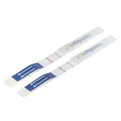
LF-018-10 Conjugation QC Lateral Flow Dipstick Kit
GO-LF-200 Gold Nanoparticle Conjugation Optimization Kit – Lateral Flow
GO-20-100 Gold Nanoparticle Conjugation Optimization Kit – 20nm
GO-15-100 Gold Nanoparticle Conjugation Optimization Kit – 15nm
GO-TEM-200 Gold Nanoparticle Conjugation Optimization Kit – TEM
GO-MEM-200 Gold Nanoparticle Conjugation Optimization Kit – Dot-Blot
GO-100-100 Gold Nanoparticle Conjugation Optimization Kit – 100nm
GO-90-100 Gold Nanoparticle Conjugation Optimization Kit – 90nm
GO-80-100 Gold Nanoparticle Conjugation Optimization Kit – 80nm
GO-70-100 Gold Nanoparticle Conjugation Optimization Kit – 70nm
GO-60-100 Gold Nanoparticle Conjugation Optimization Kit – 60nm
GO-50-100 Gold Nanoparticle Conjugation Optimization Kit – 50nm
GO-40-100 Gold Nanoparticle Conjugation Optimization Kit – 40nm
GO-30-100 Gold Nanoparticle Conjugation Optimization Kit – 30nm
GO-10-100 Gold Nanoparticle Conjugation Optimization Kit – 10nm
GO-5-100 Gold Nanoparticle Conjugation Optimization Kit – 5nm

Cytodiagnostics NHS-activated Gold Nanoparticle Conjugation Kits have been optimized for high efficiency one-step conjugation of proteins and other primary amine-containing ligands to gold nanoparticles.

The kits contains ready-to-use pre-made mixtures. No activation or manipulation of the gold nanoparticles is required prior to conjugation, which often results in poor performing conjugates. Simply mix your protein with the pre-activated NHS ester gold nanoparticles supplied in the kit to generate your conjugate.

The precisely engineered gold surface on our NHS ester-activated gold nanoparticles results in high conjugation efficiency and stable conjugates while minimizing non-specific binding in your assay.

For quality control of antibody and antigen conjugation our Conjugation QC Lateral Flow Dipstick Kit can be used.

Features & Benefits of NHS-Activated Gold Nanoparticles

  • Results in covalently bound ligand and more stable conjugate.
  • Fast and convenient one-step conjugation reaction with no pre-activation requirements.
  • Spacer between the gold nanoparticle surface and conjugated ligand minimizes effects on the tertiary protein structure, which can lead to poor performing conjugates, a common problem seen in conjugates prepared by passive adsorption.
  • Precisely engineering surface coating that reduces non-specific protein binding in biological assays.
Catalog No. Description
LF-018-10 Conjugation QC Lateral Flow Dipstick Kit
CGN10K-100-1 100nm NHS-Activated Gold Nanoparticle Conjugation Kit (3 Reactions)
CGN10K-100-3 100nm NHS-Activated Gold Nanoparticle Conjugation Kit (MIDI Scale-Up Kit)
CGN10K-100-2 100nm NHS-Activated Gold Nanoparticle Conjugation Kit (10 Reactions)
CGN10K-90-3 90nm NHS-Activated Gold Nanoparticle Conjugation Kit (MIDI Scale-Up Kit)
CGN10K-90-2 90nm NHS-Activated Gold Nanoparticle Conjugation Kit (10 Reactions)
CGN10K-90-1 90nm NHS-Activated Gold Nanoparticle Conjugation Kit (3 Reactions)
CGN10K-80-3 80nm NHS-Activated Gold Nanoparticle Conjugation Kit (MIDI Scale-Up Kit)
CGN10K-80-2 80nm NHS-Activated Gold Nanoparticle Conjugation Kit (10 Reactions)
CGN10K-80-1 80nm NHS-Activated Gold Nanoparticle Conjugation Kit (3 Reactions)
CGN10K-70-3 70nm NHS-Activated Gold Nanoparticle Conjugation Kit (MIDI Scale-Up Kit)
CGN10K-70-2 70nm NHS-Activated Gold Nanoparticle Conjugation Kit (10 Reactions)
CGN10K-70-1 70nm NHS-Activated Gold Nanoparticle Conjugation Kit (3 Reactions)
CGN10K-60-3 60nm NHS-Activated Gold Nanoparticle Conjugation Kit (MIDI Scale-Up Kit)
CGN10K-60-2 60nm NHS-Activated Gold Nanoparticle Conjugation Kit (10 Reactions)
CGN10K-60-1 60nm NHS-Activated Gold Nanoparticle Conjugation Kit (3 Reactions)
CGN10K-50-3 50nm NHS-Activated Gold Nanoparticle Conjugation Kit (MIDI Scale-Up Kit)
CGN10K-50-2 50nm NHS-Activated Gold Nanoparticle Conjugation Kit (10 Reactions)
CGN10K-50-1 50nm NHS-Activated Gold Nanoparticle Conjugation Kit (3 Reactions)
CGN5K-40-3 40nm NHS-Activated Gold Nanoparticle Conjugation Kit (MIDI Scale-Up Kit)
CGN5K-40-2 40nm NHS-Activated Gold Nanoparticle Conjugation Kit (10 Reactions)
CGN5K-40-1 40nm NHS-Activated Gold Nanoparticle Conjugation Kit (3 Reactions)
CGN5K-30-3 30nm NHS-Activated Gold Nanoparticle Conjugation Kit (MIDI Scale-Up Kit)
CGN5K-30-2 30nm NHS-Activated Gold Nanoparticle Conjugation Kit (10 Reactions)
CGN5K-30-1 30nm NHS-Activated Gold Nanoparticle Conjugation Kit (3 Reactions)
CGN5K-20-3 20nm NHS-Activated Gold Nanoparticle Conjugation Kit (MIDI Scale-Up Kit)
CGN5K-20-2 20nm NHS-Activated Gold Nanoparticle Conjugation Kit (10 Reactions)
CGN5K-20-1 20nm NHS-Activated Gold Nanoparticle Conjugation Kit (3 Reactions)
CGN5K-15-3 15nm NHS-Activated Gold Nanoparticle Conjugation Kit (MIDI Scale-Up Kit)
CGN5K-15-2 15nm NHS-Activated Gold Nanoparticle Conjugation Kit (10 Reactions)
CGN5K-15-1 15nm NHS-Activated Gold Nanoparticle Conjugation Kit (3 Reactions)
CGN5K-10-3 10nm NHS-Activated Gold Nanoparticle Conjugation Kit (MIDI Scale-Up Kit)
CGN5K-10-2 10nm NHS-Activated Gold Nanoparticle Conjugation Kit (10 Reactions)
CGN5K-10-1 10nm NHS-Activated Gold Nanoparticle Conjugation Kit (3 Reactions)
CGN5K-10-3 5nm NHS-Activated Gold Nanoparticle Conjugation Kit (MIDI Scale-Up Kit)
CGN5K-5-2 5nm NHS-Activated Gold Nanoparticle Conjugation Kit (10 Reactions)
CGN5K-5-1 5nm NHS-Activated Gold Nanoparticle Conjugation Kit (3 Reactions)

Cytodiagnostics Maleimide-Activated Gold Nanoparticle Conjugation Kits have been optimized for high efficiency one-step conjugation of thiol-containing ligands such as oligonucleotides to gold nanoparticles.

The kits contains ready-to-use pre-made mixtures. No activation or manipulation of the gold nanoparticles is required prior to conjugation, which often results in poor performing conjugates. Simply mix your protein with the pre-activated maleimide gold nanoparticles supplied in the kit to generate your conjugate.

The precisely engineered gold surface coat on our maleimide-activated gold nanoparticles results in high conjugation efficiency and stable conjugates while minimizing non-specific binding in your assay.

Features & Benefits of Maleimide-Activated Gold Nanoparticles

  • Results in covalently bound ligand and more stable conjugate.
  • Fast and convenient one-step conjugation reaction with no pre-activation requirements.
  • Spacer between the gold nanoparticle surface and conjugated ligand minimizes effects on the tertiary protein structure, which can lead to poor performing conjugates, a common problem seen in conjugates prepared by passive adsorption.
  • Precisely engineered surface coating that reduces non-specific protein binding in biological assays.
Catalog No. Description
LF-018-10 Conjugation QC Lateral Flow Dipstick Kit
MG5K-5-1 5nm Maleimide-Activated Gold Nanoparticle Conjugation Kit (3 Reactions)
MG5K-5-2 5nm Maleimide-Activated Gold Nanoparticle Conjugation Kit (10 Reactions)
MG5K-5-3 5nm Maleimide-Activated Gold Nanoparticle Conjugation Kit (MIDI Scale-Up Kit)
MG5K-10-1 10nm Maleimide-Activated Gold Nanoparticle Conjugation Kit (3 Reactions)
MG5K-10-2 10nm Maleimide-Activated Gold Nanoparticle Conjugation Kit (10 Reactions)
MG5K-10-3 10nm Maleimide-Activated Gold Nanoparticle Conjugation Kit (MIDI Scale-Up Kit)
MG5K-15-1 15nm Maleimide-Activated Gold Nanoparticle Conjugation Kit (3 Reactions)
MG5K-15-2 15nm Maleimide-Activated Gold Nanoparticle Conjugation Kit (10 Reactions)
MG5K-15-3 15nm Maleimide-Activated Gold Nanoparticle Conjugation Kit (MIDI Scale-Up Kit)
MG5K-20-1 20nm Maleimide-Activated Gold Nanoparticle Conjugation Kit (3 Reactions)
MG5K-20-2 20nm Maleimide-Activated Gold Nanoparticle Conjugation Kit (10 Reactions)
MG5K-20-3 20nm Maleimide-Activated Gold Nanoparticle Conjugation Kit (MIDI Scale-Up Kit)
MG5K-30-1 30nm Maleimide-Activated Gold Nanoparticle Conjugation Kit (3 Reactions)
MG5K-30-2 30nm Maleimide-Activated Gold Nanoparticle Conjugation Kit (10 Reactions)
MG5K-30-3 30nm Maleimide-Activated Gold Nanoparticle Conjugation Kit (MIDI Scale-Up Kit)
MG5K-40-1 40nm Maleimide-Activated Gold Nanoparticle Conjugation Kit (3 Reactions)
MG5K-40-2 40nm Maleimide-Activated Gold Nanoparticle Conjugation Kit (10 Reactions)
MG5K-40-3 40nm Maleimide-Activated Gold Nanoparticle Conjugation Kit (MIDI Scale-Up Kit)
MG10K-50-1 50nm Maleimide-Activated Gold Nanoparticle Conjugation Kit (3 Reactions)
MG10K-50-2 50nm Maleimide-Activated Gold Nanoparticle Conjugation Kit (10 Reactions)
MG10K-50-3 50nm Maleimide-Activated Gold Nanoparticle Conjugation Kit (MIDI Scale-Up Kit)
MG10K-60-1 60nm Maleimide-Activated Gold Nanoparticle Conjugation Kit (3 Reactions)
MG10K-60-2 60nm Maleimide-Activated Gold Nanoparticle Conjugation Kit (10 Reactions)
MG10K-60-3 60nm Maleimide-Activated Gold Nanoparticle Conjugation Kit (MIDI Scale-Up Kit)
MG10K-70-1 70nm Maleimide-Activated Gold Nanoparticle Conjugation Kit (3 Reactions)
MG10K-70-2 70nm Maleimide-Activated Gold Nanoparticle Conjugation Kit (10 Reactions)
MG10K-70-3 70nm Maleimide-Activated Gold Nanoparticle Conjugation Kit (MIDI Scale-Up Kit)
MG10K-80-1 80nm Maleimide-Activated Gold Nanoparticle Conjugation Kit (3 Reactions)
MG10K-80-2 80nm Maleimide-Activated Gold Nanoparticle Conjugation Kit (10 Reactions)
MG10K-80-3 80nm Maleimide-Activated Gold Nanoparticle Conjugation Kit (MIDI Scale-Up Kit)
MG10K-90-1 90nm Maleimide-Activated Gold Nanoparticle Conjugation Kit (3 Reactions)
MG10K-90-2 90nm Maleimide-Activated Gold Nanoparticle Conjugation Kit (10 Reactions)
MG10K-90-3 90nm Maleimide-Activated Gold Nanoparticle Conjugation Kit (MIDI Scale-Up Kit)
MG10K-100-1 100nm Maleimide-Activated Gold Nanoparticle Conjugation Kit (3 Reactions)
MG10K-100-2 100nm Maleimide-Activated Gold Nanoparticle Conjugation Kit (10 Reactions)
MG10K-100-3 100nm Maleimide-Activated Gold Nanoparticle Conjugation Kit (MIDI Scale-Up Kit)

Gold Conjugation Kits for Thiolated Oligonucleotides

Cytodiagnostics unique OligoREADY™ gold conjugation kits have been optimized for high efficiency one-step conjugation of thiolated oligonucleotides directly to the gold surface of particles with diameters up to 100nm.

The kit contains ready-to-use pre-made mixtures. No activation, manipulation, or time consuming “salt-aging” steps are require for conjugation. Simply mix your reduced thiol-modified oligonucleotide with the pre-activated gold nanoparticles supplied with the kit. Conjugation of the oligonucleotide is achieved by the formation of a strong and stable gold-thiol bond without any additional linkers.

Features & Benefits

  • Allows conjugation of oligonucleotides to gold nanoparticles with sizes between 5nm-100nm.
  • Fast and convenient one-step conjugation reaction with no pre-activation requirements or manipulation of the gold nanoparticles.
  • Does not require time-consuming “salt-aging” procedures.
  • Results in a thiol-oligo conjugated directly to the gold surface without any linkers
Catalog No. Description
OGC-5-1 5nm OligoREADY Gold Nanoparticle Conjugation Kit (3 reactions)
OGC-5-2 5nm OligoREADY Gold Nanoparticle Conjugation Kit (10 reactions)
OGC-10-1 10nm OligoREADY Gold Nanoparticle Conjugation Kit (3 reactions)
OGC-10-2 10nm OligoREADY Gold Nanoparticle Conjugation Kit (10 reactions)
OGC-15-1 15nm OligoREADY Gold Nanoparticle Conjugation Kit (3 reactions)
OGC-15-2 15nm OligoREADY Gold Nanoparticle Conjugation Kit (10 reactions)
OGC-20-1 20nm OligoREADY Gold Nanoparticle Conjugation Kit (3 reactions)
OGC-20-2 20nm OligoREADY Gold Nanoparticle Conjugation Kit (10 reactions)
OGC-30-1 30nm OligoREADY Gold Nanoparticle Conjugation Kit (3 reactions)
OGC-30-2 30nm OligoREADY Gold Nanoparticle Conjugation Kit (10 reactions)
OGC-40-1 40nm OligoREADY Gold Nanoparticle Conjugation Kit (3 reactions)
OGC-40-2 40nm OligoREADY Gold Nanoparticle Conjugation Kit (10 reactions)
OGC-50-1 50nm OligoREADY Gold Nanoparticle Conjugation Kit (3 reactions)
OGC-50-2 50nm OligoREADY Gold Nanoparticle Conjugation Kit (10 reactions)
OGC-60-1 60nm OligoREADY Gold Nanoparticle Conjugation Kit (3 reactions)
OGC-60-2 60nm OligoREADY Gold Nanoparticle Conjugation Kit (10 reactions)
OGC-70-1 70nm OligoREADY Gold Nanoparticle Conjugation Kit (3 reactions)
OGC-70-2 70nm OligoREADY Gold Nanoparticle Conjugation Kit (10 reactions)
OGC-80-1 80nm OligoREADY Gold Nanoparticle Conjugation Kit (3 reactions)
OGC-80-2 80nm OligoREADY Gold Nanoparticle Conjugation Kit (10 reactions)
OGC-90-1 90nm OligoREADY Gold Nanoparticle Conjugation Kit (3 reactions)
OGC-90-2 90nm OligoREADY Gold Nanoparticle Conjugation Kit (10 reactions)
OGC-100-1 100nm OligoREADY Gold Nanoparticle Conjugation Kit (3 reactions)
OGC-100-2 100nm OligoREADY Gold Nanoparticle Conjugation Kit (10 reactions)

Cytodiagnostics carboxyl functionalized gold nanoparticles have precisely engineered surface chemistry that enables coupling to various moieties that react with carboxylic acids. One of the most popular applications of this interaction is EDC/NHS coupling. The EDC activates the carboxylic acid so that it can react with NHS and acquire new functionality. The introduction of the NHS group then allows it to react with primary amines, which enables covalent coupling to proteins and antibodies with exposed amine-containing residues such as lysine. This permanent linkage has thus provided a gold nanoparticle label to the molecule in question and allows easier monitoring and localization within cells.

Cytodiagnostics carboxyl gold nanoparticles are available with diameters ranging from 5nm – 100nm and are also available with 2 different spacer lengths (3 kDa and 5 kDa). This provides the customer with the necessary tools fully optimize their experiment.

Catalog No. Description
CGC3K-5 5nm Carboxyl (carboxyl-PEG3000-SH) Gold Nanoparticles
CGC5K-5 5nm Carboxyl (carboxyl-PEG5000-SH) Gold Nanoparticles
CGC3K-10 10nm Carboxyl (carboxyl-PEG3000-SH) Gold Nanoparticles
CGC5K-10 10nm Carboxyl (carboxyl-PEG5000-SH) Gold Nanoparticles
CGC3K-15 15nm Carboxyl (carboxyl-PEG3000-SH) Gold Nanoparticles
CGC5K-15 15nm Carboxyl (carboxyl-PEG5000-SH) Gold Nanoparticles
CGC3K-20 20nm Carboxyl (carboxyl-PEG3000-SH) Gold Nanoparticles
CGC5K-20 20nm Carboxyl (carboxyl-PEG5000-SH) Gold Nanoparticles
CGC3K-30 30nm Carboxyl (carboxyl-PEG3000-SH) Gold Nanoparticles
CGC5K-30 30nm Carboxyl (carboxyl-PEG5000-SH) Gold Nanoparticles
CGC3K-40 40nm Carboxyl (carboxyl-PEG3000-SH) Gold Nanoparticles
CGC5K-40 40nm Carboxyl (carboxyl-PEG5000-SH) Gold Nanoparticles
CGC3K-50 50nm Carboxyl (carboxyl-PEG3000-SH) Gold Nanoparticles
CGC5K-50 50nm Carboxyl (carboxyl-PEG5000-SH) Gold Nanoparticles
CGC3K-60 60nm Carboxyl (carboxyl-PEG3000-SH) Gold Nanoparticles
CGC5K-60 60nm Carboxyl (carboxyl-PEG5000-SH) Gold Nanoparticles
CGC3K-70 70nm Carboxyl (carboxyl-PEG3000-SH) Gold Nanoparticles
CGC5K-70 70nm Carboxyl (carboxyl-PEG5000-SH) Gold Nanoparticles
CGC3K-80 80nm Carboxyl (carboxyl-PEG3000-SH) Gold Nanoparticles
CGC5K-80 80nm Carboxyl (carboxyl-PEG5000-SH) Gold Nanoparticles
CGC3K-90 90nm Carboxyl (carboxyl-PEG3000-SH) Gold Nanoparticles
CGC5K-90 90nm Carboxyl (carboxyl-PEG5000-SH) Gold Nanoparticles
CGC3K-100 100nm Carboxyl (carboxyl-PEG3000-SH) Gold Nanoparticles
CGC5K-100 100nm Carboxyl (carboxyl-PEG5000-SH) Gold Nanoparticles

Cytodiagnostics amine functionalized gold nanoparticles have precisely engineered surface chemistry that allows for simple and efficient binding to moieties that react with primary amines. An example of such a reaction would be a conjugation to a protein or antibody that has been tagged NHS, which would yield a covalent link between the gold nanoparticle surface and the protein. This permanent linkage allows for further downstream applications such as monitoring the distribution of the now-tagged molecule.

Cytodiagnostics amine gold nanoparticles are available with diameters ranging from 5nm – 100nm and are also available with 2 different spacer lengths (3 kDa and 5 kDa). This provides the customer with the necessary tools to fully optimize their experiment.

Catalog No. Description
CGA3K-5 5nm Amine (amine-PEG3000-SH) Gold Nanoparticles
CGA5K-5 5nm Amine (amine-PEG5000-SH) Gold Nanoparticles
CGA3K-10 10nm Amine (amine-PEG3000-SH) Gold Nanoparticles
CGA5K-10 10nm Amine (amine-PEG5000-SH) Gold Nanoparticles
CGA3K-15 15nm Amine (amine-PEG3000-SH) Gold Nanoparticles
CGA5K-15 15nm Amine (amine-PEG5000-SH) Gold Nanoparticles
CGA3K-20 20nm Amine (amine-PEG3000-SH) Gold Nanoparticles
CGA5K-20 20nm Amine (amine-PEG5000-SH) Gold Nanoparticles
CGA3K-30 30nm Amine (amine-PEG3000-SH) Gold Nanoparticles
CGA5K-30 30nm Amine (amine-PEG5000-SH) Gold Nanoparticles
CGA3K-40 40nm Amine (amine-PEG3000-SH) Gold Nanoparticles
CGA5K-40 40nm Amine (amine-PEG5000-SH) Gold Nanoparticles
CGA3K-50 50nm Amine (amine-PEG3000-SH) Gold Nanoparticles
CGA5K-50 50nm Amine (amine-PEG5000-SH) Gold Nanoparticles
CGA3K-60 60nm Amine (amine-PEG3000-SH) Gold Nanoparticles
CGA5K-60 60nm Amine (amine-PEG5000-SH) Gold Nanoparticles
CGA3K-70 70nm Amine (amine-PEG3000-SH) Gold Nanoparticles
CGA5K-70 70nm Amine (amine-PEG5000-SH) Gold Nanoparticles
CGA3K-80 80nm Amine (amine-PEG3000-SH) Gold Nanoparticles
CGA5K-80 80nm Amine (amine-PEG5000-SH) Gold Nanoparticles
CGA3K-90 90nm Amine (amine-PEG3000-SH) Gold Nanoparticles
CGA5K-90 90nm Amine (amine-PEG3000-SH) Gold Nanoparticles
CGA3K-100 100nm Amine (amine-PEG3000-SH) Gold Nanoparticles
CGA5K-100 100nm Amine (amine-PEG5000-SH) Gold Nanoparticles

Cytodiagnostics biotin functionalized gold nanoparticles are available with diameters in the range of 5nm – 100nm.  These particles have a precisely engineered surface for optimal streptavidin binding while minimizing non-specific binding to other proteins present in your sample.

The gold nanoparticle label allows for convenient binding and detection of streptavidin in applications such as TEM, ELISA and Immuno dot-blots among others.

Figure 1. Cytodiagnostics Biotin gold nanoparticles bound to an avidin-coated surface substrate. Courtesy of Trevor Olsen, University of Alberta. Images taken at the NINT Electron Microscopy Facility.

Catalog No. Description
CGB5K-5 5nm Biotin Gold Nanoparticles
CGB5K-10 10nm Biotin Gold Nanoparticles
CGB5K-15 15nm Biotin Gold Nanoparticles
CGB5K-20 20nm Biotin Gold Nanoparticles
CGB5K-30 30nm Biotin Gold Nanoparticles
CGB5K-40 40nm Biotin Gold Nanoparticles
CGB5K-50 50nm Biotin Gold Nanoparticles
CGB5K-60 60nm Biotin Gold Nanoparticles
CGB5K-70 70nm Biotin Gold Nanoparticles
CGB5K-80 80nm Biotin Gold Nanoparticles
CGB5K-90 90nm Biotin Gold Nanoparticles
CGB5K-100 100nm Biotin Gold Nanoparticles

Cytodiagnostics methoxy gold nanoparticles have precisely engineered surface chemistry that renders them inert in most applications. This inert property allows these nanoparticles to be used as excellent control samples when in combination with one of our many other functionalized nanoparticles. In applications where the nanoparticles are not being used as controls, the surface chemistry enables them to be used in solutions with much higher salt concentrations relative to the corresponding standard or stabilized nanoparticles.
Cytodiagnostics methoxy gold nanoparticles are available with diameters ranging from 5nm – 100nm and are also available with 2 different spacer lengths (2 kDa and 5 kDa), allowing for better optimization.

Catalog No. Description
CGM2K-5 5nm Methylated (methoxy-PEG2000-SH) Gold Nanoparticles
CGM5K-5 5nm Methylated (methoxy-PEG5000-SH) Gold Nanoparticles
CGM2K-10 10nm Methylated (methoxy-PEG2000-SH) Gold Nanoparticles
CGM5K-10 10nm Methylated (methoxy-PEG5000-SH) Gold Nanoparticles
CGM2K-15 15nm Methylated (methoxy-PEG2000-SH) Gold Nanoparticles
CGM5K-15 15nm Methylated (methoxy-PEG5000-SH) Gold Nanoparticles
CGM2K-20 20nm Methylated (methoxy-PEG2000-SH) Gold Nanoparticles
CGM5K-20 20nm Methylated (methoxy-PEG5000-SH) Gold Nanoparticles
CGM2K-30 30nm Methylated (methoxy-PEG2000-SH) Gold Nanoparticles
CGM5K-30 30nm Methylated (methoxy-PEG5000-SH) Gold Nanoparticles
CGM2K-40 40nm Methylated (methoxy-PEG2000-SH) Gold Nanoparticles
CGM5K-40 40nm Methylated (methoxy-PEG5000-SH) Gold Nanoparticles
CGM2K-50 50nm Methylated (methoxy-PEG2000-SH) Gold Nanoparticles
CGM5K-50 50nm Methylated (methoxy-PEG5000-SH) Gold Nanoparticles
CGM2K-60 60nm Methylated (methoxy-PEG2000-SH) Gold Nanoparticles
CGM5K-60 60nm Methylated (methoxy-PEG5000-SH) Gold Nanoparticles
CGM2K-70 70nm Methylated (methoxy-PEG2000-SH) Gold Nanoparticles
CGM5K-70 70nm Methylated (methoxy-PEG5000-SH) Gold Nanoparticles
CGM2K-80 80nm Methylated (methoxy-PEG2000-SH) Gold Nanoparticles
CGM5K-80 80nm Methylated (methoxy-PEG5000-SH) Gold Nanoparticles
CGM2K-90 90nm Methylated (methoxy-PEG2000-SH) Gold Nanoparticles
CGM5K-90 90nm Methylated (methoxy-PEG5000-SH) Gold Nanoparticles
CGM2K-100 100nm Methylated (methoxy-PEG2000-SH) Gold Nanoparticles
CGM5K-100 100nm Methylated (methoxy-PEG5000-SH) Gold Nanoparticles

Cytodiagnostics Ni-NTA gold nanoparticles have precisely engineered surface chemistry consisting of a PEG spacer terminated with a nitrilotriacetic acid (NTA) group chelated to a Ni2+ ion. This specific functionality binds to His tags, which are a common modification added to the terminal end of proteins or antibodies, consisting of 6 sequential histidine residues. The interaction between Ni-NTA and His tags enables many applications when coupled to our gold nanoparticles: preparation of gold conjugates, protein purification and isolation, cell localization and uptake studies, use in assays, and more.

Cytodiagnostics Ni-NTA gold nanoparticles are available in 12 different sizes ranging from 5nm – 100nm, are more than 95% spherical and have a uniform size distribution (CV <12%). Products are provided as suspensions at 50 OD.

Catalog No. Description
CGNTA-5 5nm Ni-NTA Gold Nanoparticles
CGNTA-10 10nm Ni-NTA Gold Nanoparticles
CGNTA-15 15nm Ni-NTA Gold Nanoparticles
CGNTA-20 20nm Ni-NTA Gold Nanoparticles
CGNTA-30 30nm Ni-NTA Gold Nanoparticles
CGNTA-40 40nm Ni-NTA Gold Nanoparticles
CGNTA-50 50nm Ni-NTA Gold Nanoparticles
CGNTA-60 60nm Ni-NTA Gold Nanoparticles
CGNTA-70 70nm Ni-NTA Gold Nanoparticles
CGNTA-80 80nm Ni-NTA Gold Nanoparticles
CGNTA-90 90nm Ni-NTA Gold Nanoparticles
CGNTA-100 100nm Ni-NTA Gold Nanoparticles

Cytodiagnostics azide functionalized gold nanoparticles are precisely engineered for covalent conjugation to any molecule tagged with an alkyne group. With the use of a copper(I) catalyst, the azide group on the gold and the alkyne-tagged molecule undergo a cycloaddition to form a stable and permanent linkage. The reaction between these 2 functional groups typically occurs rapidly, in high yield, and with no by-products, meaning that the customer will always have a stable conjugate and enough of it to use in subsequent downstream applications such as lateral flow assays and imaging. These reactions are also especially useful in applications involving oligonucleotides due to the higher prevalence of alkyne modifications.

Cytodiagnostics azide functionalized gold nanoparticles are available in 12 different sizes ranging from 5nm – 100nm, are more than 95% spherical and have a uniform size distribution (CV <12%). Products are provided as suspensions at 50 OD.

Catalog No. Description
CGAZ-5 5nm Azide Gold Nanoparticles
CGAZ-10 10nm Azide Gold Nanoparticles
CGAZ-15 15nm Azide Gold Nanoparticles
CGAZ-20 20nm Azide Gold Nanoparticles
CGAZ-30 30nm Azide Gold Nanoparticles
CGAZ-40 40nm Azide Gold Nanoparticles
CGAZ-50 50nm Azide Gold Nanoparticles
CGAZ-60 60nm Azide Gold Nanoparticles
CGAZ-70 70nm Azide Gold Nanoparticles
CGAZ-80 80nm Azide Gold Nanoparticles
CGAZ-90 90nm Azide Gold Nanoparticles
CGAZ-100 100nm Azide Gold Nanoparticles

Cytodiagnostics alkyne functionalized gold nanoparticles are precisely engineered for covalent conjugation to any molecule tagged with an azide group. With the use of a copper(I) catalyst, the alkyne group on the gold and the azide-tagged molecule undergo a cycloaddition to form a stable and permanent linkage. The reaction between these 2 functional groups typically occurs rapidly, in high yield, and with no by-products, meaning that the customer will always have a stable conjugate and enough of it to use in subsequent downstream applications such as lateral flow assays and imaging. These reactions are also especially useful in applications involving oligonucleotides due to the higher prevalence of azide modifications.

Cytodiagnostics alkyne functionalized gold nanoparticles are available in 12 different sizes ranging from 5nm – 100nm, are more than 95% spherical and have a uniform size distribution (CV <12%). Products are provided as suspensions at 50 OD.

Catalog No. Description
CGALK-5 5nm Alkyne Gold Nanoparticles
CGALK-10 10nm Alkyne Gold Nanoparticles
CGALK-15 15nm Alkyne Gold Nanoparticles
CGALK-20 20nm Alkyne Gold Nanoparticles
CGALK-30 30nm Alkyne Gold Nanoparticles
CGALK-40 40nm Alkyne Gold Nanoparticles
CGALK-50 50nm Alkyne Gold Nanoparticles
CGALK-60 60nm Alkyne Gold Nanoparticles
CGALK-70 70nm Alkyne Gold Nanoparticles
CGALK-80 80nm Alkyne Gold Nanoparticles
CGALK-90 90nm Alkyne Gold Nanoparticles
CGALK-100 100nm Alkyne Gold Nanoparticles

Cytodiagnostics dibenzocyclooctyne (DBCO) functionalized gold nanoparticles are suitable for covalent conjugation of any azide-tagged molecule through click chemistry. The rigid structure of the DBCO allows for quick reactions under more moderate conditions, and eliminates the need for a copper catalyst. This chemistry confers higher stability in aqueous conditions and the lack of copper greatly increases its overall biocompatibility, enabling a multitude of biological applications.

Our DBCO functionalized gold nanoparticles are available in 9 different sizes ranging from 20 – 100nm, are more than 95% spherical and have uniform size distribution (CV <12%).

For custom sizes, formulations or bulk quantities please contact our customer service department, info@stratech.co.uk.

Catalog No. Description
CGDBC-20 20nm DBCO Gold Nanoparticles
CGDBC-30 30nm DBCO Gold Nanoparticles
CGDBC-40 40nm DBCO Gold Nanoparticles
CGDBC-50 50nm DBCO Gold Nanoparticles
CGDBC-60 60nm DBCO Gold Nanoparticles
CGDBC-70 70nm DBCO Gold Nanoparticles
CGDBC-80 80nm DBCO Gold Nanoparticles
CGDBC-90 90nm DBCO Gold Nanoparticles
CGDBC-100 100nm DBCO Gold Nanoparticles

Gold NanoUrchins

Cytodiagnostics Gold NanoUrchins have unique optical properties compared to spherical gold nanoparticles of the same core diameter. The spiky uneven surface causes a red shift in the surface plasmon peak and a larger enhancement of the electromagnetic field at the tips of the Gold NanoUrchin spikes compared to that of a spherical particles. As an example, 100nm spherical gold nanoparticles have an SPR peak at 570nm while 100nm Gold NanoUrchins have a SPR peak at around 680nm, figure 1.

Figure 1. Left) UV-VIS spectra of 100nm Gold NanoUrchins (blue) and 100nm standard gold nanoparticles (green). Note the red-shift in the SPR-peak. Right) UV-VIS spectra of Gold NanoUrchins ranging in size from 50nm to 100nm in diameter.


Binding of ligands such as proteins to the Gold NanoUrchin surface also causes a larger shift in the surface plasmon resonance peak compared to standard spherical gold nanoparticles. This feature makes them ideal in the development of sensitive SPR-based detection assays, figure 2.

Our Gold NanoUrchins are available in 6 different sizes with a multitude of surface chemistries, see product categories below.

Figure 2. Refractive index induced shift of the lambda max (LSPR-shift) for standard gold nanoparticles and gold nanourchins upon addition of glycerol.

Gold NanoUrchins

Catalog No. Description
GU-Intro Gold NanoUrchins Introduction Kit
GU-50 50nm Gold NanoUrchins
GU-60 60nm Gold NanoUrchins
GU-70 70nm Gold NanoUrchins
GU-80 80nm Gold NanoUrchins
GU-90 90nm Gold NanoUrchins
GU-100 100nm Gold NanoUrchins

Cytodiagnostics Gold NanoUrchins have unique optical properties compared to spherical gold nanoparticles of the same core diameter. The spiky uneven surface causes a red shift in the surface plasmon peak and a larger enhancement of the electromagnetic field at the tips of the Gold NanoUrchin spikes compared to that of a spherical particles. As an example, 100nm spherical gold nanoparticles have an SPR peak at 570nm while 100nm Gold NanoUrchins have a SPR peak at around 680nm.

Reactant Free Gold NanoUrchins are supplied in 0.1mM phosphate buffered saline (PBS) and are extensively purified to be 99% free of residual reactants from manufacturing. These particles have the same high protein binding efficiency as our standard gold nanoparticle preparations for use in applications such as conjugate development, lateral flow and Surface Enhanced Raman Spectroscopy (SERS) with the added benefit of being reactant free for more sensitive applications such as cell work and nanotoxicology studies.

Catalog No. Description
RFU-50 50nm Reactant Free Gold NanoUrchins
RFU-60 60nm Reactant Free Gold NanoUrchins
RFU-70 70nm Reactant Free Gold NanoUrchins
RFU-80 80nm Reactant Free Gold NanoUrchins
RFU-90 90nm Reactant Free Gold NanoUrchins
RFU-100 100nm Reactant Free Gold NanoUrchins
RFU-Intro Reactant Free Gold NanoUrchins Introduction Kit

Our extensively purified endotoxin free gold nanourchins are ideal for sensitive applications requiring a minimal amount of contaminants, such as cellular toxicity studies, immunological studies, and aseptic assays. This product is well suited for conjugate development by passive adsorption of proteins and other ligands to the gold nanourchins surface. The resulting conjugate can then be used in downstream applications.

All of our batches go through stringent quality control prior to shipment to our customers using dynamic light scattering to guarantee size and monodispersity and lot specifications are supplied with each product. The endotoxin levels of all batches are validated by the Limulus amebocyte lysate (LAL) test and do not exceed 0.5EU/mL.

Catalog No. Description
UEF-90 90nm Endotoxin Free Gold NanoUrchins
UEF-80 80nm Endotoxin Free Gold NanoUrchins
UEF-70 70nm Endotoxin Free Gold NanoUrchins
UEF-60 60nm Endotoxin Free Gold NanoUrchins
UEF-50 50nm Endotoxin Free Gold NanoUrchins
UEF-100 100nm Endotoxin Free Gold NanoUrchins

Custom Conjugation Services – Nanoparticles

Cytodiagnostics provides an array of options for customers to do their own conjugations. Our gold and silver nanoparticles, and gold nanourchins are optimally designed for efficient passive adsorption of a multitude of proteins, antibodies, thiolated oligos, and more. For covalent conjugates, Cytodiagnostics also provides ready-to-use kits: Gold Conjugation Kits, Silver Conjugation Kits, NanoUrchin Conjugation Kits.

Let Us Do It For You!

Cytodiagnostics custom conjugation services help design and produce your very own custom conjugate. This includes most ligands ranging from proteins to enzymes to oligonucleotides, and this service extends to all the nanoparticles in our expansive catalog. You as a customer tell us the parameters and end-goal you need met, and our team of experts will work out the rest for you.

Ligand Sourcing · Sourced by Cytodiagnostics · Provided by customer
Potential Ligands · Proteins · Full Antibodies (monoclonal, polyclonal) · Fab’, F(ab’)2, Fc, and scFv fragments · Enzymes · Aptamers · Thiolated Oligonucleotides · Aminated Oligonucleotides
Potential Nanoparticles · Standard spherical gold · Standard spherical silver · Gold NanoUrchins
Nanoparticle Sizes 5nm – 400nm (standard spherical gold) · 10nm – 100nm (standard spherical silver) · 50nm – 100nm (Gold NanoUrchins)
Average Final Product Yield · 10 mL at 3 OD
Average Timeframe · 1 – 2 weeks
Additional Services · Purification of conjugate · Functionality testing

Gold NanoUrchin Conjugates and Kits

Gold nanourchin protein conjugates are a great alternative to traditional gold protein conjugates if peak absorbance is desired in the 570nm-680nm range.
Cytodiagnostics unique gold nanourchin protein conjugates can be used for a variety of applications including use as secondary detection agents in immunoblotting, lateral flow/vertical flow devices, ELISA, light microscopy and electron microscopy.

If you are interested in having your own antibody, or other protein conjugated to our gold nanourchins as a custom service please contact us.

Browse Our Available Gold NanoUrchin Conjugates

Streptavidin Gold NanoUrchin Conjugates

Cytodiagnostics Streptavidin conjugated gold nanourchins can be used for secondary detection of biotinylated probes in assays such as immunoblotting, ELISA, light microscopy and electron microscopy applications.

Streptavidin gold nanourchin conjugates can also be used for convenient and fast conjugation of any biotinylated ligand such as antibodies and oligonucleotides to the gold surface.

Anti-6X His Gold NanoUrchin Conjugates

Anti-6X His IgG is an antibody produced in mouse that binds specifically to a sequence of 6 repeating histidine residues, known as a His tag. These His tags are typically expressed at either the C- or N-terminal regions of recombinant proteins and aid in purification and isolation using immobilized metal affinity chromatography. His tags have a strong affinity for Ni2+ through chelation.

Cytodiagnostics anti-6X His conjugated gold NanoUrchins are suitable for use in applications such as lateral flow, immunoblotting, light microscopy, and electron microscopy.

Anti-FITC Gold NanoUrchin Conjugates

Anti-FITC IgG is an antibody produced in sheep that binds specifically to fluorescein isothiocyanate (FITC). FITC is a fluorochrome dye that absorbs ultraviolet light and emits a yellow-green light when excited. This antibody can be used to detect FITC-labelled antibodies and proteins.

Cytodiagnostics anti-FITC conjugated gold NanoUrchins are suitable for use in applications such as lateral flow, immunoblotting, light microscopy, and electron microscopy.

Anti-Human Gold NanoUrchin Conjugates

Goat anti-human secondary antibodies are produced in goat and specifically target the various classes of human immunoglobulins. These gold NanoUrchin conjugates are specific against human IgG, and its subclasses. This antibody-antibody interaction allows for the development of bio-assays and detection of human antibody-labelled targets.

Cytodiagnostics anti-human conjugated gold NanoUrchins are suitable for applications such as lateral flow, immunoblotting, light microscopy, and electron microscopy.

Anti-Rabbit Gold NanoUrchin Conjugates

Goat anti-rabbit secondary antibodies are produced in goat and target rabbit IgG antibodies, along with their subclasses. This antibody-antibody interaction allows for the development of bio-assays and detection of rabbit antibody-labelled targets.

Cytodiagnostics anti-rabbit conjugated gold NanoUrchins are suitable for applications such as lateral flow, immunoblotting, light microscopy, and electron microscopy.

Anti-Mouse Gold NanoUrchin Conjugates

Goat anti-mouse secondary antibodies are produced in goat and target mouse IgG antibodies, along with their subclasses. This antibody-antibody interaction allows for the development of bio-assays and detection of mouse antibody-labelled targets.

Cytodiagnostics anti-mouse conjugated gold NanoUrchins are suitable for applications such as lateral flow, immunoblotting, light microscopy, and electron microscopy.

Anti-Rat Gold NanoUrchin Conjugates

Goat anti-rat secondary antibodies are produced in goat and target rat IgG antibodies, along with their subclasses. This antibody-antibody interaction allows for the development of bio-assays and detection of rat antibody-labelled targets.

Cytodiagnostics anti-rat conjugated gold NanoUrchins are suitable for applications such as lateral flow, immunoblotting, light microscopy, and electron microscopy.

Anti-Sheep Gold NanoUrchin Conjugates

Rabbit anti-sheep secondary antibodies are produced in rabbit and target sheep IgG antibodies, along with their subclasses. This antibody-antibody interaction allows for the development of bio-assays and detection of sheep antibody-labelled targets.

Cytodiagnostics anti-sheep conjugated gold NanoUrchins are suitable for applications such as lateral flow, immunoblotting, light microscopy, and electron microscopy.

Anti-Chicken Gold NanoUrchin Conjugates

Goat anti-chicken secondary antibodies are produced in goat and target chicken IgY antibodies. This antibody-antibody interaction allows for the development of bio-assays and detection of chicken antibody-labelled targets.

Cytodiagnostics anti-chicken conjugated gold NanoUrchins are suitable for applications such as lateral flow, immunoblotting, light microscopy, and electron microscopy.

Protein A Gold NanoUrchin Conjugates

Cytodiagnostics Protein A conjugated gold nanourchins can be used for secondary detection of antibody probes in assays such as lateral flow, immunoblotting, ELISA, light microscopy and electron microscopy applications.

Protein A gold nanourchin conjugates can also be used for capturing of antibody species with affinity for Protein A to the gold nanourchin surface.

Protein G Gold NanoUrchin Conjugates

Protein G is a bacterial protein that has a high affinity for the Fc region of IgG-type antibodies. As such, Protein G gold conjugates allow for convenient and quick conjugation of Fc-containing antibodies to the gold surface, which can then be utilized further downstream.

Cytodiagnostics Protein G conjugated gold NanoUrchins are suitable for use in immunoblotting, light microscopy, electron microscopy applications, and other procedures for detection of antibody-labelled samples.

Protein L Gold NanoUrchin Conjugates

Protein L is a bacterial protein that binds to the kappa light chains of IgG-type antibodies. However, it does not bind to lambda light chains. As such, Protein L gold conjugates allow for convenient and quick conjugation of antibodies containing a kappa chain to the gold surface, which can then be utilized further downstream. An advantage of Protein L over Protein A and Protein G is that it is capable of binding to single-chain antibody fragments (scFv) and Fab fragments, allowing for finer optimization.

Cytodiagnostics Protein L conjugated gold NanoUrchins are suitable for use in immunoblotting, light microscopy, electron microscopy applications, and other procedures for detection of antibody-labelled samples.

Transferrin Gold NanoUrchin Conjugates

Transferrin is an iron-binding glycoprotein found in the blood plasma of vertebrates. It is a key component in iron transport and metabolism throughout an organism. Cytodiagnostics Transferrin conjugated gold NanoUrchins utilize holo-transferrin, which is the iron-saturated form, meaning that they can be readily taken up by cells through endocytosis. This allows for easier visualization of cellular uptake of iron using dark field microscopy, and electron microscopy.

NHS-Activated Gold NanoUrchins Conjugation

Cytodiagnostics NHS-activated Gold NanoUrchin Conjugation Kits have been optimized for high efficiency one-step conjugation of proteins and other primary amine-containing ligands to gold nanoparticles.

The kits contains ready-to-use pre-made mixtures. No activation or manipulation of the gold nanourchins is required prior to conjugation, which often results in poor performing conjugates. Simply mix your protein with the pre-activated NHS ester gold nanourchins supplied in the kit to generate your conjugate.

The precisely engineered gold surface coat on our NHS ester-activated gold nanourchins results in high conjugation efficiency and stable conjugates while minimizing non-specific binding in your assay.

For quality control of antibody and antigen conjugation our Conjugation QC Lateral Flow Dipstick Kit can be used.

Features & Benefits of NHS-Activated Gold NanoUrchins

  • Results in covalently bound ligand and more stable conjugate.
  • Fast and convenient one-step conjugation reaction with no pre-activation requirements.
  • Spacer between the gold nanourchin surface and conjugated ligand minimizes effects on the tertiary protein structure, which can lead to poor performing conjugates, a common problem seen in conjugates prepared by passive adsorption.
  • Precisely engineering surface coating that reduces non-specific protein binding in biological assays.
Catlog No. Description
LF-018-10 Conjugation QC Lateral Flow Dipstick Kit
GUN10K-50-1 50nm NHS-Activated Gold NanoUrchins Conjugation Kit (3 Reactions)
GUN10K-50-2 50nm NHS-Activated Gold NanoUrchins Conjugation Kit (10 Reactions)
GUN10K-50-3 50nm NHS-Activated Gold NanoUrchins Conjugation Kit (MIDI Scale-Up Kit)
GUN10K-60-1 60nm NHS-Activated Gold NanoUrchins Conjugation Kit (3 Reactions)
GUN10K-60-2 60nm NHS-Activated Gold NanoUrchins Conjugation Kit (10 Reactions)
GUN10K-60-3 60nm NHS-Activated Gold NanoUrchins Conjugation Kit (MIDI Scale-Up Kit)
GUN10K-70-1 70nm NHS-Activated Gold NanoUrchins Conjugation Kit (3 Reactions)
GUN10K-70-2 70nm NHS-Activated Gold NanoUrchins Conjugation Kit (10 Reactions)
GUN10K-70-3 70nm NHS-Activated Gold NanoUrchins Conjugation Kit (MIDI Scale-Up Kit)
GUN10K-80-1 80nm NHS-Activated Gold NanoUrchins Conjugation Kit (3 Reactions)
GUN10K-80-2 80nm NHS-Activated Gold NanoUrchins Conjugation Kit (10 Reactions)
GUN10K-80-3 80nm NHS-Activated Gold NanoUrchins Conjugation Kit (MIDI Scale-Up Kit)
GUN10K-90-1 90nm NHS-Activated Gold NanoUrchins Conjugation Kit (3 Reactions)
GUN10K-90-2 90nm NHS-Activated Gold NanoUrchins Conjugation Kit (10 Reactions)
GUN10K-90-3 90nm NHS-Activated Gold NanoUrchins Conjugation Kit (MIDI Scale-Up Kit)
GUN10K-100-1 100nm NHS-Activated Gold NanoUrchins Conjugation Kit (3 Reactions)
GUN10K-100-2 100nm NHS-Activated Gold NanoUrchins Conjugation Kit (10 Reactions)
GUN10K-100-3 100nm NHS-Activated Gold NanoUrchins Conjugation Kit (MIDI Scale-Up Kit)

Maleimide-Activated Gold NanoUrchins Conjugation

Cytodiagnostics Maleimide-activated Gold NanoUrchin Conjugation Kits have been optimized for high efficiency one-step conjugation of proteins and other thiol-containing ligands to gold nanoparticles.

The kits contains ready-to-use pre-made mixtures. No activation or manipulation of the gold nanourchins is required prior to conjugation, which often results in poor performing conjugates. Simply mix your protein with the pre-activated maleimide gold nanourchins supplied in the kit to generate your conjugate.

The precisely engineered gold surface coat on our maleimide-activated gold nanourchins results in high conjugation efficiency and stable conjugates while minimizing non-specific binding in your assay.

Features & Benefits of Maleimide-Activated Gold NanoUrchins

  • Results in covalently bound ligand and more stable conjugate.
  • Fast and convenient one-step conjugation reaction with no pre-activation requirements.
  • Spacer between the gold nanourchin surface and conjugated ligand minimizes effects on the tertiary protein structure, which can lead to poor performing conjugates, a common problem seen in conjugates prepared by passive adsorption.
  • Precisely engineering surface coating that reduces non-specific protein binding in biological assays.
Catlog No. Description
MUG10K-50-1 50nm Maleimide-Activated Gold NanoUrchins Conjugation Kit (3 Reactions)
MUG10K-50-2 50nm Maleimide-Activated Gold NanoUrchins Conjugation Kit (10 Reactions)
MUG10K-50-3 50nm Maleimide-Activated Gold NanoUrchins Conjugation Kit (MIDI Scale-Up Kit)
MUG10K-60-1 60nm Maleimide-Activated Gold NanoUrchins Conjugation Kit (3 Reactions)
MUG10K-60-2 60nm Maleimide-Activated Gold NanoUrchins Conjugation Kit (10 Reactions)
MUG10K-60-3 60nm Maleimide-Activated Gold NanoUrchins Conjugation Kit (MIDI Scale-Up Kit)
MUG10K-70-1 70nm Maleimide-Activated Gold NanoUrchins Conjugation Kit (3 Reactions)
MUG10K-70-2 70nm Maleimide-Activated Gold NanoUrchins Conjugation Kit (10 Reactions)
MUG10K-70-3 70nm Maleimide-Activated Gold NanoUrchins Conjugation Kit (MIDI Scale-Up Kit)
MUG10K-80-1 80nm Maleimide-Activated Gold NanoUrchins Conjugation Kit (3 Reactions)
MUG10K-80-2 80nm Maleimide-Activated Gold NanoUrchins Conjugation Kit (MIDI Scale-Up Kit)
MUG10K-80-3 80nm Maleimide-Activated Gold NanoUrchins Conjugation Kit (10 Reactions)
MUG10K-90-1 90nm Maleimide-Activated Gold NanoUrchins Conjugation Kit (3 Reactions)
MUG10K-90-2 90nm Maleimide-Activated Gold NanoUrchins Conjugation Kit (10 Reactions)
MUG10K-90-3 90nm Maleimide-Activated Gold NanoUrchins Conjugation Kit (MIDI Scale-Up Kit)
MUG10K-100-1 100nm Maleimide-Activated Gold NanoUrchins Conjugation Kit (3 Reactions)
MUG10K-100-2 100nm Maleimide-Activated Gold NanoUrchins Conjugation Kit (10 Reactions)
MUG10K-100-3 100nm Maleimide-Activated Gold NanoUrchins Conjugation Kit (MIDI Scale-Up Kit)

Cytodiagnostics biotin functionalized gold nanourchins are available with diameters in the range of 50nm – 100nm and have their surface plasmon resonance absorption and scattering characteristics in the 580-680nm range. These particles have a precisely engineered surface for optimal streptavidin binding while minimizing non-specific binding to other proteins present in your sample.

The gold nanourchin label allows for convenient binding and detection of streptavidin in applications such as Lateral Flow, TEM, ELISA and Immuno dot-blots among others.

Catlog No. Description
GUB10K-50 50nm Biotin Gold NanoUrchins
GUB10K-60 60nm Biotin Gold NanoUrchins
GUB10K-70 70nm Biotin Gold NanoUrchins
GUB10K-80 80nm Biotin Gold NanoUrchins
GUB10K-90 90nm Biotin Gold NanoUrchins
GUB10K-100 100nm Biotin Gold NanoUrchins

Carboxyl functionalized gold nanourchins are ideal for conjugation of proteins and other primary amine containing ligands using standard EDC/NHS coupling chemistry.

Our carboxylated gold nanourchins are available in 6 different sizes ranging from 50-100nm and have uniform size distribution (CV <15%).

Features

  • Precisely engineered surface with an optimized carboxyl group density for easy conjugation.
  • Available with SPR absorption maximum in the 600nm range.
  • Enhanced LSPR properties compared to spherical gold nanoparticles.

Applications

Ideal for development of gold nanourchin conjugates for use in for example:

  • Immunoblotting
  • Lateral Flow Assays
  • LSPR Assays
  • Microscopy Applications (e.g. TEM)
Catlog No. Description
GUC3K-50 50nm Carboxyl (carboxyl-PEG3000-SH) Gold NanoUrchins
GUC5K-50 50nm Carboxyl (carboxyl-PEG5000-SH) Gold NanoUrchins
GUC3K-60 60nm Carboxyl (carboxyl-PEG3000-SH) Gold NanoUrchins
GUC5K-60 60nm Carboxyl (carboxyl-PEG5000-SH) Gold NanoUrchins
GUC3K-70 70nm Carboxyl (carboxyl-PEG3000-SH) Gold NanoUrchins
GUC5K-70 70nm Carboxyl (carboxyl-PEG5000-SH) Gold NanoUrchins
GUC3K-80 80nm Carboxyl (carboxyl-PEG3000-SH) Gold NanoUrchins
GUC5K-80 80nm Carboxyl (carboxyl-PEG5000-SH) Gold NanoUrchins
GUC3K-90 90nm Carboxyl (carboxyl-PEG3000-SH) Gold NanoUrchins
GUC5K-90 90nm Carboxyl (carboxyl-PEG5000-SH) Gold NanoUrchins
GUC3K-100 100nm Carboxyl (carboxyl-PEG3000-SH) Gold NanoUrchins
GUC5K-100 100nm Carboxyl (carboxyl-PEG5000-SH) Gold NanoUrchins

Cytodiagnostics methoxy gold nanourchins have precisely engineered surface chemistry that renders them inert in most applications. This inert property allows these nanoparticles to be used as excellent control samples when in combination with one of our many other functionalized nanourchins. In applications where the nanourchins are not being used as controls, the surface chemistry enables them to be used in solutions with much higher salt concentrations relative to the corresponding standard nanourchins.
Cytodiagnostics methoxy gold nanourchins are available with diameters ranging from 50nm – 100nm and are also available with 2 different spacer lengths (2 kDa and 5 kDa), allowing for better optimization.

Catlog No. Description
GUM2K-50 50nm Methylated (methoxy-PEG2000-SH) Gold NanoUrchins
GUM5K-50 50nm Methylated (methoxy-PEG5000-SH) Gold NanoUrchins
GUM2K-60 60nm Methylated (methoxy-PEG2000-SH) Gold NanoUrchins
GUM5K-60 60nm Methylated (methoxy-PEG5000-SH) Gold NanoUrchins
GUM2K-70 70nm Methylated (methoxy-PEG2000-SH) Gold NanoUrchins
GUM5K-70 70nm Methylated (methoxy-PEG5000-SH) Gold NanoUrchins
GUM2K-80 80nm Methylated (methoxy-PEG2000-SH) Gold NanoUrchins
GUM5K-80 80nm Methylated (methoxy-PEG5000-SH) Gold NanoUrchins
GUM2K-90 90nm Methylated (methoxy-PEG2000-SH) Gold NanoUrchins
GUM5K-90 90nm Methylated (methoxy-PEG5000-SH) Gold NanoUrchins
GUM2K-100 100nm Methylated (methoxy-PEG2000-SH) Gold NanoUrchins
GUM5K-100 100nm Methylated (methoxy-PEG5000-SH) Gold NanoUrchins

Silver Nanoparticles

Cytodiagnostics spherical silver nanoparticles are available with core sizes of 10nm – 100nm.

Our unique silver nanoparticles synthesis and purification protocols consistently produce high quality monodisperse silver nanoparticles with a narrow size distribution (CV <15%) and high purity. Cytodiagnostics nano silver products are ideal for a wide range of applications including photovoltaics, biological sensor development and nanotoxicology studies.

Spherical Silver Nanoparticles

Cytodiagnostics spherical silver nanoparticles are available with core sizes of 10nm – 100nm.

Our unique silver nanoparticles synthesis and purification protocols consistently produce high quality monodisperse silver nanoparticles with a narrow size distribution (CV <15%) and high purity. Cytodiagnostics nano silver products are ideal for a wide range of applications including photovoltaics, biological sensor development and nanotoxicology studies.

Catalog No. Description
S-10 10nm Silver Nanoparticles
S-20 20nm Silver Nanoparticles
S-30 30nm Silver Nanoparticles
S-40 40nm Silver Nanoparticles
S-50 50nm Silver Nanoparticles
S-60 60nm Silver Nanoparticles
S-80 80nm Silver Nanoparticles
S-100 100nm Silver Nanoparticles
S-Size-01 Silver Nanoparticles Introduction Kit (10nm-50nm)
S-Size-02 Silver Nanoparticles Introduction Kit (20nm-100nm)

Cytodiagnostics unique Reactant Free Silver Nanoparticles have the same properties as our Standard Silver Nanoparticles with the added benefit of higher purity (99%).

The reactant free formulation is ideal when performing for example toxicity studies by isolating the effects of the nanoparticles while minimizing those from contaminating reactants remaining from the manufacturing process.

Catalog No. Description
SRF-10 10nm Reactant Free Silver Nanoparticles
SRF-20 20nm Reactant Free Silver Nanoparticles
SRF-30 30nm Reactant Free Silver Nanoparticles
SRF-40 40nm Reactant Free Silver Nanoparticles
SRF-50 50nm Reactant Free Silver Nanoparticles
SRF-60 60nm Reactant Free Silver Nanoparticles
SRF-80 80nm Reactant Free Silver Nanoparticles
SRF-100 100nm Reactant Free Silver Nanoparticles
SRF-Size-01 Silver Nanoparticles Introduction Kit (10nm-50nm, reactant free)
SRF-Size-02 Silver Nanoparticles Introduction Kit (20nm-100nm, reactant free)

Custom Conjugation Services – Nanoparticles

Cytodiagnostics provides an array of options for customers to do their own conjugations. Our gold and silver nanoparticles, and gold nanourchins are optimally designed for efficient passive adsorption of a multitude of proteins, antibodies, thiolated oligos, and more. For covalent conjugates, Cytodiagnostics also provides ready-to-use kits: Gold Conjugation Kits, Silver Conjugation Kits, NanoUrchin Conjugation Kits.

Let Us Do It For You!

Cytodiagnostics custom conjugation services help design and produce your very own custom conjugate. This includes most ligands ranging from proteins to enzymes to oligonucleotides, and this service extends to all the nanoparticles in our expansive catalog. You as a customer tell us the parameters and end-goal you need met, and our team of experts will work out the rest for you.

Ligand Sourcing · Sourced by Cytodiagnostics · Provided by customer
Potential Ligands · Proteins · Full Antibodies (monoclonal, polyclonal) · Fab’, F(ab’)2, Fc, and scFv fragments · Enzymes · Aptamers · Thiolated Oligonucleotides · Aminated Oligonucleotides
Potential Nanoparticles · Standard spherical gold · Standard spherical silver · Gold NanoUrchins
Nanoparticle Sizes 5nm – 400nm (standard spherical gold) · 10nm – 100nm (standard spherical silver) · 50nm – 100nm (Gold NanoUrchins)
Average Final Product Yield · 10 mL at 3 OD
Average Timeframe · 1 – 2 weeks
Additional Services · Purification of conjugate · Functionality testing

Silver Nanoparticle Conjugates and Kits

Silver nanoparticle protein conjugates are a great alternative to traditional gold protein conjugates if peak absorbance is desired in the 400-500nm range.

Cytodiagnostics silver nanoparticle protein conjugates can be used for a variety of applications such as secondary detection agents in immunoblotting, lateral flow/vertical flow devices, ELISA, light microscopy and electron microscopy.

If you are interested in having your own antibody, or other protein conjugated to our silver nanoparticles as a custom service please contact us.

Browse Our Available Silver Nanoparticle Conjugates

Streptavidin Silver Conjugates

Cytodiagnostics Streptavidin conjugated silver nanoparticles can be used for secondary detection of biotinylated probes in assays such as immunoblotting, ELISA, light microscopy and electron microscopy applications.

Streptavidin silver conjugates can be used for convenient and fast conjugation of any biotinylated ligand such as antibodies and oligonucleotides to the silver surface.

Anti-Human Silver Conjugates

Goat anti-human secondary antibodies are produced in goat and specifically target the various classes of human immunoglobulins. These silver nanoparticle conjugates are specific against human IgG, and its subclasses. This antibody-antibody interaction allows for the development of bio-assays and detection of human antibody-labelled targets.

Cytodiagnostics anti-human conjugated silver nanoparticles are suitable for applications such as lateral flow, immunoblotting, light microscopy, and electron microscopy.

Anti-Mouse Silver Conjugates

Goat anti-mouse secondary antibodies are produced in goat and target mouse IgG antibodies, along with their subclasses. This antibody-antibody interaction allows for the development of bio-assays and detection of mouse antibody-labelled targets.

Cytodiagnostics anti-mouse conjugated silver nanoparticles are suitable for applications such as lateral flow, immunoblotting, light microscopy, and electron microscopy.

Anti-Rabbit Silver Conjugates

Goat anti-rabbit secondary antibodies are produced in goat and target rabbit IgG antibodies, along with their subclasses. This antibody-antibody interaction allows for the development of bio-assays and detection of rabbit antibody-labelled targets.

Cytodiagnostics anti-rabbit conjugated silver nanoparticles are suitable for applications such as lateral flow, immunoblotting, light microscopy, and electron microscopy.

Protein A Silver Conjugates

Protein A is a bacterial protein that has a high affinity for the Fc region of IgG-type antibodies. As such, Protein A silver conjugates allow for convenient and quick conjugation of Fc-containing antibodies to the silver surface, which can then be utilized further downstream.

Cytodiagnostics Protein A conjugated silver nanoparticles are suitable for use in immunoblotting, light microscopy, electron microscopy applications, and other procedures for detection of antibody-labelled samples.

Protein G Silver Conjugates

Protein G is a bacterial protein that has a high affinity for the Fc region of IgG-type antibodies. As such, Protein A silver conjugates allow for convenient and quick conjugation of Fc-containing antibodies to the silver surface, which can then be utilized further downstream.

Cytodiagnostics Protein G conjugated silver nanoparticles are suitable for use in immunoblotting, light microscopy, electron microscopy applications, and other procedures for detection of antibody-labelled samples.

Activated Silver Nanoparticle Conjugation Kits

Two different strategies are commonly used for conjugation of proteins to silver nanoparticles, i.e. classic passive adsorption, and covalent conjugation. Cytodiagnostics offers convenient kits for conjugation using either one of these strategies, see product categories below.

The classic passive adsorption strategy utilizes the property of non-functionalized (e.g. citrate coated) standard silver nanoparticles to readily bind proteins when the pH of the solution is at or close to the isoelectric point of the protein to be conjugated. This binding is “non-specific” and is governed by ionic, van der Waals, and hydrophobic forces. Although fairly straightforward, this strategy has several drawbacks such as desorption of the conjugated protein over time, and in some cases, partial denaturation or disturbances to the tertiary protein structure which can impact the gold conjugate functionality.

In contrast, NHS-activated silver nanoparticles allows for covalent conjugation of a protein while also minimizing many of the drawbacks encountered with the passive conjugation strategy discussed above. In many cases, a silver conjugate with an increased functionality and stability is thus obtained.

Covalent conjugation to NHS-activated silver nanoparticles is achieved by the reaction between primary amines available on proteins and the N-hydroxysuccinimide ester functionalized silver surface. Conjugation is quick and straightforward and only requires mixing of your protein of interest with the pre-activated silver nanoparticles supplied with our kits.

Catalog No. Description
SO-40-100 Silver Nanoparticle Conjugation Optimization Kit – 40nm
SO-30-100 Silver Nanoparticle Conjugation Optimization Kit – 30nm
SO-20-100 Silver Nanoparticle Conjugation Optimization Kit – 20nm
SO-10-100 Silver Nanoparticle Conjugation Optimization Kit – 10nm

NHS-Activated Silver Nanoparticles Conjugation

Cytodiagnostics NHS-activated Silver Nanoparticle Conjugation Kits have been optimized for high efficiency one-step conjugation of proteins and other primary amine-containing ligands to silver nanoparticles.

The kits contains ready-to-use pre-made mixtures. No activation or manipulation of the silver nanoparticles is required prior to conjugation, which often results in poor performing conjugates. Simply mix your protein with the pre-activated NHS ester silver nanoparticles supplied in the kit to generate your conjugate.

The precisely engineered silver surface coat on our NHS ester-activated silver nanoparticles results in high conjugation efficiency and stable conjugates while minimizing non-specific binding in your assay.

Features & Benefits of NHS-Activated Silver Nanoparticles

  • Results in covalently bound ligand and more stable conjugate.
  • Fast and convenient one-step conjugation reaction with no pre-activation requirements.
  • Spacer between the silver nanoparticle surface and conjugated ligand minimizes effects on the tertiary protein structure, which can lead to poor performing conjugates, a common problem seen in conjugates prepared by passive adsorption.
  • Precisely engineering surface coating that reduces non-specific protein binding in biological assays.
Catlog No. Description
SN5K-10-1 10nm NHS-Activated Silver Nanoparticle Conjugation Kit (3 Reactions)
SN5K-10-2 10nm NHS-Activated Silver Nanoparticle Conjugation Kit (10 Reactions)
SN5K-10-3 10nm NHS-Activated Silver Nanoparticle Conjugation Kit (MIDI Scale-Up Kit)
SN5K-20-1 20nm NHS-Activated Silver Nanoparticle Conjugation Kit (3 Reactions)
SN5K-20-2 20nm NHS-Activated Silver Nanoparticle Conjugation Kit (10 Reactions)
SN5K-20-3 20nm NHS-Activated Silver Nanoparticle Conjugation Kit (MIDI Scale-Up Kit)
SN5K-30-1 30nm NHS-Activated Silver Nanoparticle Conjugation Kit (3 Reactions)
SN5K-30-2 30nm NHS-Activated Silver Nanoparticle Conjugation Kit (10 Reactions)
SN5K-30-3 30nm NHS-Activated Silver Nanoparticle Conjugation Kit (MIDI Scale-Up Kit)
SN5K-40-1 40nm NHS-Activated Silver Nanoparticle Conjugation Kit (3 Reactions)
SN5K-40-2 40nm NHS-Activated Silver Nanoparticle Conjugation Kit (10 Reactions)
SN5K-40-3 40nm NHS-Activated Silver Nanoparticle Conjugation Kit (MIDI Scale-Up Kit)
SN5K-50-1 50nm NHS-Activated Silver Nanoparticle Conjugation Kit (3 Reactions)
SN5K-50-2 50nm NHS-Activated Silver Nanoparticle Conjugation Kit (10 Reactions)
SN5K-50-3 50nm NHS-Activated Silver Nanoparticle Conjugation Kit (MIDI Scale-Up Kit)
SN5K-60-1 60nm NHS-Activated Silver Nanoparticle Conjugation Kit (3 Reactions)
SN5K-60-2 60nm NHS-Activated Silver Nanoparticle Conjugation Kit (10 Reactions)
SN5K-60-3 60nm NHS-Activated Silver Nanoparticle Conjugation Kit (MIDI Scale-Up Kit)
SN5K-80-1 80nm NHS-Activated Silver Nanoparticle Conjugation Kit (3 Reactions)
SN5K-80-2 80nm NHS-Activated Silver Nanoparticle Conjugation Kit (10 Reactions)
SN5K-80-3 80nm NHS-Activated Silver Nanoparticle Conjugation Kit (MIDI Scale-Up Kit)
SN5K-100-1 100nm NHS-Activated Silver Nanoparticle Conjugation Kit (3 Reactions)
SN5K-100-2 100nm NHS-Activated Silver Nanoparticle Conjugation Kit (10 Reactions)
SN5K-100-3 100nm NHS-Activated Silver Nanoparticle Conjugation Kit (MIDI Scale-Up Kit)

Carboxyl functionalized silver nanoparticles are ideal for conjugation of proteins and other primary amine containing ligands using standard EDC/NHS coupling chemistry.

Our carboxylated silver nanoparticles are available in 8 different sizes ranging from 10-100nm, are more than 95% spherical and have uniform size distribution (CV <18%). In addition, the choice of two different length PEG surface spacer allows control of the distance between a conjugated molecule and the silver surface.

Features

  • Superior size distribution compared to the leading competitor; available from 10nm to 100nm.
  • Precisely engineered surface with an optimized carboxyl group density for easy conjugation.

Applications

  • Ideal for development of silver conjugates for use in for example:
  • Immunoblotting
  • Lateral Flow Assays
  • LSPR Assays
  • Microscopy Applications (e.g. TEM, Darkfield)
Catlog No. Description
SC3K-10 10nm Carboxylated (carboxyl-PEG3000-SH) Silver Nanoparticles
SC5K-10 10nm Carboxylated (carboxyl-PEG5000-SH) Silver Nanoparticles
SC3K-20 20nm Carboxylated (carboxyl-PEG3000-SH) Silver Nanoparticles
SC5K-20 20nm Carboxylated (carboxyl-PEG5000-SH) Silver Nanoparticles
SC3K-30 30nm Carboxylated (carboxyl-PEG3000-SH) Silver Nanoparticles
SC5K-30 30nm Carboxylated (carboxyl-PEG5000-SH) Silver Nanoparticles
SC3K-40 40nm Carboxylated (carboxyl-PEG3000-SH) Silver Nanoparticles
SC5K-40 40nm Carboxylated (carboxyl-PEG5000-SH) Silver Nanoparticles
SC3K-50 50nm Carboxylated (carboxyl-PEG3000-SH) Silver Nanoparticles
SC5K-50 50nm Carboxylated (carboxyl-PEG5000-SH) Silver Nanoparticles
SC3K-60 60nm Carboxylated (carboxyl-PEG3000-SH) Silver Nanoparticles
SC5K-60 60nm Carboxylated (carboxyl-PEG5000-SH) Silver Nanoparticles
SC3K-80 80nm Carboxylated (carboxyl-PEG3000-SH) Silver Nanoparticles
SC5K-80 80nm Carboxylated (carboxyl-PEG5000-SH) Silver Nanoparticles
SC3K-100 100nm Carboxylated (carboxyl-PEG3000-SH) Silver Nanoparticles
SC5K-100 100nm Carboxylated (carboxyl-PEG5000-SH) Silver Nanoparticles

Cytodiagnostics methoxy silver nanoparticles have precisely engineered surface chemistry that renders them inert in most applications. This inert property allows these nanoparticles to be used as excellent control samples when in combination with one of our many other functionalized nanoparticles. In applications where the nanoparticles are not being used as controls, the surface chemistry enables them to be used in solutions with much higher salt concentrations relative to the corresponding standard nanoparticles.
Cytodiagnostics methoxy silver nanoparticles are available with diameters ranging from 10nm – 100nm and are also available with 2 different spacer lengths (2 kDa and 5 kDa), allowing for better optimization.

Catlog No. Description
SM2K-10 10nm Methylated (methoxy-PEG2000-SH) Silver Nanoparticles
SM5K-10 10nm Methylated (methoxy-PEG5000-SH) Silver Nanoparticles
SM2K-20 20nm Methylated (methoxy-PEG2000-SH) Silver Nanoparticles
SM5K-20 20nm Methylated (methoxy-PEG5000-SH) Silver Nanoparticles
SM2K-30 30nm Methylated (methoxy-PEG2000-SH) Silver Nanoparticles
SM5K-30 30nm Methylated (methoxy-PEG5000-SH) Silver Nanoparticles
SM2K-40 40nm Methylated (methoxy-PEG2000-SH) Silver Nanoparticles
SM5K-40 40nm Methylated (methoxy-PEG5000-SH) Silver Nanoparticles
SM2K-50 50nm Methylated (methoxy-PEG2000-SH) Silver Nanoparticles
SM5K-50 50nm Methylated (methoxy-PEG5000-SH) Silver Nanoparticles
SM2K-60 60nm Methylated (methoxy-PEG2000-SH) Silver Nanoparticles
SM5K-60 60nm Methylated (methoxy-PEG5000-SH) Silver Nanoparticles
SM2K-80 80nm Methylated (methoxy-PEG2000-SH) Silver Nanoparticles
SM5K-80 80nm Methylated (methoxy-PEG5000-SH) Silver Nanoparticles
SM2K-100 100nm Methylated (methoxy-PEG2000-SH) Silver Nanoparticles
SM5K-100 100nm Methylated (methoxy-PEG5000-SH) Silver Nanoparticles

Cytodiagnostics biotin functionalized silver nanoparticles are available with diameters in the range of 10nm – 100nm and have surface plasmon resonance absorption and scattering characteristics in the 400nm-500nm range. These particles have a precisely engineered surface for optimal streptavidin binding while minimizing non-specific binding to other proteins present in your sample.

The silver nanoparticle label allows for convenient binding and detection of streptavidin in applications such as TEM, ELISA and Immuno dot-blots among others.

Catlog No. Description
SB5K-10 10nm Biotin Silver Nanoparticles
SB5K-20 20nm Biotin Silver Nanoparticles
SB5K-30 30nm Biotin Silver Nanoparticles
SB5K-40 40nm Biotin Silver Nanoparticles
SB5K-50 50nm Biotin Silver Nanoparticles
SB5K-60 60nm Biotin Silver Nanoparticles
SB5K-80 80nm Biotin Silver Nanoparticles
SB5K-100 100nm Biotin Silver Nanoparticles

Gold Nanorods

In addition to our functionalized spherical gold nanoparticles Cytodiagnostics is proud to offer functionalized gold nanorods. These particles are available with either methyl (methoxy), carboxyl or amine terminated groups and in three different aspect ratios. The available aspect ratios give rise to longitudinal major surface plasmon absorption peaks of 650nm, 700nm and 770nm, respectively.

Our amine and carboxyl coated gold nanorods can conveniently be used for conjugation of biological molecules such as antibodies and other proteins while methyl terminated rods are ideal in applications requiring minimal non-specific adsorption to the gold nanorod surface.

Cytodiagnostics gold nanorods are extensively purified after synthesis resulting in particles with only very minor contaminants from manufacturing that might interfere with downstream applications.

Cytodiagnostics carboxyl functionalized gold nanorods have precisely engineered surface chemistry that enables coupling to various moieties that react with carboxylic acids. One of the most popular applications of this interaction is EDC/NHS coupling. The EDC activates the carboxylic acid so that it can react with NHS and acquire new functionality. The introduction of the NHS group then allows it to react with primary amines, which enables covalent coupling to proteins and antibodies with exposed amine-containing residues such as lysine. This permanent linkage has thus provided a gold nanoparticle label to the molecule in question and allows easier monitoring and localization within cells.

Cytodiagnostics carboxyl gold nanorods are available in 3 different aspect ratios, with absorption maxima of 650nm, 700nm or 770nm, and are also available with 2 different spacer lengths (3 kDa and 5 kDa). This provides the customer with the necessary tools fully optimize their experiment.

Catalog No. Description
GRC3K-10-770 Carboxyl Gold Nanorods (carboxyl-PEG3000-SH), 10nm diameter, absorption max 770nm
GRC3K-15-700 Carboxyl Gold Nanorods (carboxyl-PEG3000-SH), 15nm diameter, absorption max 700nm
GRC5K-25-650 Carboxyl Gold Nanorods (carboxyl-PEG5000-SH), 25nm diameter, absorption max 650nm
GRC5K-10-770 Carboxyl Gold Nanorods (carboxyl-PEG5000-SH), 10nm diameter, absorption max 770nm
GRC5K-15-700 Carboxyl Gold Nanorods (carboxyl-PEG5000-SH), 15nm diameter, absorption max 700nm
GRC3K-25-650 Carboxyl Gold Nanorods (carboxyl-PEG3000-SH), 25nm diameter, absorption max 650nm

Cytodiagnostics amine functionalized gold nanorods have precisely engineered surface chemistry that allows for simple and efficient binding to moieties that react with primary amines. An example of such a reaction would be a conjugation to a protein or antibody that has been tagged NHS, which would yield a covalent link between the gold nanoparticle surface and the protein. This permanent linkage allows for further downstream applications such as monitoring the distribution of the now-tagged molecule.

Cytodiagnostics amine gold nanorods are available with 3 different aspect ratios, with maxima of 650nm, 700nm, or 770nm, and are also available with 2 different spacer lengths (3 kDa and 5 kDa). This provides the customer with the necessary tools to fully optimize their experiment.

Catalog No. Description
GRA5K-25-650 Amine Gold Nanorods (amine-PEG5000-SH), 25nm diameter, absorption max 650nm
GRA5K-10-770 Amine Gold Nanorods (amine-PEG5000-SH), 10nm diameter, absorption max 770nm
GRA3K-10-770 Amine Gold Nanorods (amine-PEG3000-SH), 10nm diameter, absorption max 770nm
GRA5K-15-700 Amine Gold Nanorods (amine-PEG5000-SH), 15nm diameter, absorption max 700nm
GRA3K-15-700 Amine Gold Nanorods (amine-PEG3000-SH), 15nm diameter, absorption max 700nm
GRA3K-25-650 Amine Gold Nanorods (amine-PEG3000-SH), 25nm diameter, absorption max 650nm

Cytodiagnostics methoxy gold nanorods have precisely engineered surface chemistry that renders them inert in most applications. This inert property allows these nanorods to be used as excellent control samples when in combination with one of our other functionalized nanorods. In applications where the nanorods are not being used as controls, the surface chemistry enables them to be used in solutions with much higher salt concentrations.
Cytodiagnostics methoxy gold nanorods are available in 3 different aspect ratios, with absorption maxima of 650nm, 700nm or 770nm, and are also available with 2 different spacer lengths (2 kDa and 5 kDa), allowing for better optimization.

Catalog No. Description
GRM2K-25-650 Methyl Gold Nanorods (methoxy-PEG2000-SH), 25nm diameter, absorption max 650nm
GRM5K-25-650 Methyl Gold Nanorods (methoxy-PEG5000-SH), 25nm diameter, absorption max 650nm
GRM2K-10-770 Methyl Gold Nanorods (methoxy-PEG2000-SH), 15nm diameter, absorption max 700nm
GRM5K-15-700 Methyl Gold Nanorods (methoxy-PEG5000-SH), 15nm diameter, absorption max 700nm
GRM2K-10-770 Methyl Gold Nanorods (methoxy-PEG2000-SH), 10nm diameter, absorption max 770nm
GRM5K-10-770 Methyl Gold Nanorods (methoxy-PEG5000-SH), 10nm diameter, absorption max 770nm

Quantum Dots

Cytodiagnostics Cadmium Selenide/Zinc Sulfide (CdSe/ZnS) core/shell quantum dots are functionalized with either a small alkyl ligand (organic-soluble) or with a carboxylic acid ligand (water-soluble).
Our quantum dots are available with emissions from 520nm-650nm.
These fluorescent nanoparticles are ideal for applications in e.g. nanoelectronics and opto-electronics or for further functionalization with various polymers and ligands.

Catalog No. Description
QD-450-A Cadmium Selenide Zinc Sulfide Quantum Dots 450nm – Alkyl
QD-525-A Cadmium Selenide Zinc Sulfide Quantum Dots 525nm – Alkyl
QD-540-A Cadmium Selenide Zinc Sulfide Quantum Dots 540nm – Alkyl
QD-575-A Cadmium Selenide Zinc Sulfide Quantum Dots 575nm – Alkyl
QD-630-A Cadmium Selenide Zinc Sulfide Quantum Dots 630nm – Alkyl
QD-650-A Cadmium Selenide Zinc Sulfide Quantum Dots 650nm – Alkyl
Catalog No. Description
QD-450-C Cadmium Selenide Zinc Sulfide Quantum Dots 450nm – Carboxyl
QD-525-C Cadmium Selenide Zinc Sulfide Quantum Dots 525nm – Carboxyl
QD-540-C Cadmium Selenide Zinc Sulfide Quantum Dots 540nm – Carboxyl
QD-575-C Cadmium Selenide Zinc Sulfide Quantum Dots 575nm – Carboxyl
QD-630-C Cadmium Selenide Zinc Sulfide Quantum Dots 630nm – Carboxyl
QD-650-C Cadmium Selenide Zinc Sulfide Quantum Dots 650nm – Carboxyl

Iron Oxide Nanoparticles

Superparamagnetic iron oxide (Fe3O4, magnetite) nanoparticles are one of the most widely studied nanomaterials today. They are colloidally stable as there is no net magnetic moment and magnetic coupling between particles. However, the intrinsic ferromagnetic character of the materials leads to the high magnetic moment of these nanoparticles.

Applications include medical diagnostics, catalysis, probing agents to indirectly study the structure of mixed SAMs, solid phase extraction, anode materials for Li-ion batteries, and electromagnetic interference (EMI) shielding materials.

Cytodiagnostics iron oxide nanocrystals are available in both water soluble and non-polar solvent soluble formats and with 5 nm, 10 nm, and 20 nm diameters.

Catalog No. Description
IO-O20 20nm Iron Oxide Nanoparticles in Toluene
IO-O10 10nm Iron Oxide Nanoparticles in Toluene
IO-O5 5nm Iron Oxide Nanoparticles in Toluene
Catalog No. Description
IO-A20 20nm Iron Oxide Nanoparticles in Water
IO-A5 5nm Iron Oxide Nanoparticles in Water
IO-A10 10nm Iron Oxide Nanoparticles in Water

Lateral Flow Assay Products

The lateral flow immunoassay test, also known as immunochromatography assay, or strip test is an extremely versatile and fast method for visual detection of antigen in a sample. Lateral flow immunoassays are essentially immunoassays adapted to operate along a single axis to suit the test strip format but can also be operated in a vertical flow format.

The probes used in lateral flow and other similar assays such as vertical flow are commonly based on gold nanoparticle antibody conjugates. Due to the optical properties of noble metal nanoparticles such as gold, detection with the naked eye can be achieved with excellent sensitivity. The assay can also be adapted to run both in non-competitive and competitive mode.

For more information on rapid flow tests please read our vertical flow immunoassay or lateral flow immunoassays technical note or browse our products below.

Lateral Flow Assay

While rapid assay methods have made a major impact on a variety of diagnostic testing over the last twenty years only a handful of development can make the claim to have taken testing out of the laboratory.

One that can, and is in widespread use as a result, is the lateral flow immunoassay test, also known as the immunochromatography assay, or strip test. Like many of the best ideas, lateral flow immunoassays take clever and sophisticated technology and turn it into something so simple to operate that almost anyone can use it.

The Technology

The basic technology that underlies lateral flow immunoassays was first described in the 1960s, but the first real commercial application was Unipath’s Clearview home pregnancy test launched in 1988. Since then, this technology has been employed to develop a wide and ever-growing range of assays for clinical, veterinary, agricultural, food industry, bio-defence and environmental applications.

Strip assays are extremely versatile and are available for an enormous range of analytes from blood proteins to mycotoxins and from viral pathogens to bacterial toxins. Assays has even been developed for wine producers to assess the amount of botrytis rot in newly harvested grapes as well as for use in the clinical lab identifying cardiac markers. This shows the vast range that this technology can be applied too.

Lateral flow immunoassays are essentially immunoassays adapted to operate along a single axis to suit the test strip format. There are a number of variations of the technology that have been developed into commercial products one being Vertical Flow Technology, but they all operate using the same basic principle.

Figure 1. Advantages of lateral flow assays.

How Does a Lateral Flow Assay Work?

A typical lateral flow rapid test strip consist of the following components:

Sample pad – an adsorbent pad onto which the test sample is applied.

Conjugate or reagent pad – this contains antibodies specific to the target analyte conjugated to coloured particles (usually colloidal gold nanoparticles, or latex microspheres).

Reaction membrane – typically a nitrocellulose or cellulose acetate membrane onto which anti-target analyte antibodies are immobilized in a line that crosses the membrane to act as a capture zone or test line (a control zone will also be present, containing antibodies specific for the conjugate antibodies).

Wick or waste reservoir – a further absorbent pad designed to draw the sample across the reaction membrane by capillary action and collect it.

The components of the strip are usually fixed to an inert backing material and may be presented in a simple dipstick format or within a plastic casing with a sample port and reaction window showing the capture and control zones.

Figure 2. Principle of lateral flow assay.

Cytodiagnostics manufactures a full product line of gold nanoparticles (colloidal gold) for use in a variety of lateral flow assays. Our diverse product line of different type of nanoparticles offers you products with a narrow size distribution (CV of less than 12%), exceptional adsorption and conjugation properties and with greater than 95% spherical particles. In addition, our batch to batch variability is extremely low (+/- 2nm), which assures that you our customer will always end up with a product within the specified size range that you ordered.

The high shape uniformity of our colloidal gold will minimize the variability within your assay by e.g. allowing control over the available surface area while absorbing or covalently conjugating proteins to our gold nanoparticles. It will also ensure a more uniform flow rate across your membrane for improved reproducibility and overall results.

Background

Vertical flow immunoassays rely on the same basic principles as the more common lateral flow immunoassay format with some modifications. The most apparent difference between the two methods being the vertical and lateral flow of fluid. However, vertical flow technology has several advantages over traditional lateral flow assays with the most significant being the reduced assay time (<5 minutes), table I.

Vertical flow immunoassays can be used for rapid detection of an antigen(s) in a sample of interest. Samples can come from a wide range of application areas such as clinical, veterinary, agricultural, food, bio-defence and environmental industries. Detection sensitivity is generally in the lower nanogram per ml range even in complex sample matrices.

Table I. Features and benefit of rapid vertical flow technology compared to lateral flow and other rapid flow type tests.

How does a Vertical Flow Assay Work

As with lateral flow, vertical flow immunoassays rely on the immobilization of a capture antibody on a reagent pad to which the sample of interest (with or without antigen to be detected) is applied. Detection of the bound antigen is subsequently achieved through the binding of an antigen specific antibody gold conjugate. This step completes a sandwich consisting of a capture antibody, an antigen and finally the gold conjugate and results in a direct and permanent visually detectable red coloured dot indicating the presence of the antigen, figure 1.

Of interest is that alternative colour of detection can easily be achieved by using a different type of nanoparticle conjugate probe for detection. One example being antibody conjugated gold nanourchins, which will yield a blue coloured dot upon positive detection in the assay.

Figure 1. Illustration of the appearance of the Miriad™ vertical flow cartridge after applying a gold conjugate (A) or a gold nanourchin conjugate (B) when target is present (A, B) and not present (C) in the sample analyzed.

Multiplexing Capabilities in Vertical Flow Immunoassays
Vertical flow technology also allows for easy multiplexing. In the Miriad™ vertical flow test cartridges distributed by Cytodiagnostics Inc., the presence of 4 targets can be evaluated simultaneously in a single sample in 5 minutes or less.

Multiplexing is easily achieved by spotting capture antibodies against different antigens at pre-determined locations and/or patterns on the membrane. For easier visualization this multiplexing can also be coupled with nanoparticle probes of different colours, e.g. using a gold conjugate and a gold nanourchin conjugate with red and blue detection colours, respectively.

Figure 2. Multiplex detection in a vertical flow immunoassay. Left image shows positive detection of 2 out of 3 antigens (red dots) and the control. The right image shows positive detection of two different antigens using two nanoparticle probes with different colours (red: gold nanoparticles, blue: gold nanourchins.

Browse Available Product Categories Below

Catalog No. Description
LF-004 Protein G Gold Conjugate Impregnated Conjugate Pads for Lateral Flow
LF-001 Protein A Gold Conjugate Impregnated Conjugate Pads for Lateral Flow
Catalog No. Description
LF-019 Universal Lateral Flow Assay Kit
LF-018 Conjugation QC Lateral Flow Dipstick Kit
LF-015 Mouse IgG Fc Lateral Flow Dipstick Assay Kit
LF-016 Human IgG Fc Lateral Flow Dipstick Assay Kit
LF-014 Rabbit IgG Fc Lateral Flow Dipstick Assay Kit

ELISA Kits & Reagents

What is an ELISA (enzyme-linked immunosorbent assay)?

An ELISA (enzyme-linked immunosorbent assay) is a plate-based assay for detecting and quantifying soluble molecules such as antibodies, proteins, peptides, haptens and drugs.

In an ELISA, the antigen (molecule to be detected and quantified) is immobilized onto a solid surface (microplate) and then complexed with an antibody that is conjugated to a reporter enzyme (e.g., Horseradish Peroxidase). These complexes are then incubated with the appropriate substrate for the reporter enzyme to produce a measurable product. The most essential element of any ELISA is a highly specific antibody-antigen interaction.

Background

The enzyme linked immunosorbent assay (ELISA) is a highly sensitive method for detecting and quantifying a specific protein in a complex mixture such as plasma or serum. First developed by Engvall and Perlmann in 1971, the ELISA method allows for proteins to be immobilized onto wells of a microplate using specific antibodies and detected with an enzyme-conjugated antibody. ELISAs are typically performed in 96-well microtiter plates to which antibodies or proteins have been passively adsorbed. This immobilization and binding of reagents makes ELISAs easy to both design and perform. Having the components of the ELISA immobilized to the surface of a microplate makes it easy to remove and wash away non-bound material during the assay. This makes ELISA a powerful tool for the specific measurement of low abundance targets within a complex biological sample.

All ELISAs are composed of the same basic elements:

1) Coating/capture of antigens to the surface of microplate wells.

2) Plate blocking – to bind and cover all of the unsaturated surface-binding sites of the microplate wells not occupied by the capture antibody/protein.

3) Detection – incubation with an antigen-specific antibody that will bind specifically to the antigen.

4) Measurement of the signal – the signal generated by the reporter enzyme conjugated to the detection antibody is measured on a microtiter plate reader. The most commonly used enzyme labels are horseradish peroxidase (HRP) and alkaline phosphatase (AP). The choice of substrate to be used depends upon the sensitivity required to detect the antigen of interest as well as the instrumentation available, i.e. spectrophotometer, fluorometer, etc.

Cytodiagnostics, Inc. offers high-quality and fully optimized ELISA kits that allow you to reliably and consistently measure target-specific antibodies with confidence. A complete, ready-to-use kit for the detection of Rabbit IgG is currently available with other in development. In addition to the ready-to-use ELISA format, services such as: lot reservation, bulk packaging and custom target development are available to provide convenience to assist with your research needs.

References

Engvall, Eva, and Peter Perlmann. Enzyme-linked immunosorbent assay (ELISA) quantitative assay of immunoglobulin G. Immunochemistry 8.9 (1971): 871-874.

Products

Catlog No. Description
EL-001 Rabbit IgG ELISA Kit
EL-002 Mouse IgG ELISA Kit
EL-003 Human IgG ELISA Kit
EL-004 Human VEGF ELISA Kit
BU-001 ELISA Stabilizer and Blocking Solution

Sample Analysis

Catlog No. Description
EL-001 Rabbit IgG ELISA Kit
EL-002 Mouse IgG ELISA Kit
EL-003 Human IgG ELISA Kit
EL-004 Human VEGF ELISA Kit
BU-001 ELISA Stabilizer and Blocking Solution

Antibody Detection Dipsticks

Catalog No. Description
EL-002 Mouse IgG ELISA Kit
LF-015-10 Mouse IgG Fc Lateral Flow Dipstick Assay Kit
EL-003 Human IgG ELISA Kit
LF-016-10 Human IgG Fc Lateral Flow Dipstick Assay Kit
EL-001 Rabbit IgG ELISA Kit
LF-014-10 Rabbit IgG Fc Lateral Flow Dipstick Assay Kit
LF-019-10 Universal Lateral Flow Assay Kit
EL-004 Human VEGF ELISA Kit

Vertical Flow Products

Cytodiagnostics rapid test development kits are ideal for customers who want to improve the development flow of a rapid test kit from inception to proof of concept stage in a research setting, and finally to a commercial product.

Based on patented Rapid Vertical Flow Technology (MedMira Inc.), these products offer a comparable but faster alternative to common lateral flow based strategies.

Our Rapid Test Development Kits are supplied with off-the-shelf assembled components that can immediately be used for straightforward detection of both antibodies and antigens in a sample. No sourcing of additional hardware components is required. As a researcher you only need to provide the capture antigen or antibody, sample to be tested, and in the case of antigen detection, a primary antibody that can be conjugated to our gold nanoparticles supplied with several of our kits.

Detection of an antibody in a sample can be done in four simple steps:

1. Spot your antigen

2. Let it dry

3. Apply your sample

4. Detect by applying gold conjugate


If you are interested in Cytodiagnostics helping you with your assay development please contact us to discuss your project.

Catalog No. Description
VF-1-01 Miriad RVF Cartridges
VF-5-01 Miriad RVF Antigen Detection Kit (Gold NanoUrchins)
VF-4-01 Miriad RVF Antibody Detection Kit (Gold NanoUrchins)
VF-3-01 Miriad RVF Antigen Detection Kit
VF-2-01 Miriad RVF Antibody Detection Kit

Immunoblotting Products

Catalog No. Description
SR-07-01 Gold Protein Detection Kit for Membranes
Catalog No. Description
SR-07-01 Silver Enhancement Kit for Microscopy
SR-01-02 Silver Enhancement Kit for Membranes

Services

Cytodiagnostics offers flexible solutions for development and manufacturing of lateral flow assays based on our noble-metal nanoparticles (standard gold nanoparticles and our unique gold nanourchins).

By leveraging years of experience in nanoparticle manufacturing and assay development we can reduce the time from project inception to a finished product. Whether it is a smaller project or full-scale assay development project with the goal of launching a commercial product, Cytodiagnostics is here to help.

Common service and project types:

  • Antibody conjugation and screening
  • New assay development
  • Transfer and optimization of existing proof-of-concept assay into a flow based rapid test setting.
  • Deposition of your gold conjugate on conjugate pads

If you are interested in our custom assay development services please contact us and we would be delighted to discuss your project.

Our ELISA Research and Development team has over 20 years of experience with immunoassay development, sample handling, data analysis, and proteomics. We employ high-quality validation and testing procedures to ensure all custom assays we develop perform according to desired specifications.

Is a suitable or affordable assay not commercially available for your target of interest?

Read below for more information on Cytodiagnostics ELISA development services or contact us today to discuss your project.

Custom Immunoassay Development and Services We Offer

  • Development of a unique ELISA specific to your target, with defined project milestones and progress reports to suit your needs.
  • QC testing
  • Bulk manufacturing

Required materials and reagents to begin assay development:

  1. Analyte
  2. Antibodies
  3. Analyte negative samples
  4. Analyte positive samples

Typical ELISA Development Project Phases

Step 1: Antibody Pair Identification and Assay Titration

  1. Selection of compatible capture and detection antibody pairs.
  2. Determination of an optimal capture and detection antibody conjugate pair providing optimal signal-to-noise ratio: titration experiments.
  3. Development and optimization of ELISA plate coating procedures for optimal antigen capture: titration experiment.

Step 2: Assay Optimization

  1. Selection of proper sample diluent for signal generation and detection.
  2. Assessment and elimination of sample matrix effects, i.e., interference or non-specific binding.
  3. Construction of a standard curve that reflects the characteristics of the sample matrix: Parallelism.
  4. Development of an assay protocol that can detect the analyte of interest within the target sensitivity range.

Step 3: Assay Validation

  1. Determining sensitivity and precision of the assay.
  2. Determining performance of the assay in various sample matrices: Spike-in and recovery and Linearity of dilution.
  3. Optimizing assay components and incubation protocols to meet final detection requirements.

Step 4. Production of Finished Kits

  1. Quality control assessment of final components.
  2. Assembly, packaging, and delivery of ELISA kit.

If you are interested in learning more about the services we offer or want to discuss your custom ELISA development project please contact us.

Product Literature


Technical Notes

Contact UsView All Products Browse our Available Tech Notes Below Our technical literature is made available in an effort to Read More...

Frequently Asked Questions

Contact UsView All Products Below are answers to the most frequently asked questions about our products and services. If you Read More...

First for Antibodies

Stratech Scientific have been successful in a recent antibodies tender from the Southern Universities Purchasing Consortium. In the award criterion Read More…

Gold Nanoparticle Handling and Storage

Storage Store product away from direct sunlight at 4-25°C. Lower temperature prolongs the shelf life of the product. Do NOT Read More…

Silver Nanoparticle Handling and Storage

  Storage Store silver nanoparticles at 2-8°C and protected from light. Do NOT freeze. If frozen, the silver nanoparticles will Read More…

Conjugation of Proteins to Zepto Ultra Express Fluorescent Microspheres

Zepto™ Ultra Express Microspheres are metal-coated fluorescent polystyrene beads designed for fast surface functionalization with molecules containing primary amines, tiol Read More…

Covalent Conjugation of Proteins to Carboxylated Gold Nanoparticles

Background Gold nanoparticle conjugates have been widely used in biological research and biosensing applications. For example, gold nanoparticles can function Read More…

Conjugation of Proteins to Silver Nanoparticles

Background Silver nanoparticle conjugates are useful for a wide range of biological applications including bio-sensing and bio-imaging. Stable silver nanoparticle Read More…

Introduction to Gold Nanoparticle Characterisation

Background Gold nanoparticles present distinct optical and physical properties, which are dependent upon their size (diameter), shape, surface structure and Read More…

Cytodiagnostics vs Leading Competitor

Colloidal gold is a versatile nanomaterial that has been used as an essential component in many biomedical applications, from diagnostic Read More…

Silver Nanoparticle Properties

Introduction Silver nanoparticles (colloidal silver) have unique optical, electronic, and antibacterial properties, and are widely used in areas such as Read More…

Gold Nanoparticle Properties

Background Gold nanoparticles (colloidial gold) have been extensively used for applications both in biology (e.g. bio-imaging) and technology (e.g. photonics) Read More…

Lateral Flow Immunoassays

While rapid assay methods have made a major impact on a variety of diagnostic testing over the last twenty years Read More…

Vertical Flow Immunoassays

Background Vertical flow immunoassays rely on the same basic principles as the more common lateral flow immunoassay format with some Read More…

Sharper Emission Peaks

Fluorescent Nanocrystals

Their range of semiconductor grade fluorescent nanocrystals/quantum dots  has the potential to be used in numerous applications in the field Read More…