Nucleotides in Cell Biology

Nucleotides for Application in Cell Cycle & Proliferation

Cell proliferation is characterized by de novo DNA synthesis during the S-Phase of the cell cycle as well as increased transcriptional activity accompanied by de novo RNA synthesis.

A common approach for monitoring global DNA and RNA synthesis relies on the incorporation of 5-labeled (deoxy) uridine analogs instead of their structural analogs (RNA: uracil; DNA: thymidine) into nascent DNA or RNA strands by cellular DNA and RNA polymerases, respectively. Non-phosphorylated labeled (deoxy)uridines (5-X-(d)U) are cell permeable and thus suitable for in vivo labeling (living cells)[1-5]. On the contrary, labeled (deoxy)nucleotide triphosphates (5-X-(d)UTP) are not cell permeable and can be used for in vitro DNA/RNA synthesis analysis (cell free extracts)[6,7] or needs to be delivered into cells for in vivo studies e.g. by lipofection or microinjection[8].

Traditional approaches rely on the incorporation of Bromine-labeled (deoxy)uridine (5-Br(d)U) or (deoxy)uridine triphosphates (5-Br(d)UTP) that are subsequently detected by BrdU specific antibodies (Tab. 1). There are however harsh permeabilization procedures required to achieve a sufficient staining efficiency which is due to the high molecular weight of the antibody (around 150.000 kDa) that limits the rate of antibody diffusion into the cell.

The incorporation of ethynyl-labeled (deoxy)uridine (5-E(d)U) provides a novel alternative for in vivo monitoring of DNA and RNA synthesis. The detection of 5-E(d)U is achieved by covalent conjugation of an azide-containing fluorophore → Click Chemistry via Cu(I) catalyzed alkyne-azide click chemistry (CuAAC) (Tab. 1) that are readily incorporated into cells under mild permeabilization conditions due their significantly lower size (<1000 kDa).

Table 1: Nucleotide selection guide for monitoring of global DNA and RNA synthesis.

Measurement of de novo… Cell permeable nucleosides Non cell permeable nucleotides Detection
…DNA synthesis 5-BrdUTP[2] BrdU specific antibody
5-EdU[3,4] Azides of fluorescent dyes → Click Chemistry
…RNA synthesis 5-BrUTP[6,7] BrdU specific antibody
5-EU[8] Azides of fluorescent dyes → Click Chemistry

Further alkyne modified uridine analogs → Click Chemistry such as 5-EdUTP or EUTP are available as well.


Name Cat. No. Size
5-Bromo-UTP NU-121S 50 μl (10 mM)
5-Bromo-UTP NU-121L 5 x 50 μl (10 mM)
5-Bromo-dUTP NU-122S 50 μl (10 mM)
5-Bromo-dUTP NU-122L 5 x 50 μl (10 mM)
5-Ethynyl-uridine (5-EU) CLK-N002-10 10 mg
5-Ethynyl-2′-deoxyuridine (5-EdU) CLK-N001-25 25 mg
5-Ethynyl-2′-deoxyuridine (5-EdU) CLK-N001-100 100 mg
5-Ethynyl-2′-deoxyuridine (5-EdU) CLK-N001-500 500 mg
5-Ethynyl-2′-deoxyuridine (5-EdU) CLK-N001-5000 5 g
5-Ethynyl-UTP (5-EUTP) CLK-T08-S 5 μl (100 mM)
5-Ethynyl-UTP (5-EUTP) CLK-T08-L 5 x 5 μl (100 mM)
5-Ethynyl-UTP (5-EUTP) CLK-T08-XL 50 μl (100 mM)
5-Ethynyl-dUTP (5-EdUTP) CLK-T07-S 5 μl (100 mM)
5-Ethynyl-dUTP (5-EdUTP) CLK-T07-L 5 x 5 μl (100 mM)
5-Ethynyl-dUTP (5-EdUTP) CLK-T07-XL 50 μl (100 mM)

Selected References

[1] Vogel et al. (1986) Detection of bromodeoxyuridine-incorporation in mammalian chromosomes by a bromodeoxyuridine-antibody. Human Genetics 72 (2):129.
[2] Salic et al. (2008) A chemical method for fast and sensitive detection of DNA synthesis in vivo. Proc. Natl. Acad. Sci. USA 105:2415.
[3] Li et al. (2010) Fluorogenic “click” reaction for labeling and detection of DNA in proliferating cells. Biotechniques 49 (1):525.
[4] Halicka et al. (2010) Segregation of RNA and separate packaging of DNA and RNA in apoptotic bodies during apoptosis. Exp. Cell Res. 269:248.
[5] Jao et al. (2008) Exploring RNA transcription and turnover in vivo by using click chemistry. Proc. Natl. Acad. Sci. USA 105 (41):15779.
[6] Pasero et al. (1999) In vitro DNA replication in yeast nuclear extracts. Methods 18 (3):368.
[7] Javed et al. (2004) In situ immunofluorescence analysis analyzing RNA synthesis by 5-Bromouridine-5’-Triphosphate labeling. Methods in Molecular Biology 285 (1):29.
[8] Haukenes et al. (1997) Labeling of RNA transcripts of eukaryotic cells in culture with BrUTP using a liposome transfection reagent (DOTAP). Biotechniques 22:308.

Nucleotides for Application in Apoptosis (TUNEL assay)

Apoptosis is the process of an intracellular death program leading to characteristic biochemical and morphological changes within a cell that consequently result in cell death[1]. A failure of cells to undergo apoptosis is a common feature of many cancers [2] thus investigation of apoptosis inducing mechanisms is of particular importance for cancer research and requires suitable detection methods.

A hallmark of late apoptosis is extensive genomic DNA fragmentation that generates a multitude of DNA double-strand breaks (DSBs) with accessible 3′-hydroxyl (3′-OH) groups. This characteristic forms the basis for a well-established apoptosis detection methodTerminal deoxynucleotidyl transferase dUTP Nick End Labeling (TUNEL) assay[3].

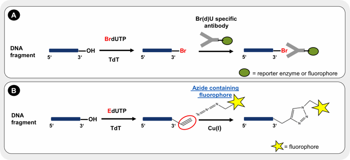
TUNEL assays identify apoptotic cells by the terminal deoxynucleotidyl transferase (TdT)-mediated addition of labeled (X) deoxyuridine triphosphate nucleotides (X-dUTPs) to the 3’-OH end of DNA strand breaks that are subsequently visualized depending on the introduced label (Fig. 1), thus serving as parameter for the percentage of apoptotic cells within the analyzed cell population.

The assay sensitivity strongly depends on the incorporation efficiency of the modified dUTP that is influenced by size/bulkiness of the attached label. A number of fluorescently labeled dUTPs → Probes & Epigenetics, biotinylated dUTPs → Probes & Epigenetics or digoxigenylated dUTPs → Probes & Epigenetics are generally suitable substrates for TdT, but dUTP labeled with smaller labels such as bromine (BrdUTP) or an alkyne group (EdUTP) have been demonstrated to exhibit a higher incorporation efficiency and thus higher sensitivity in TUNEL assays probably due to minimal sterical hindrance[3,4,5].

While incorporation of BrdUTP is detected by specific fluorophore or reporter enzyme labeled antibodies (Fig. 1A), detection of EdUTP is achieved by covalent conjugation of an azide-containing fluorophore → Click Chemistry via Cu(I) catalyzed alkyne-azide click chemistry (CuAAC) (Fig. 1B).

Figure 1: The principle of TUNEL assay relies on terminal deoxynucleotidyl transferase (TdT)-mediated addition of a modified dUTP (X-dUTP) to 3’-OH ends of DNA fragments that are generated as a result of apoptosis induction. To avoid the loss of fragmented DNA and to allow enzyme and nucleotide entrance, cells need to be fixed and subsequently permeabilized prior to the labeling reaction. Incorporated bromoylated dUTP (BrdUTP) is detected by specific antibody conjugates with a reporter enzyme or fluorescent dye (A). The incorporation of alkyne-containing dUTP (EdUTP) is visualized by Cu(I)-catalyzed alkyne-azide click chemistry (CuAAC) with an azide containing fluorphore → Click Chemistry (B).


Name Cat. No. Size
5-Bromo-dUTP NU-122S 50 μl (10 mM)
5-Bromo-dUTP NU-122L 5 x 50 μl (10 mM)
5-Ethynyl-dUTP (5-EdUTP) CLK-T07-S 5 μl (100 mM)
5-Ethynyl-dUTP (5-EdUTP) CLK-T07-L 5 x 5 μl (100 mM)
5-Ethynyl-dUTP (5-EdUTP) CLK-T07-XL 50 μl (100 mM)

Selected References

[1] Kerr et al. (1972) Apoptosis: a basic biological phenomenon with wide-ranging implications in tissue kinetics. Br. J. Cancer 26 (4):239.
[2] Lowe et al. (2000) Apoptosis in Cancer. Carcinogenesis 21 (3):485.
[3] Darzynkiewicz et al. (2008) Analysis of apoptosis by cytometry using TUNEL assay. Methods 44 (3):250.
[4] Li et al. (1995) Labelling DNA strand breaks with BrdUTP: Detection of apoptosis and cell proliferation. Cell proliferation 28 (11):571.
[5] Li et al. (1995) Single–step procedure for labeling DNA strand breaks with fluorescein- or BODIPY-conjugated deoxynucleotides: Detection of apoptosis and bromdeoxyuridine incorporation. Cytometry 20:172.

Nucleotides for Application in Protein-DNA/-RNA interaction (EMSA)

A variety of cellular processes are triggered by sequence-specific binding of a protein to a nucleic acid (DNA or RNA) e.g. regulation of gene expression by transcription factor binding to specific DNA sequences or coordination of translation by RNA-binding proteins.

Electromobility shift assays (EMSA) are a prominent technique to detect the affinity of a protein to a known DNA or RNA sequence in vitro[1]. Briefly, a labeled RNA or DNA probe is incubated with a protein source (crude cell extract or recombinant protein) and subsequently separated via non-denaturing polyacrylamide gel electrophoresis. End labeling of DNA and RNA probes is generally preferred over random labeling methods due to minimized interference with protein binding. Protein-nucleic acid complexes exhibit a slower mobility compared to free nucleic acid probes thus complex formation can be identified by the altered migration pattern (shift).

Visualization of a protein-nucleic acid complex depends on type of label used for DNA/RNA probe labeling. Traditional labeling approaches rely on the enzymatic incorporation of radioisotope labels however, several non-radioactively labeled nucleotides have been successfully used for EMSA probe labeling (Tab. 1). Further enzymatically incorporable labeled nucleotides are available within our DNA Labeling → Probes & Epigenetics and RNA Labeling → Probes & Epigenetics section.

Table 1: Nucleotide selection guide for nonradioactive EMSA probe labeling.

DIG: digoxigenin; ATTO680: fluorescent dye (exc.: 680 nm /em.: 700 nm); DY776: fluorescent dye (exc.: 771 nm /em.: 801 nm).

Generation of… Method Labeling site Enzyme Labeled nucleotide Incorporated nucleotides Detection
…DNA probe 3′-End Labeling 3′-OH Terminal Deoxynucleotidyl Transferase (TdT) Biotin-11-UTP[2] 1-3 Indirect via reporter enzyme or fluorophore conjugated streptavidin
DIG-11-ddUTP[3] 1 Indirect via reporter enzyme or fluorophore conjugated Digoxigenin antibody
5′-End Labeling 5′-OH T4 Polynucleotide Kinase (T4 PNK) ATPγS[4] 1 Indirect via Iodacetamide modified biotin or fluorophores
…RNA probe In vitro transcription random T7 RNA Polymerase UTP-ATTO680[5,6] multiple Direct measurement of fluorescence
UTP-DY776[6] multiple Direct measurement of fluorescence
5′-End Labeling 5′-OH T4 Polynucleotide Kinase (T4 PNK) ATPγS[4] 1 Indirect via Iodacetamide modified biotin or fluorophores
Name Cat. No.
ATPyS  NU-406
Biotin-11-UTP, Biotin-X-(5-aminoallyl)-UTP NU-821-BIOX
Biotin-11-UTP – high concentration, Biotin-X-(5-aminoallyl)-UTP NU-821-BIOX-HC
Aminoallyl-UTP-ATTO-680 NU-821-680
Aminoallyl-UTP-DY-776 NU-821-776
DIG-11-ddUTP NU-1619-DIGX


Selected References

[1] Hellmann et al. (2007) Electrophoretic mobility shift assay (EMSA) for detecting protein–nucleic acid interactions. Nature Protocols 2:1849.
[2] Yan et al. (2006) Characterization of the human intestinal CD98 promoter and its regulation by interferon-γ. Am. J Physiol. Gastrointest. Liver Physiol. 292:G535.
[3] Kass et al. (2000) Non-radioactive electrophoretic mobility shift assay using digoxigenin-ddUTP labeled
probes. Dros. Inf. Serv. 83:185.
[4] Pagano et al. (2011) Quantitative approaches to monitor protein-nucleic acid interactions using fluorescent probes. RNA 17:14.
[5] Besse et al. (2009) Drosophila PTB promotes formation of high-order RNP particles and represses oskar translation. Genes Dev. 23:195.
[6] Kohn et al. (2010) Near-infrared (NIR) dye-labeled RNAs identify binding of ZBP1 to the noncoding Y3-RNA. RNA 16 (7):1420.

Nucleotides for Application in Epigenetics

Epigenetics is the study of mechanisms that induce heritable changes in gene expression without changing the DNA sequence.

The most extensively studied epigenetic mechanisms so far are DNA methylation and histone modifications that regulate gene expression by chromatin remodeling which subsequently results in increased or decreased DNA accessibility for the transcriptional machinery depending on the specific epigenetic modification pattern[1].

In addition, RNA methylation has most recently gained attention as an epigenetic modification in mammalian cells as well[2].

Several nucleotide analogs (Tab. 1) are suitable for epigenetic modification analysis. Please refer to the corresponding references for detailed application data.

Table 1: Selection guide for epigenetic modification analysis.

Epigenetic modification Nucleotides and Nucleosides References
DNA Methylation N6-Methyl-dATP
5-Carboxy-2′-dC
5-Formyl-2′-dC
5-Methyl-2′-dC
5-Hydroxymethyl-2′-dC
5-Aza-dCTP
5-Methyl-dCTP [3,4]
5-Hydroxymethyl-dCTP
RNA Methylation N6-Methyl-ATP [2]
2’OMe-ATP [2]
5-Methyl-CTP [5]
5-Hydroxy-UTP
4SedTTP [6]
Name Cat. No. Size
N6-Methyl-dATP NU-949S 10 μl (100 mM)
N6-Methyl-dATP NU-949L 5 x 10 μl (100 mM)
5-Carboxy-dC N-1067-5 5 mg
5-Formyl-dC N-1069-5 5 mg
5-Hydroxymethyl-dC N-1070-100 100 mg
5-Aza-dCTP NU-1118 20 mg
5-Methyl-dCTP NU-1125S 10 μl (100 mM)
5-Methyl-dCTP NU-1125L 5 x 10 μl (100 mM)
5-Hydroxymethyl-dCTP NU-932S 100 μl (100 mM)
5-Hydroxymethyl-dCTP NU-932L 5 x 100 μl (100 mM)
N6-Methyl-ATP NU-1101S 10 μl (100 mM)
N6-Methyl-ATP NU-1101L 5 x 10 μl (100 mM)
2’OMe-ATP NU-1184S 50 μl (100 mM)
2’OMe-ATP NU-1184L 5 x 50 μl (100 mM)
5-Methyl-CTP NU-1138S 10 μl (100 mM)
5-Methyl-CTP NU-1138L 5 x 10 μl (100 mM)
5-Hydroxy-UTP NU-883S 10 μl (100 mM)
5-Hydroxy-UTP NU-883L 5 x 10 μl (100 mM)
4SedTTP NU-979 1 mg

Selected References

[1] Portela et al. (2010) Epigenetic modifications and human disease. Nature Biotechnology 28 (10):1057.
[2] Meyer et al. (2012) Comprehensive Analysis of mRNA Methylation Reveals Enrichment in 3′ UTRs and near Stop Codons. Cell 149 (7):1635.
[3] Frauer et al. (2009) A versatile non-radioactive assay for DNA methyltransferase activity and DNA binding. Nucleic Acids Research 37 (3):e22.
[4] Kaito et al. (2001) Activation of the maternally preset program of apoptosis by microinjection of 5-aza-2′-deoxycytidine and 5-methyl-2′-deoxycytidine-5′-triphosphate in Xenopus laevis embryos. Development, Growth & Differentiation 43 (4):383.
[5] Karikó et al. (2005) Suppression of RNA Recognition by Toll-like Receptors: The Impact of Nucleoside Modification and the Evolutionary Origin of RNA. Immunity 23:165.
[6] Hong et al. (2018) Precise Antibody-Independent m6A Identification via 4SedTTP-Involved and FTO-Assisted Strategy at Single-Nucleotide Resolution. J. Am. Chem. Soc. 140 (18):5886.

SUPPORT

outstanding technical support

PRODUCT

we offer a full product guarantee

DELIVERY

we offer free delivery to UK universities and non profit organisations