A tumor marker is a substance produced, or induced, by cancer cells that can be detected in body fluids or tumor tissues. The identification of a single marker, or a combination of different tumor markers, can reveal the presence of a particular cancer, its stage, and provide a prognostic outlook following treatment or during an extended therapeutic course. Please view the slideshow video clip below that highlights some of our tumor marker antibodies for different cancers. The table lists specific GeneTex products for key tumor markers recognized by the U.S. National Institutes of Health (NIH).
Tumor Markers

Pancreatic cancer is the third leading cause of cancer-related death in the United States. With a predicted five-year relative survival rate of 9%, pancreatic cancer continues to be clinically challenging due to its resistance to even the most aggressive combinations of surgery, chemotherapy, and radiotherapy. Because of indistinct symptoms and the absence of reliable diagnostic biomarkers, diagnoses are usually delayed and thus made when the disease is already advanced (which decreases five-year survival to less than 3%). Nevertheless, extensive research has highlighted the importance of several biomarkers and reagents that can significantly improve early detection.
KRAS has the distinction of being a preeminent oncoprotein as it is mutated in nearly all pancreatic ductal adenocarcinomas (PDAC), with KRAS G12D and G12V mutations representing 75% of these changes (1). GeneTex’s RAS (G12D Mutant) monoclonal antibody [HL10] (GTX635362) is the first commercially available recombinant rabbit antibody that demonstrates exceptional sensitivity and specificity for the RAS G12D mutant protein in samples from human pancreatic cancer cell lines and a mouse model of PDAC by western blot (WB) and immunohistochemistry (IHC-P), respectively (Figure 1).

Figure 1. GeneTex’s recombinant rabbit RAS (G12D mutant) antibody [HL10] (GTX635362) is sensitive and specific for the RAS G12D mutation by WB and IHC-P in human pancreatic cancer cell lines and a PDAC mouse model.
Carbohydrate antigen 19-9 (CA19-9), also known as sialyl-LewisA, is a tetrasaccharide with the sequence Neu5Acα2-3Galβ1-3[Fucα1-4]GlcNAcβ. It is used clinically to assist in the diagnosis of pancreatic cancer, but is more widely used to monitor therapy and detect recurrences in diagnosed cases. CA19-9 is also elevated in many other diseases, including cholangiocarcinoma, colorectal cancer, hepatocellular carcinoma, and cirrhosis (2). GeneTex’s CA19-9 monoclonal antibody [GT174] (GTX635388) showed superior sensitivity for WB, IHC-P, and immunocytochemistry (ICC/IF) in comparative testing against other well-known antibodies (Figures 2 & 3).

Figure 2. GeneTex’s CA19-9 monoclonal antibody [GT174] (GTX635388) generates a robust signal for IHC-P and WB in comparison to other antibodies.
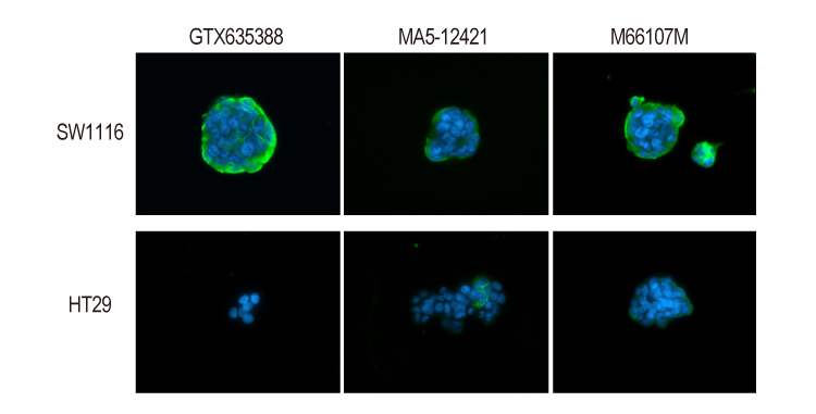
Figure 3. GeneTex’s CA19-9 monoclonal antibody [GT174] (GTX635388) demonstrates superior sensitivity for ICC/IF on SW1116 cells (high CA19-9 expression) and HT29 cells (low CA19-9 expression) in comparison to other antibodies.
Thrombospondin 2 (THBS2) is a secreted glycoprotein that can be expressed together with CA19-9 in patients with early pancreatic ductal adenocarcinoma (PDAC) (3). GeneTex offers a selection of antibody reagents for THBS2 research. Several of these are able to detect secreted protein in conditioned media from human AsPC-1 pancreatic tumor cells by WB. In addition, some of the antibodies clearly recognize THBS2 in human pancreatic tumor tissue samples by IHC-P (Figure 4).

Figure 4. GeneTex’s Thrombospondin 2 polyclonal antibody (GTX134554) is validated for both WB and IHC-P (left), while the monoclonal antibody [GT7511] (GTX635392) is validated for WB (right).
Glypican-1 expression is upregulated in many human malignancies, including pancreatic cancer (4). There is ongoing research into the potential clinical utility of assessing enriched glypican-1 expression on circulating exosomes as an early cancer marker. GeneTex proudly offers an outstanding, well-cited antibody specific for glypican-1 that has been thoroughly validated for WB, ICC/IF, IHC-P, FACS, IP, and ELISA (Figure 5).

Figure 5. Glypican-1 antibody [N3C3] (GTX104557) detects glycosylated glypican-1 protein in various cell lines by WB and performs well in FACS.
Though CA19-9 is the most familiar tumor marker associated with pancreatic cancer, there is ongoing interest in developing pancreatic cancer marker panels that include other proteins, glycoproteins, and microRNAs.
| Cat. No. | Product Name | Reactivity | Applications |
| GTX635362 | RAS (G12D Mutant) antibody [HL10] | Hu, Ms | WB, ICC/IF, IHC-P |
| GTX635388 | CA19-9 antibody [GT174] | Hu | WB, ICC/IF, IHC-P |
| GTX134554 | Thrombospondin 2 antibody | Hu | WB, IHC-P |
| GTX104557 | Glypican-1 antibody [N3C3] | Hu, Ms, Rat | WB, ICC/IF, IHC-P, FACS, IP, ELISA |
| GTX70214 | p53 antibody [DO1] | Hu, Ms | WB, ICC/IF, IHC-P, IHC-Fr, FACS, IP, ELISA |
| GTX112980 | SMAD4 antibody | Hu, Ms, Rat | WB, ICC/IF, IHC-P |
| GTX15677 | CEA antibody [1106] | Hu | WB, ICC/IF, FACS, IP, ELISA, RIA |
| GTX100459 | MUC1 antibody | Hu | WB, ICC/IF, IHC-P, IP |
| GTX100664 | MUC2 antibody [C3], C-term | Hu, Ms | ICC/IF, IHC-P, IHC-Fr, IHC |
| GTX628832 | Pancreatic Polypeptide antibody [GT6512] | Hu, Ms | WB, ICC/IF, IHC-P |
| GTX132480 | RAS antibody | Hu, Rat, Zfsh, Frg, Squirrel | WB, IHC-P |
| GTX108598 | N-RAS antibody | Hu, Ms, Rat | WB |
| GTX116041 | H-Ras antibody | Hu, Ms, Rat | WB, ICC/IF, IHC-P, IP |
| GTX100509 | Her2 / ErbB2 antibody | Hu | WB, IHC-P |
| GTX109294 | MST1 antibody | Hu, Ms | WB, ICC/IF, IHC-P, IP |

Breast cancer is the most common malignancy among women, but also occurs in men (1). Risk factors include female gender, age, family history, smoking, metabolic syndrome and genetic predisposition, among others. There are various types of breast cancer, with ductal carcinoma in situ (DCIS) and invasive ductal carcinoma (IDC) being the most common. Triple-negative breast cancer, with malignant cells having low or absent HER2, estrogen receptor, and progesterone receptor expression, accounts for 10-15% of cases and is particularly aggressive.
HER2, also known as proto-oncogene Neu or ERBB2, is a member of the human epidermal growth factor receptor family of tyrosine kinases. GeneTex’s HER2 / ERBB2 antibody (GTX100509) specifically detects HER2 protein by WB and generates the expected expression pattern in HER2-positive breast tumor sections by IHC-P (Figure 1).

Figure 1. GeneTex’s HER2 / ERBB2 antibody (GTX100509) is sensitive and specific for HER2 expression (left). A side-by-side comparison between this antibody and a competitor’s reagent was performed for IHC-P at 1:500 and 1:100 dilutions, respectively (right).
Estrogen Receptor Alpha (ERα) and Beta (ERβ) have distinct cellular distributions, regulate separate sets of genes, and oppose each other in breast cancer (2). ERα and ERβ have been extensively studied and targeted for the development of therapeutics against hormone-positive breast cancer. GeneTex’s Estrogen Receptor Alpha antibody [1F3] (GTX70171) is a mouse monoclonal antibody whose sensitivity and specificity have been stringently validated by GeneTex and in multiple publications. The Estrogen Receptor Beta antibody [14C8] (GTX70174) is one of the most commonly used reagents to detect ERβ with more than one hundred citations (Figure 2).

The progesterone receptor (PR) is a well-known estrogen receptor (ER) target gene that is expressed in over two-thirds of ER-positive breast cancers (3). It acts as a transcriptional regulator and activator of signal transduction pathways (i.e., MAPK, PI3K/Akt, and c-Src) that can drive proliferative signaling in breast cancer cells (4). GeneTex’s Progesterone Receptor antibody [N1], N-term (GTX116051) was developed for IHC-P. It demonstrates distinct staining differences on PR-negative and PR-positive breast tumor tissue sections (Figure 3).

Figure 3. GeneTex’s Progesterone Receptor antibody [N1], N-term (GTX116051) gives negative staining of PR-negative breast cancer tissue sections (left) and robust nuclear staining in PR-positive sections (right).
Some inherited mutations in BRCA1 are established drivers of breast cancer. GeneTex’s BRCA1 antibody [17F8] (GTX70111) is a highly cited monoclonal antibody validated for various applications including WB, ICC/IF, IHC-P, IP, ELISA, and ChIP (Figure 4).

Figure 4. GeneTex’s BRCA1 antibody [17F8] (GTX70111) was validated for WB using knockout cell lysate (left) and for IHC on carcinoma tissue sections (right).
Table: Markers associated with breast cancer
| Cat. No. | Product Name | Reactivity | Applications |
| GTX70111 | BRCA1 antibody [17F8] – ChIP grade | Hu, Ms | WB, ICC/IF, IHC-P, IP, ELISA, ChIP assay, IHC |
| GTX70123 | BRCA2 antibody [5F6] | Hu | WB, IHC-P, IHC-Fr |
| GTX70171 | Estrogen Receptor alpha antibody [1F3] | Hu, Rb | WB, IHC-Fr, FACS, IP |
| GTX70174 | Estrogen Receptor beta antibody [14C8] | Hu, Ms, Mk | WB, ICC/IF, IHC-P, IHC-Fr, FACS, Dot, ChIP assay, IHC |
| GTX116051 | Progesterone Receptor antibody [N1], N-term | Hu | ICC/IF, IHC-P |
| GTX100509 | Her2 / ErbB2 antibody | Hu | WB, IHC-P |
| GTX79597 | uPA antibody | Hu, Ms, Rat | WB, IHC-P, IHC-Fr, ELISA |
| GTX60537 | PAI-1 antibody [1D5] | Hu, Ms | WB, IHC-P, FACS, ELISA |
| GTX20697 | CA125 antibody [OV185:1] | Hu | IHC-P |
| GTX17254 | CEA antibody [Col-1] | Hu | IHC-P |
| GTX70214 | p53 antibody [DO1] | Hu, Ms | WB, ICC/IF, IHC-P, IHC-Fr, FACS, IP, ELISA |
| GTX16667 | Ki67 antibody [SP6] | Hu, Ms, Rat, Chk | WB, ICC/IF, IHC-P, IHC-Fr, FACS, IHC |

Ovarian cancer is the second most common gynecological cancer in the United States. It is diagnosed primarily in post-menopausal women though it can also occur in younger patients, particularly in the context of certain genetic mutations (e.g., in BRCA1, BRCA2, and TP53). Most ovarian tumors develop in the epithelium of the ovary, representing more than 95 percent of disease cases (1). Other rarer malignancies include germ cell and stromal cell ovarian cancers, though there are at least 30 types of ovarian cancer described.
Patients with ovarian cancer often have an elevated level of Cancer Antigen 125 (CA 125, also known as MUC16), a member of the high-molecular-weight mucin family of glycoproteins. CA 125 is thought to regulate tumor cell growth, invasion, and metastasis (2). GeneTex’s mouse monoclonal CA 125 antibody [OV185:1] (GTX20697) offers high sensitivity and batch consistency to reliably detect CA 125 expression in ovarian tumor samples by IHC-P (Figure 1) and by WB.
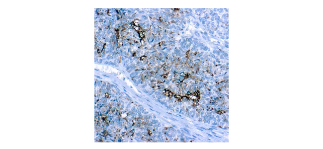
Figure 1. GeneTex’s mouse monoclonal CA 125 antibody [OV185:1] (GTX20697) shows high signal-to-noise ratio on an ovarian tumor section by IHC-P.
Human Epididymis Protein 4 (HE4) is frequently overexpressed in ovarian tumors and is known to be a tumor biomarker for ovarian cancer (3-5). Recent studies have reported that the combined measurement of HE4 and CA 125 improves the diagnostic sensitivity and specificity of ovarian cancer (6-8). GeneTex’s HE4 antibody [JF62-09] (GTX01063) is a recombinant rabbit monoclonal antibody thoroughly validated for WB, ICC/IF, IHC-P, and FACS, as shown in Figure 2.
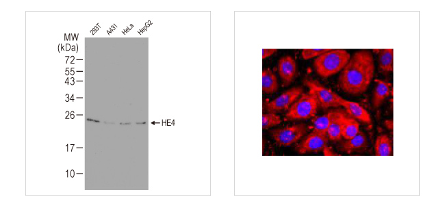
Figure 2. GeneTex’s recombinant rabbit monoclonal HE4 antibody [JF62-09] (GTX01063) detects endogenous HE4 protein by WB and reveals robust HE4 expression in SKOV-3, an ovarian cancer cell line, by ICC/IF.
References:
- World J Transl Med. 2014 Apr 12;3(1):1-8.
- Gynecol Oncol. 2011 Jun 1;121(3):434-43.
- Cancer Res. 2003 Jul 1;63(13):3695-700.
- Gynecol Oncol. 2016 May;141(2):303-311.
- Tumour Biol. 2012 Feb;33(1):141-8.
- J Ovarian Res. 2019 Mar 27;12(1):28.
- Med Oncol. 2014 Jan;31(1):808.
- Int J Gynecol Cancer. 2012 Nov;22(9):1474-82.

Primary liver cancer is a leading cause of cancer mortality worldwide, with hepatocellular carcinoma (HCC) being the most common type affecting almost one million individuals annually (1). The risk of HCC is higher in people with chronic liver disease and cirrhosis due to viral hepatitis (i.e., HBV and HCV), alcohol overconsumption, or nonalcoholic steatohepatitis (NASH) related to diabetes and obesity, among other causes. While early detection of HCC is associated with 5‐year survival rates up to 70%, most patients are diagnosed at an advanced stage for which effective therapeutic options are limited (2).
Measurement of serum alpha-fetoprotein (AFP) combined with abdominal ultrasound examination are widely used in the clinical diagnosis of liver cancer. AFP overexpression is commonly found in HCC. Although several studies suggest that AFP may promote tumor cell proliferation via inhibition of apoptotic signals, the specific mechanism for its oncogenic activity remains unclear (3). GeneTex’s mouse monoclonal AFP antibody [2A9] (GTX84954) is validated for multiple applications, including WB, ICC/IF, IHC-P, and FACS (Figure 1). Interestingly, this antibody was cited in a recent Scientific Reports paper that examined stem cell reprogramming (4).
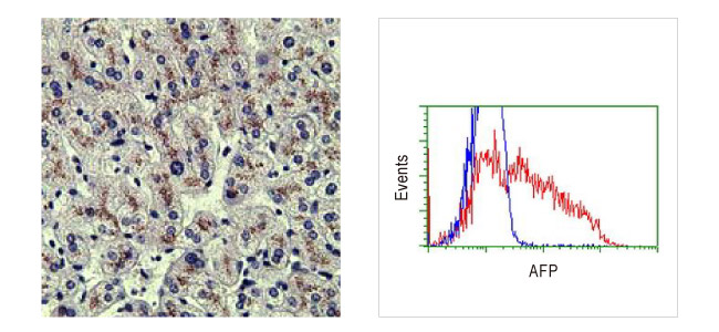
Figure 1. GeneTex’s AFP antibody [2A9] (GTX84954) used for IHC-P on human liver tissue and for FACS analysis of Jurkat cells.
Glypican-3 (GPC3) is a glycoprotein attached to the cell membrane. GPC3 overexpression is found in HCC, but not in healthy adult liver. Studies have shown GPC3 to be a reliable serological and immunohistochemical marker for HCC diagnosis, and a potential target for immunotherapies (5-6). GeneTex’s Glypican-3 antibody [GT2473] (GTX633410) is a cited mouse monoclonal antibody validated for WB, ICC/IF, and IHC-P. It generates the expected cell membrane staining by ICC/IF and can detect both glycosylated and non-glycosylated forms of GPC3 by WB (Figure 2).
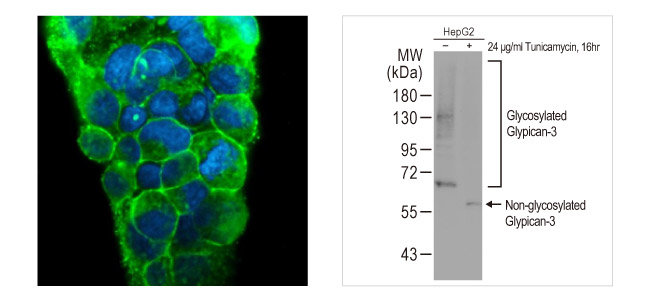
Figure 2. GeneTex’s Glypican-3 antibody [GT2473] (GTX633410) detects GPC3 protein on the HepG2 cell membrane by ICC/IF and various forms of GPC3 by WB of HepG2 whole cell extracts -/+ tunicamycin treatment.
Golgi membrane protein 1, also known as GP73, GOLM1, and GOLPH2, is a type II Golgi membrane glycoprotein often overexpressed in the hepatocytes of patients with hepatitis and HCC. Reports indicate that GOLPH2 is a promising serum marker for the diagnosis of HCC (7-8). GeneTex has two well-cited, knockout (KO) cell extract-validated GOLPH2 polyclonal antibodies (GTX107702 and GTX116154) that stain the Golgi apparatus very distinctively by ICC/IF (Figure 3).
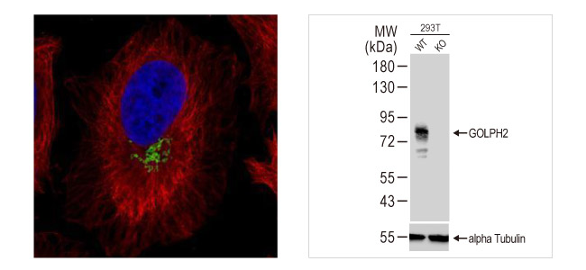
Figure 3. GeneTex’s GOLPH2 antibody [C3], C-term (GTX107702) used for ICC/IF on HeLa cells and for WB with both WT and GOLPH2-KO 293T cell extracts.

Colorectal cancer (CRC) is one of the most commonly diagnosed cancers worldwide, causing more than half a million deaths annually. The overall five-year survival rate is around 65 percent, but varies depending on clinical stage (1). However, like many other malignancies, early stage disease may evoke no cancer-specific symptoms. Therefore, regular stool occult blood testing and colonoscopy screening are essential. Biomarkers for CRC have been extensively studied, with carcinoembryonic antigen (CEA) and CA 19-9 being the most well-established prognostic factors.
Carcinoembryonic antigen (CEA) is a glycoprotein expressed in embryonic entodermal tissue that forms the epithelial lining of the digestive and respiratory tracts. Though usually detected at low levels in healthy adults, it can be elevated in smokers and in patients with various non-neoplastic conditions. However, increased CEA is also seen in certain malignancies, including CRC, pancreatic cancer, and specific tumors of the endocervix and ovary. Depending on the actual serum CEA value, physicians should suspect malignant or metastatic disease. Data supports the use of this tumor marker for detection of CRC recurrence, prognosis, and response to therapy. CEA antibodies are commonly used in immunohistochemistry (IHC) to study tumor progression and for tumor characterization. GeneTex’s CEA antibody [COL-1] is a cited monoclonal antibody whose utility for IHC was demonstrated on human colon carcinoma tissue (Figure 1).
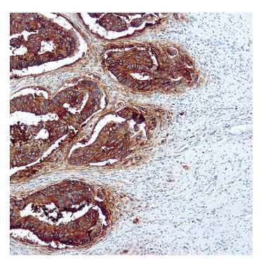
Figure 1. GeneTex’s CEA antibody [Col-1] (GTX17254) used for IHC-P on a human colon carcinoma tissue section.
Table: Markers associated with colorectal cancer
| Product Name | Reactivity | Applications | Cat. No. |
| CEA antibody [Col-1] | Hu | IHC-P | GTX17254 |
| CA19-9 antibody [GT933] | Hu | WB, ICC/IF, IHC-P, FACS, ELISA, Glycan Array | GTX635389 |
| B-Raf (V600E Mutant Specific) antibody [RM8] | Hu | WB, ICC/IF, IHC-P, ELISA | GTX33595 |
| RAS (G12D Mutant) antibody [HL10] | Hu, Ms | WB, ICC/IF, IHC-P | GTX635362 |
| Cytokeratin 7 antibody [N1C2] | Hu | WB, ICC/IF, IHC-P, IHC-Fr | GTX109723 |
| Cytokeratin 20 antibody [N2C2], Internal | Hu, Ms | WB, ICC/IF, IHC-P | GTX110600 |
References:
- World J Gastroenterol. 2016 Feb 7; 22(5): 1745–1755.

Melanoma is the deadliest form of skin cancer that develops from melanocytes, a pigment-producing cell. The incidence of melanoma has doubled over the last three decades in the United States (1). With treatment, the five-year survival rate is more than 90% for cases contained at the primary site, and less than 30% once there is metastasis. Early detection is therefore crucial to prevent local invasion and distant spread.
S100B, a member of the S100 family of calcium-binding cytosolic proteins, is an independent prognostic marker for melanoma (2-4). Elevated S100B and lactate dehydrogenase (LDH) levels are clinically useful for the detection of tumor progression and metastasis (5). GeneTex’s S100 beta antibody (GTX129573) is a well-cited antibody validated for western blot, immunohistochemistry, and immunostaining in various species, including human, mouse, and rat (Figure 1).

Figure 1. GeneTex’s S100 beta antibody (GTX129573) detects S100B protein in mouse retina by immunohistochemistry (green) and by western blot in A375 cell lysates.
LDHA facilitates glycolytic flux by converting pyruvate to lactate and NADH to NAD+. It is primarily cytoplasmic, but is also found in mitochondria and the nucleus. In addition to its metabolic function, LDHA is also a single‐stranded DNA‐binding protein participating in DNA duplication and transcription. Elevated expression of LDHA is observed in various cancers. In melanoma, LDHA regulates mitochondrial ROS accumulation, tropomyosin oxidation, and cytoskeletal remodeling to control tumor metastasis. Blockade of LDHA resulted in improved efficacy of PD-1/PD-L1-related immunotherapy (6). GeneTex’s LDHA antibody (GTX101416) is a well-cited polyclonal antibody validated for multiple applications and species (Figure 2).
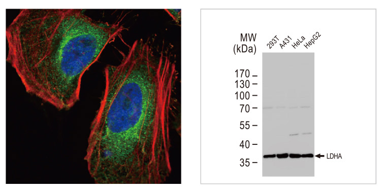
Figure 2. GeneTex’s LDHA antibody (GTX101416) exhibits an expected staining pattern in ICC/IF and generates a robust band at the predicted molecular weight in various human cell lines by western blot.
MelanA, also known as MART1, is a melanocyte lineage-specific marker. The mouse monoclonal antibody [A103] was raised against recombinant Melan A and is positive in most primary melanomas as well as being a useful tool to detect metastatic disease [7]. Melanoma-associated Antigen is recognized by the mouse monoclonal antibody [HMB-45], which is positive in 90-100% of primary melanomas (Figure 3).
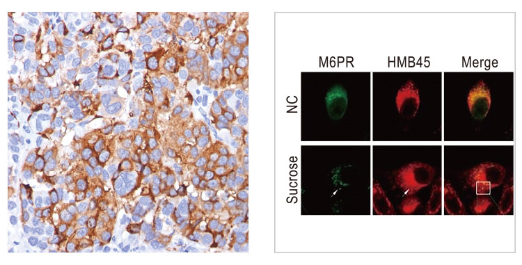
Table: Markers associated with Melanoma
| Product Name | Reactivity | Applications | Cat. No. |
| S100 beta antibody | Hu, Ms, Rat, Squirrel | WB, ICC/IF, IHC-P, IHC-Fr | GTX129573 |
| LDHA antibody | Hu, Ms, Rat | WB, ICC/IF, IHC-P | GTX101416 |
| Melan A antibody [A103] | Hu, Ms, Rat, Dog | WB, ICC/IF, IHC-P, FACS | GTX34832 |
| Melanoma associated antigen antibody [HMB45] | Hu | WB, ICC/IF, IHC-P | GTX71957 |
| Melanoma gp100 antibody [PMEL/2037] | Hu | IHC-P, ELISA, Protein Array | GTX17772 |
| MITF antibody | Hu, Ms, Chk, Xenopus | WB, ICC/IF, IHC-P, IHC-Fr, ChIP assay | GTX00738 |
References:
- “SEER Stat Fact Sheets: Melanoma of the Skin”.
- Oncology. 2005;68(4-6):341-9.
- Tumori. Nov-Dec 2004;90(6):607-10.
- J Clin Oncol. 2009 Jan 1; 27(1): 38–44.
- Pathol Oncol Res. 2002;8(3):183-7.
- Cancers (Basel). 2019 Apr; 11(4): 450.
- Am J Clin Pathol. 2002 Dec;118(6):930-6.

Worldwide, lung cancer is the most common cancer in men and the third most in women, with two million cases diagnosed in 2018. About 90 percent of cases are the result of smoking in men, and 80 percent in women. There are two main forms: Non-small cell lung carcinoma (NSCLC) representing almost 90% of cases, and small cell lung carcinoma (SCLC), which usually grows and spreads faster. Several factors can be found at high levels in patient serum, including CEA, NSE, CYFRA21-1, SCC, ProGRP, and CA 125 (1), but their value for monitoring clinical course remains under study.
NSE is a 78 kDa gamma-homodimer expressed as a dominant enolase-isoenzyme in neuronal and neuroendocrine tissues. Elevated levels of serum NSE are often associated with neuronal disorders and neural crest-derived tumors. Up to 70 percent of patients with small cell lung carcinoma (SCLC) have high levels of serum NSE, as SCLC cells can express neuronal gene programs (2). GeneTex’s NSE antibody (GTX101553) is a well-cited polyclonal antibody that recognizes NSE in brain tissue sections and demonstrates a typical membrane distribution in a human lung cancer cell line xenograft (Figure 1).
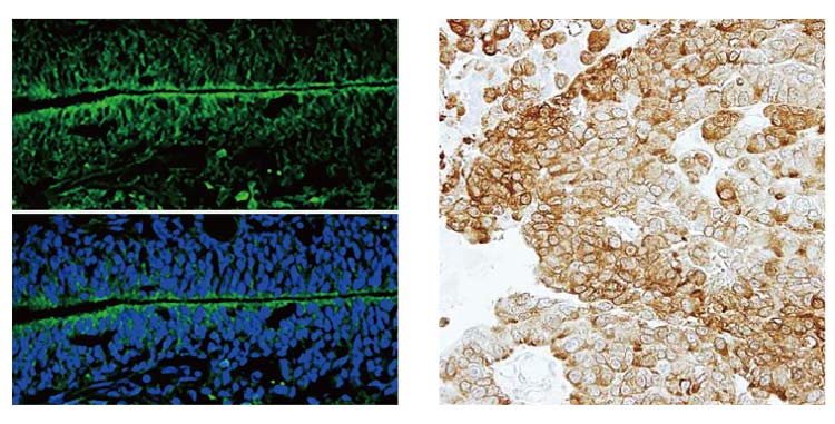
Figure 1. GeneTex’s NSE antibody (GTX101553) detects NSE protein by immunohistochemistry in a rat brain tissue section (left) and in a human lung cancer cell xenograft section (right).
Table: Markers associated with lung cancer
| Product Name | Reactivity | Applications | Cat. No. |
| NSE antibody [N1C1] | Hu, Ms, Rat | WB, IHC-P, IHC-Fr | GTX101553 |
| PD-L1 antibody | Hu | WB, ICC/IF, IHC-P, IHC-Fr | GTX104763 |
| IDH1 antibody [GT1521] | Hu, Ms, Rat | WB, ICC/IF, IP | GTX629818 |
| EGFR antibody [GT133] | Hu | WB, ICC/IF, IHC-P, IP | GTX628887 |
| FAM83B antibody [N1N2], N-term | Hu | WB, IHC-P | GTX107222 |
References:
- Gan To Kagaku Ryoho. 2001 Dec;28(13):2089-93.
- J Thorac Dis. 2019 Apr;11(4):1182-1189.
Leukemias develop when blood cells, primarily white blood cells, undergo neoplastic transformation. They can be acute or chronic. Acute leukemias are characterized by rapid disease progression due to expansion of immature lymphocytes or myeloid cells, and are commonly diagnosed in children. In contrast, chronic leukemias involve aberrant hematopoietic development, and can generate extremely high white blood cell counts. CD (cluster of differentiation) factors are the most common leukemia markers. They are membrane proteins predominantly expressed on the cell surface, and are used to classify disease type.
CD33, also known as Siglec-3 or gp67, is a 67 kDa glycosylated cell surface receptor that mediates cell-cell interactions and maintains immune cells in a resting state. Although CD33 is found on some lymphoid subsets, it is highly expressed on myeloid lineage cells, and is commonly used for the diagnosis of acute myeloid leukemia (AML). CD33 is an established target for therapy, with CD33-positive blasts detected in almost 90 percent of patients presenting with AML (1). GeneTex’s CD33 antibody [WM53] (GTX00477) is a well-known mouse monoclonal antibody that is available with different conjugates, including FITC (GTX00477-06), PE (GTX00477-08), and APC (GTX00477-07), among others. These antibodies are all useful for flow cytometry and validated with human peripheral blood samples (Figure 1).
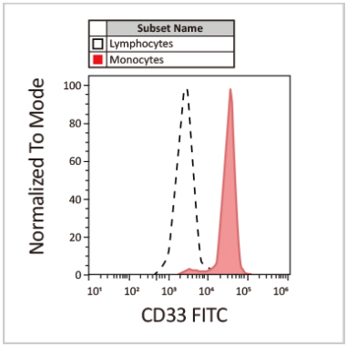
Figure 1. GeneTex’s CD33 antibody [WM53] (FITC) (GTX00477-06) used for FACS on a human peripheral blood sample.
CD4 and CD8 are glycoproteins expressed on the surface of immune cells, particularly T cells. They function as co-receptors with T cell receptors (TCRs) to assist interaction with MHC molecules on antigen-presenting cells, resulting in activation of the adaptive immune response. The CD4/CD8 ratio is used clinically to monitor disease progression of chronic lymphocytic leukemia (CLL), with a higher CD8 count being associated with longer median survival (2). GeneTex’s CD4 antibody [GK1.5] (GTX44531) is validated for multiple applications including IHC and FACS, while the CD8 antibody [SK1] (FITC) (GTX01468-06) is validated for FACS (Figure 2).
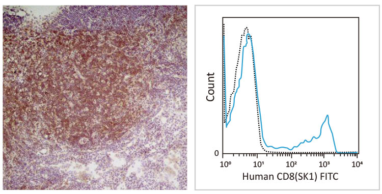
Figure 2. GeneTex’s CD4 antibody [GK1.5] (GTX44531) stains CD4-positive lymphocytes in mouse lymph node (left), while the CD8 antibody [SK1] (FITC) (GTX01468-06) demonstrates robust detection of CD8-positive cells in human peripheral blood (right).
CD44 is a multifunctional transmembrane glycoprotein involved in cell migration, proliferation, differentiation, and signaling pathways that mediate cell survival. It is involved in many malignancies as well as being recognized as a cancer stem cell marker. Importantly, it is expressed in leukocytes and is a marker of leukemia-initiating cells, being found to be critical in the pathogenesis of T cell acute lymphoblastic leukemia (T-ALL) (3). GeneTex’s CD44 antibody [GT462] (GTX628895) is a cited mouse monoclonal antibody validated by knockout lysate and ideal for multiple applications, including WB, IHC-P, FACS, and IP (Figure 3).
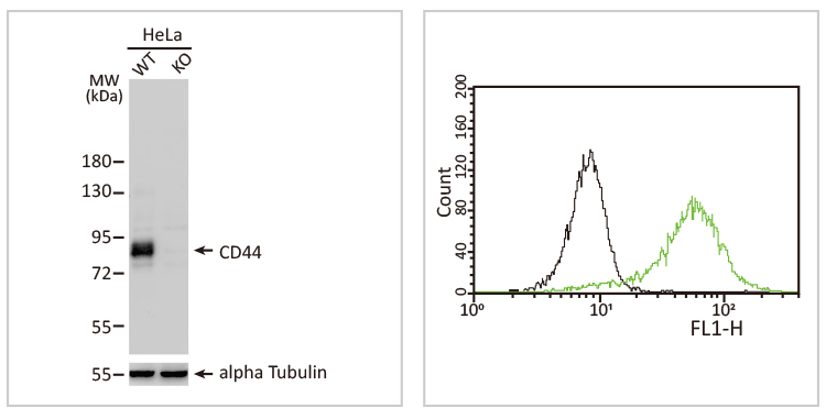
Figure 3. GeneTex’s CD44 antibody [GT462] (GTX628895) was validated using HeLa knockout lysate (left), and performs in flow cytometry analyzing the human acute promyelocytic leukemia cell line HL-60 (right).
Table: Markers associated with Leukemia
| Product Name | Reactivity | Applications | Cat. No. |
| CD33 antibody [WM53] | Hu, Primate | WB, ICC/IF, IHC-Fr, FACS, IP, MS | GTX00477 |
| CD4 antibody [GK1.5] | Hu, Ms | WB, ICC/IF, IHC-P, IHC-Fr, FACS, IP, ELISA | GTX44531 |
| CD8 antibody [SK1] (FITC) | Hu, AGMK, Bb, Chmp, ClMK | FACS | GTX01468-06 |
| CD44 antibody | Hu, Ms, Rat, Rb | WB, ICC/IF, IHC-P, IHC-Fr, IP, IHC | GTX102111 |
| CD138 antibody [1A3H4] | Hu, Ms | WB, ICC/IF, IHC-P, FACS, ELISA | GTX00451 |
| CD52 antibody | Ms | WB, ICC/IF, IHC-P | GTX00743 |
References:
- Blood Cancer J. 2014 Jun; 4(6): e218.
- Leuk Lymphoma. 2010 Oct;51(10):1829-36.
- Front Oncol. 2018 Oct 31;8:488