Signosis is a leader of bioassay provider over a decade, dedicated to the development and commercialization of plate-based analysis products in the life sciences community. We focus on many regulatory key molecules: transcription factors, cytokines, miRNAs, and disease markers. Our mission is to provide excellent products and service worldwide, which include scientists in pharmaceutical companies and biotech industry, academic and research institutes in many countries. We object to develop innovative assay products with high sensitivity, high throughput and cost-effectiveness.

At Signosis, we also offer a wide-selection of validated luciferase reporter cell lines that measure transcription factor activity as a read-out for various signaling pathways in many cell lines and provide a wide spectrum of over-expression stable cell lines, including a series of EGFR mutant over-expression stable cell lines.

In addition, Signosis provides custom services in making custom assay kits, or/and performing the assay and data analysis, generating custom stable cell lines to facilitate greatly scientists research projects.

Signosis has distributors all around the world, including Germany, United Kingdom, France, Sweden, Norway, Denmark, Finland, Italy, Australia, Egypt, Israel, Korea, Japan, Taiwan and China. If your country is not on this list, please contact our USA headquarters for more information. Our headquarter is located in the sunny Silicon Valley.


Product Categories

Stable Cell Lines

Gene function analysis, target discovery and validation, assay development, and compound screening often need cell-based assays. Stable cell lines that are engineered to express a gene of interest via transgene integrations into the host genome provide an efficient approach to conduct such analysis. However, because establishment of such cell lines are tedious in procedures, time-consuming in development process, and expensive in terms of labor, reagents, and materials, as well as post-transfection validation of individual clones, it is always debatable whether is worthy of developing such stable cell lines in individual laboratories.

Signosis now provides a series of gene expression- or luciferase reporter-ready stable cell lines in order to facilitate the studies.

Grow Your Research Vision

Research Partners’ Success Stories of Leveraging Stable Cell Line

Our key research kits

Firefly Luciferase Reporter Cell Lines for Monitoring
TF Binding and Activation

Simple, rapid, and sensitive Firefly Reporter system

Gene function analysis, target discovery and validation, assay development, and compound screening often need cell-based assays. Stable cell lines that are engineered to express a gene of interest via transgene integrations into the host genome provide an efficient approach to conduct such analysis.

At Signosis, we help researchers save time and labor in generating and validating stable cell lines so scientists can spend more effort on solving the big questions. We offer a wide selection of validated Firefly luciferase reporter cell lines that measure transcription factor activity as a read-out for various signaling pathways.

Principle

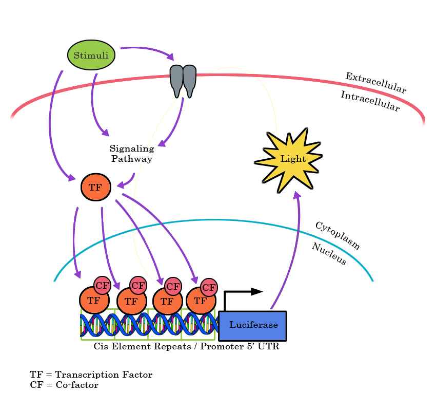
TF luciferase reporter stable cell line utilizes artificial promoter constructs to drive luciferase expression. The promoter region can consists of multiple repeats of a cis-element TF binding site, a DNA fragment from the promoter region of a known TF downstream gene, or a DNA fragment containing putative/known TF binding sites. There are several ways that a TF can be activated, such as through extracellular stimuli or through intracellular signaling pathways. Once activated, the TF translocates to the nucleus and often interacts with relevant co-factors to drive gene expression. Once luciferase is expressed, it can generate light in an enzymatic assay and the amount of light measured is positively correlated with the level of TF activation

Benefits

High Sensitivity & Responsiveness
Each cell line is validated to induce a strong reporter signal in response to stimuli, and is highly responsive to our Firefly Luciferase Substrate.
Consistent

​TF reporter construct is stably integrated into the genome to avoid experimental/cell-to-cell variations.

Time-Saving

Cell line can be used for experiments right away to study different signaling pathways.

Routine Mycoplasma Testing

All cell lines tested negative for mycoplasma.

Signosis Firefly Luciferase Substrate

We also offer our own Firefly Luciferase Substrate which can generate a strong light signal that is comparable to our competitor’s, but at a more cost-effective price.

Signaling Pathways – Cell Lines

Description Catalog No.
NFkB Secreted Luciferase Reporter HeLa Stable Cell Line SL-4001
NFkB Secreted Luciferase Reporter HEK293 Stable Cell Line SL-4012
NFkB Luciferase Reporter HepG2 Stable Cell Line SL-0017
ELK-TAD Luciferase Reporter HEK293 Stable Cell Line SL-0040
TCF/LEF Luciferase Reporter HEK293 Stable Cell Line SL-0015
Stat1 Luciferase Reporter Hela Stable Cell Line SL-0004
NFkB Luciferase Reporter HEK293 Stable Cell Line SL-0012
NFkB Luciferase Reporter MCF7 Stable Cell Line SL-0013
Estrogen Receptor Luciferase Reporter T47D Stable Cell Line SL-0002
NFkB Luciferase Reporter HeLa Stable Cell Line SL-0001
p53 Luciferase Reporter Hela Stable Cell Line SL-0011
SMAD/TGFbeta Luciferase Reporter HepG2 Stable Cell Line SL-0016
AR Luciferase Reporter MDA-MB-453 Stable Cell Line SL-0008
NRF2/ARE Luciferase Reporter MCF7 Stable Cell Line SL-0010
NFkB Luciferase Reporter NIH 3T3 Stable Cell Line SL-0006
GR Luciferase Reporter MDA-MB-453 Stable Cell Line SL-0009
p53 Luciferase Reporter RKO Stable Cell Line SL-0007
HIF Luciferase Reporter NIH3T3 Stable Cell Line SL-0005 SL-0005
NFkB Luciferase Reporter A549 Stable Cell Line SL-0014
Control HepG2 Luciferase Reporter Stable Cell Line SL-0039
Control Hela Luciferase Reporter Stable Cell Line SL-0038
SMAD/BMP Responsive Luciferase Reporter HEK293 Stable Cell Line SL-0051
CREB Luciferase Reporter HEK293 Stable Cell Line SL-0020
Glucocorticoid Receptor Luciferase Reporter Hela Stable Cell Line SL-0021
FXR Luciferase Reporter HepG2 Stable Cell Line SL-0055
NFkB Luciferase Reporter MEF Stable Cell Line SL-0033
NRF2/ARE Luciferase Reporter HepG2 Stable Cell Line SL-0046
MRF Luciferase Reporter HEK293 Stable Cell Line SL-0053
NFAT Luciferase Reporter Hela Stable Cell Line SL-0018
TCF/LEF Luciferase Reporter Hela Stable Cell Line SL-0022
HIF Luciferase Reporter Neuro2a Stable Cell Line SL-0027
NFkB Luciferase Reporter Jurkat Stable Cell Line SL-0050
NRF2/ARE Luciferase Reporter NIH3T3 Stable Cell Line SL-0047
ELK-TAD Luciferase Reporter Hela Stable Cell Line SL-0041
CHOP Luciferase Reporter Mia_Paca2 Stable Cell Line SL-0025
NRF2/ARE Luciferase Reporter HEK293 Stable Cell Line SL-0042
IRF Luciferase Reporter HEK293 Stable Cell Line SL-0035
ATF6 Luciferase Reporter CHO-K1 Stable Cell Line SL-0024
NFkB Luciferase Reporter MDA-MB-231 Stable Cell Line SL-0043
TCF/LEF Luciferase Reporter CHO-K1 Stable Cell Line SL-0028
NFkB Luciferase Reporter Neuro2a Stable Cell Line SL-0026
CREB Luciferase Reporter NIH/3T3 Stable Cell Line SL-0031
IFNa-Luciferase Reporter HeLa Stable Cell Line SL-0052
HIF Luciferase Reporter Hela Stable Cell Line SL-0023
SMAD/TGFbeta Luciferase Reporter NIH/3T3 Stable Cell Line SL-0030
IRF Luciferase Reporter HepG2 Stable Cell Line SL-0049
HIF Luciferase Reporter Cos-7 Stable Cell Line SL-0034
NFAT Luciferase Reporter Jurkat Stable Cell Line SL-0032
AP-1 Luciferase Reporter Hela Stable Cell Line SL-0019
NFAT Luciferase Reporter NIH 3T3 Stable Cell Line SL-0029
ATF4 Luciferase Reporter HEK293 Stable Cell Line SL-0080
ATF6 Luciferase Reporter HEK293 Stable Cell Line SL-0084
Amino acid deprivation-AARE Luciferase Reporter 293 Stable Cell Line SL-0066
Amino acid deprivation-AARE Luciferase Reporter MEF Stable Cell Line SL-0065
ER Stress-ERSE Luciferase Reporter 293 Stable Cell Line SL-0068
NFAT Luciferase Reporter HEK293 Stable Cell Line SL-0078
ELK Luciferase Reporter HEK293 Stable Cell Line SL-0077
AP1 Luciferase Reporter HEK293 Stable Cell Line SL-0085
XRE Luciferase Reporter HEK293 Stable Cell Line SL-0075
Stat3 Luciferase Reporter HEK293 Stable Cell Line SL-0071
GAS/Stat1 Luciferase Reporter HCT116 Stable Cell Line SL-0074
ATF4 Luciferase Reporter HepG2 Stable Cell Line SL-0079
ATF6 Luciferase Reporter HepG2 Stable Cell Line SL-0082
ER Stress Luciferase Reporter HepG2 Stable Cell Line SL-0069
Stat3 Luciferase Reporter HepG2 Stable Cell Line SL-0081
ER Stress-ERSE Luciferase Reporter MEF Stable Cell Line SL-0067
CHOP Luciferase Reporter HEK293 Stable Cell Line SL-0083
p53 Luciferase Reporter HCT116 Stable Cell Line SL-0072

Gaussia Luciferase Reporter Cell Lines for Monitoring
TF Binding and Activation

Simple and Nonlethal Secreted Gaussia Luciferase Reporter system

Gene function analysis, target discovery and validation, assay development, and compound screening often need cell-based assays. Stable cell lines that are engineered to express a gene of interest via transgene integrations into the host genome provide an efficient approach to conduct such analysis.

At Signosis, we help researchers save time and labor in generating and validating stable cell lines so scientists can spend more effort on solving the big questions. We offer a wide selection of validated luciferase reporter cell lines that measure transcription factor activity as a read-out for various signaling pathways.

Principle

TF luciferase reporter stable cell line utilizes artificial promoter constructs to drive luciferase expression. The promoter region can consists of multiple repeats of a cis-element TF binding site, a DNA fragment from the promoter region of a known TF downstream gene, or a DNA fragment containing putative/known TF binding sites. There are several ways that a TF can be activated, such as through extracellular stimuli or through intracellular signaling pathways. Once activated, the TF translocates to the nucleus and often interacts with relevant co-factors to drive Gaussia Luciferase gene expression.

The Gaussia Luciferase is a gene from the copepod Gaussia princeps, which is a small, naturally secreted luciferase of approximately 20kDa. This luciferase is naturally secreted from the cells, which allows for the measurement of reporter activity in the media without harming the cells.

Gaussia luciferase requires a coelenterazine-based luciferase substrate for reading and should not be used with firefly luciferase substrate.

Benefits

High Sensitivity & Responsiveness

Each cell line is validated to induce a strong reporter signal in response to stimuli, and is highly responsive to our Gaussia Luciferase Substrate.

Naturally Secreted Luciferase

Gaussia Luciferase is naturally secreted from the cells. This also means readings can be taken without killing or lysing the cells.

Consistent

​TF reporter construct is stably integrated into the genome to avoid experimental/cell-to-cell variations.

Time-Saving

Cell line can be used for experiments right away to study different signaling pathways.

Routine Mycoplasma Testing
All cell lines tested negative for mycoplasma.
Signosis Gaussia Luciferase Substrate

We offer our own 1 step Gaussia Luciferase Substrate that is simple and easy to use while being comparable to our competitor’s substrates in price.

Products


Description Catalog No.
NFkB Secreted Luciferase Reporter HeLa Stable Cell Line SL-4001
NFkB Secreted Luciferase Reporter HEK293 Stable Cell Line SL-4012

Related Products

Description Catalog No.
Gaussia Luciferase Substrate (10mL for 200 reactions) GLUC010
Gaussia Luciferase Substrate (50mL for 1000 reactions) GLUC050

GAL4 Reporter Stable Cell Lines

The GAL4-UAS system is a powerful technique for studying gene expression and function. It has also been adapted to study nuclear receptor chemical-binding functions. Signosis GAL4-UAS-luc Reporter Stable Cell Lines is a reliable, rapid, and sensitive method for analyzing the expression of your target gene upon exposure to related ligands or other stimuli.

Principle

Nuclear hormone receptors (NHRs) are a group of ligand-binding transcription factors (TFs). More than 350 NHRs are available in the protein data bank. Like other TFs, they can regulate gene expression by binding to specific DNA regulatory elements, but their activities are modulated by the corresponding ligands.

Although DNA regulatory element-luciferase reporter cell lines can be used to study endogenous or exogenous receptors and analyze the receptor signaling pathway in a native biological context, the NHR DNA element-luciferase reporter cell lines lack the specificity and uniqueness due to the similarity in binding sequences among their entire family members.

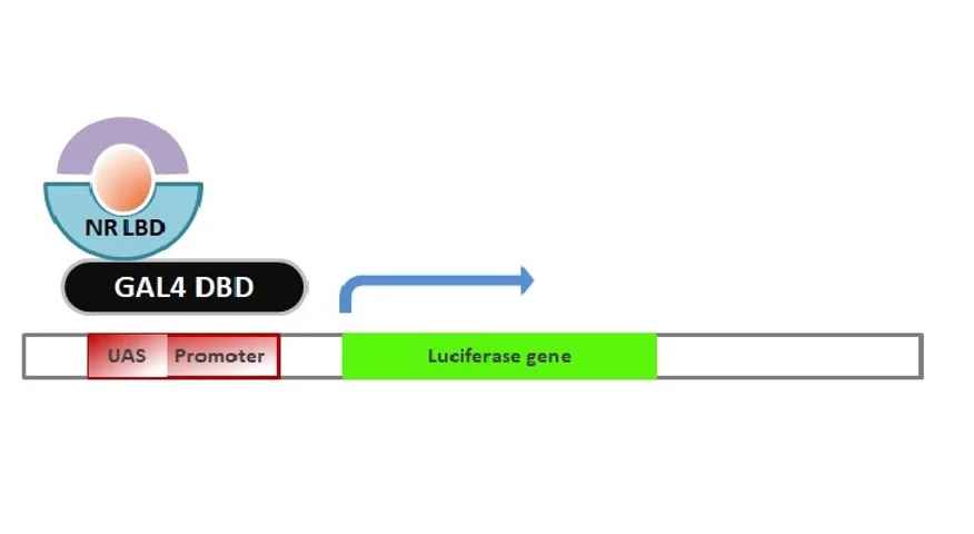
Signosis has developed ligand binding domain(LBD) -driven GAL4 reporter stable cell lines by delivering two vectors, one expressing 8 copies of GAL4 upstream activator sequences (UAS) in front of the luciferase reporter gene, another expressing Gal4 DNA Binding Domain (DBD) bound to a ligand binding domain (LBD) of your choice. Upon addition of a corresponding ligand, the DBD-LBD fusion protein is activated, which binds to GAL promoter binding site that will drive forward the expression of luciferase. With this method, our GAL4 based cell line assays will have minimal cross-reactivity with other nuclear receptor, leading to the assays having high sensitivity and specificity, while also having low toxicity of the chimeric receptors to the cells when overexpressed. These cell lines can be used to screen drug compounds for agonist or antagonist.

​Stable clones were selected with both hygromycin and G418, and a functional assay was subsequently conducted where clones with the highest induction fold chosen.

Benefits

Highly Specific

The responsiveness of this stable cell line is driven by the specific ligand-binding domain with low/no cross-reactivity with other members of the nuclear receptor.

Less Toxic

The chimeric receptors display less toxic effects on the cells.

Highly Sensitive

More than 50 fold induction in response to the corresponding stimuli.

Routine Mycoplasma Testing

All cell lines tested negative for mycoplasma.

Signosis Firefly Luciferase Substrate

We also offer our own Firefly Luciferase Substrate which can generate a strong light signal that is comparable to our competitor’s, but at a more cost-effective price.

NR LBD-Driven GAL4 Cell Lines

Signosis has developed ready-to-use Nuclear Receptor Ligand Binding Domain driven GAL4 reporter cell lines.

These cell lines are a reliable, rapid, and sensitive method for analyzing the intracellular status of NRs upon exposure to drug candidates or other stimuli. These functional assays allow for measurement of receptor activation or inhibition by compounds and can be used to determine compound potency and selectivity.

Description Catalog No.
GR LBD-driven GAL4 reporter HEK 293 Stable Cell Line SL-3001
PPAR-gamma LBD-Driven GAL4 Reporter HEK 293 Stable Cell Line SL-3002
PPAR-alpha LBD-Driven GAL4 Reporter HEK 293 Stable Cell Line SL-3003
PPAR-delta LBD-Driven GAL4 Reporter HEK 293 Stable Cell Line SL-3004

Ultra Sensitive NR LBD-DRIVEN GAL 4 Cell Lines

The Ultra Sensitive LBD-driven GAL4 reporter cell lines has been optimized with an enhanced promoter upstream of the luciferase gene to maximize signal output in response to extremely low concentrations of inducers. These cell line not only yields the enhanced basal signal suitable for any instrument detection, but also produces the higher induction to monitor the specific pathways in an ultra sensitive manner.

Description Catalog No.
Ultra Sensitive PPARdelta LBD-Driven GAL4 Reporter HEK293 Stable Cell Line SL-3004-HS
Ultra Sensitive PPARalpha LBD-Driven GAL4 Reporter HEK293 Stable Cell Line SL-3003-HS
Ultra Sensitive PPARgamma LBD-Driven GAL4 Reporter HEK293 Stable Cell Line SL-3002-HS
Ultra Sensitive GR LBD-driven GAL4 reporter HEK 293 stable cell line SL-3001-HS

GAL4-UAS-Luc Cell Lines

Signosis has developed the GAL4-UAS-Luc Reporter Stable Cell Lines as a reliable, rapid, and sensitive method for analyzing the expression of your target gene of interest upon exposure to related ligands or other stimuli. These cell lines are transfected only with the GAL4-UAS-LUC vector, and are ready to be transfected with your customized GAL4 vector containing your NR of interest.

Description Catalog No.
UAS Luciferase Reporter HEK293 Stable Cell Line SL-3000
UAS Luciferase Reporter HeLa Stable Cell Line SL-4000
UAS Luciferase Reporter CHO Stable Cell Line SL-5000

Research Application

GAL4 LBD-driven GAL4 Reporter HEK 293

  • Studying the effects of ligand binding on GAL4-mediated transcriptional activity
  • Identifying ligands that activate or inhibit the target receptors by monitoring GAL4-driven luciferase expression
  • Screening for potential therapeutic compounds that modulate the target receptors by measuring changes in luciferase activity
  • Investigating the mechanisms underlying the regulation of the target receptors by analyzing the GAL4-mediated transcriptional response to various stimuli
  • Comparing the activity and selectivity of different compounds on the target receptors by using the GAL4 reporter assay
  • Studying the interaction between the target receptors and other signaling pathways or co-regulators by analyzing the changes in GAL4-mediated transcriptional activity upon co-expression of different genes or treatments.

PPAR-gamma LBD-driven GAL4 reporter HEK 293

  • Studying the effects of ligand binding on PPAR-gamma-mediated transcriptional activity
  • Identifying compounds that activate or inhibit PPAR-gamma activity by monitoring GAL4-driven luciferase expression
  • Investigating the role of PPAR-gamma in adipogenesis and glucose homeostasis by analyzing the GAL4-mediated transcriptional response to various stimuli
  • Screening for potential therapeutic compounds that target PPAR-gamma for the treatment of diabetes and other metabolic disorders
  • Studying the interaction between PPAR-gamma and other signaling pathways or co-regulators by analyzing the changes in GAL4-mediated transcriptional activity upon co-expression of different genes or treatments.

PPAR-alpha and PPAR-delta LBD-driven GAL4 reporter HEK 293

  • Studying the effects of ligand binding on PPAR-alpha or PPAR-delta-mediated transcriptional activity
  • Identifying compounds that activate or inhibit PPAR-alpha or PPAR-delta activity by monitoring GAL4-driven luciferase expression
  • Investigating the roles of PPAR-alpha and PPAR-delta in lipid metabolism and energy homeostasis by analyzing the GAL4-mediated transcriptional response to various stimuli
  • Screening for potential therapeutic compounds that target PPAR-alpha or PPAR-delta for the treatment of metabolic disorders, including dyslipidemia and obesity
  • Studying the interaction between PPAR-alpha or PPAR-delta and other signaling pathways or co-regulators by analyzing the changes in GAL4-mediated transcriptional activity upon co-expression of different genes or treatments.

Related Products

Description Catalog No.
Firefly Luciferase Substrate (100mL) for 2000 reactions LUC100
Firefly Luciferase Substrate (15mL) for 300 reactions LUC015
Firefly Luciferase Lysis Buffer 5X (10mL) LS-001

Receptor Tyrosine Kinase & HER2, Mutants Stable Cell Line

Gene function analysis, target discovery and validation, assay development, and compound screening often need cell-based assays. Stable cell lines that are engineered to express a gene of interest via transgene integrations into the host genome provide an efficient approach to conduct such analysis.

At Signosis, we help researchers save time and labor in generating and validating stable cell lines so scientists can spend more effort on solving the big questions. We offer a wide selection of validated luciferase reporter cell lines that measure transcription factor activity as a read-out for various signaling pathways.

Principle

BaF3, a murine interleukin-3 dependent pro-B cell line has been well-known as a model system for assessing downstream signaling of kinase oncogenes, and the ability of small-molecule kinase inhibitors to block kinase activity. Even though BaF3 is suitable as a transfection or transduction host, the viral delivery system is still the main approach. Signosis has been engineered as a non-viral vector-based electroporation delivery system to establish a panel of EGFR, Flt3 and Her2 and mutants BaF3 stable cell lines. The cell lines have been primarily selected with GFP, and subsequently validated with PCR-sequencing and finally confirmed western blot. The clone with the strongest expression level has been launched for facilitating drug screening and associated researches.

Benefits

Non Viral Involvement

Our engineered expression vector has been delivered into the cells with electroporation.

Double Selection

​The single clones have been selected with GFP and hygromycin.

Solid Validation

The selected clones have been validated with PCR-sequencing and Western blot analysis.

Functional Analysis

​The cell lines have been conducted the proliferation test with the corresponding inhibitors.

Receptor Kinase & Mutant Stable Cell Line

EGFR Mutants

EGFR Stably Expressing Cell Lines

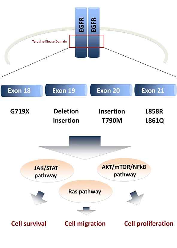
EGFR mutations occur in many types of cancers including non-small cell lung cancer. Common mutations found in these patients are deletions in exon 19 (Del19) and L858R substitution in exon 21. Patients with these mutations are sensitive to first (gefitinib or erlotinib) and second (afatinib) generation EGFR tyrosine kinase inhibitors (TKIs), whereas patients with wild type EGFR are not. However, eventually, all patients become resistant to first and/or second-generation EGFR TKIs, and acquired T790M secondary mutation in exon 20 accounts for more than 50% of the acquired resistance. Cells expressing EGFR with either L858R+ T790M or Del19+T790M double mutations are resistant to induced apoptosis in the presence of first or second-generation EGFR TKIs.

Eight cell lines stably expressing HA-tagged EGFR have been established in the IL3-dependent Ba/F3 cell lines —WT EGFR, L858R EGFR mutant, L858R/T790M EGFR double mutant, Del19 (del747-752) EGFR mutant, Del19/T790M EGFR mutant, three ins20 (D770_N771insSVD, A763_Y764insFQEA, A767_dupASV) EGFR mutants —and Ba/F3 cell line expressing empty vector. Also, two cell lines stably expressing HA-tagged EGFR have been established in HCC827 cell lines —Exon19del EGFR mutant, Exon19del/T790M EGFR double mutant—and HCC827 cell line expressing empty vector.

Due to their proliferation upon EGFR activation, sensitivity to TKIs, or resistance to induced apoptosis, these cell lines can be used to study the molecular mechanism underlying susceptibility of tumors to the drugs as well as screening and validating new TKIs.

Description Catalog No.
EGFR (Del19 (Del746-750)) BaF3 Stable Cell Line EL-021
EGFR( D770_N771insSVD ) BaF3 Stable Cell Line EL-008
EGFR (A767_dupASV) BaF3 Stable Cell Line EL-010
EGFR (Del19-T790M) BaF3 Stable Cell Line EL-014
EGFR (Del19 (del747-753InsS)) BaF3 Stable Cell Line EL-011
EGFR (E19del+T790M) HCC827 Stable Cell Line EL-006
EGFR (L858R) Ba/F3 Stable Cell Line EL-003
EGFR (E19del) HCC827 Stable Cell Line EL-005
EGFR (L858R+T790M) Ba/F3 Stable Cell Line EL-004
EGFR (M766_A767insAI) BaF3 Stable Cell Line EL-018
EGFR (Y764_V765insHH) BaF3 Stable Cell Line EL-020
EGFR WT Ba/F3 Stable Cell Line EL-002
EGFR (H773_V774insH) BaF3 Stable Cell Line EL-019
EGFR (A763_Y764insFQEA) BaF3 Stable Cell Line EL-009
EGFR (L858R) CHO Stable Cell Line EL-016
EGFR (T790M) CHO Stable Cell Line EL-017

HER2 Mutants

HER2 (ERBB2) Stably Expressing Cell Lines

The proto-oncogene human epidermal growth factor receptor 2 (HER2) gene also known as ERBB2 is a member of the ERBB/HER RTK family which is comprised of EGFR (EGFR/HER1/ERBB1), HER3/ERBB3, and HER4/ERBB4. This gene encodes for a transmembrane glycoprotein receptor with an intracellular domain with tyrosine kinase activity which is responsible for the activation of intracellular signal transduction pathways controlling epithelial cell growth, differentiation, and angiogenesis. Aberrations of the HER2 gene which usually leads to increased receptor signaling have been observed in many different types of cancer.

Description Catalog No.
HER2 (WT) BaF3 Stable Cell Line EL-0031
HER2 (S310F) BaF3 Stable Cell Line EL-0032
HER2 (A775_G776insYVMA) BaF3 Stable Cell Line EL-0033
HER2 (P780_Y781insGSP) BaF3 Stable Cell Line EL-0034
HER2 (P95) BaF3 Stable Cell Line EL-0035
HER2 (A775_G776insYVMA, C805S) BaF3 Cells EL-0044

FLT3 Mutants

FLT3 Stably Expressing Cell Lines

The FMS-like tyrosine kinase 3 (FLT3) also known as cluster of differentiation antigen 135 (CD135) or fetal liver kinase-2 (Flk2) is a class III receptor tyrosine kinase (RTK) that plays a key role in controlling survival, proliferation, and differentiation of hematopoietic cells and it widely expresses in acute lymphoblastic leukemia (ALL) and acute myeloid leukemia (AML). Mutations in FLT3 (both internal tandem duplications (ITD) and point mutations) are among the most common somatic mutations in AML patients and are associated with poor prognosis and outcome in those patients

Description Catalog No.
FLT3 D835Y Ba/F3 Stable Cell Line SL-0113
FLT3 D835V Ba/F3 Stable Cell Line SL-0114
FLT3 D835E Ba/F3 Stable Cell Line SL-0115
FLT3 D835N Ba/F3 Stable Cell Line SL-0116
FLT3 D835F Ba/F3 Stable Cell Line SL-0117
FLT3 D835H Ba/F3 Stable Cell Line SL-0118
FLT3 WT Ba/F3 Stable Cell Line SL-0119

Research Applications

EGFR mutants

  • Investigating the effects of EGFR mutations on downstream signaling pathways and cellular processes, such as cell proliferation, differentiation, and survival.
  • Identifying novel therapeutic strategies for targeting EGFR mutants in cancer by testing the efficacy of various inhibitors or combination therapies.
  • Screening for small molecule compounds or antibodies that selectively target EGFR mutants over wild-type EGFR.
  • Developing in vitro and in vivo models to study the mechanisms of resistance to EGFR inhibitors and to test novel combination therapies.
  • Evaluating the potential of EGFR mutants as diagnostic and prognostic biomarkers for specific types of cancer.

HER2 (ERBB2) and mutants

  • Investigating the effects of HER2 mutations on downstream signaling pathways and cellular processes, such as cell proliferation, differentiation, and survival.
  • Identifying novel therapeutic strategies for targeting HER2 and its mutants in cancer by testing the efficacy of various inhibitors or combination therapies.
  • Screening for small molecule compounds or antibodies that selectively target HER2 mutants over wild-type HER2.
  • Developing in vitro and in vivo models to study the mechanisms of resistance to HER2 inhibitors and to test novel combination therapies.
  • Evaluating the potential of HER2 and its mutants as diagnostic and prognostic biomarkers for specific types of cancer.

FLT3 and mutants

  • Investigating the effects of FLT3 mutations on downstream signaling pathways and cellular processes, such as cell proliferation, differentiation, and survival.
  • Identifying novel therapeutic strategies for targeting FLT3 and its mutants in cancer by testing the efficacy of various inhibitors or combination therapies.
  • Screening for small molecule compounds or antibodies that selectively target FLT3 mutants over wild-type FLT3.
  • Developing in vitro and in vivo models to study the mechanisms of resistance to FLT3 inhibitors and to test novel combination therapies.
  • Evaluating the potential of FLT3 and its mutants as diagnostic and prognostic biomarkers for specific types of cancer.

Baf3 Stable Cell Lines

  • Studying the signaling pathways and cellular processes that are regulated by specific receptors or mutants expressed in the Baf3 stable cell lines.
  • Identifying and characterizing novel ligands or compounds that modulate the activity of the receptors or mutants expressed in the Baf3 stable cell lines.
  • Evaluating the efficacy and selectivity of various inhibitors or combination therapies targeting the receptors or mutants expressed in the Baf3 stable cell lines.
  • Developing in vitro and in vivo models to study the effects of specific mutations or treatments on cell proliferation, differentiation, and survival in the context of the Baf3 stable cell lines.
  • Investigating the mechanisms of resistance to various inhibitors or combination therapies in the Baf3 stable cell lines, and testing novel strategies to overcome resistance.
Description Catalog No.
Firefly Luciferase Substrate (100mL) for 2000 reactions LUC100
Firefly Luciferase Substrate (15mL) for 300 reactions LUC015
Firefly Luciferase Lysis Buffer 5X (10mL) LS-001

EGFR Vectors

pMSCV-EGF-GFP Vectors

pCMV-EGFR Vectors

pLenti-MSCV-EGFR-GFP Vectors

Epidermal growth factor receptor (EGFR) is a transmembrane tyrosine kinase receptor that plays a critical role in regulating cell division and death. Mutations in EGFR gene that lead to overexpression of the protein have been associated with a number of different cancers. The EGFR gene is present on chromosome 7p11.2 and has 28 coding exons in which exons 5–7 and 13–16 code for the ligand-binding domain and exons 18–24 code for the TK domain.

Downstream effects of EGFR activation include modulation of three major pathways of RAS-RAF-MAPK, PI3K/AKT, and JAK/STAT pathways which can lead to cell survival, proliferation, and migration of cancer cells.

​We offer a collection of unique cloning and expression cDNA vectors contain WT and mutated EGFR gene. These mutations are clinically approved and are under further investigation by many researchers. Our high-quality vectors are fully sequenced and are ready to use. We also offer custom cloning services for your specific construct needs.

pMSCV-EGF-GFP Vectors Research Application

  • ​Overexpressing EGF in cells to study the effects of EGF on cell proliferation, differentiation, and survival.
  • Generating stable cell lines expressing EGF for long-term studies of EGF signaling pathways and downstream effects.
  • Investigating the mechanisms of resistance to EGF inhibitors or combination therapies in cells that overexpress EGF.
  • Developing animal models of EGF-driven cancers by transfecting cells with pMSCV-EGF-GFP vectors and injecting them into animals.
  • Evaluating the efficacy of various inhibitors or combination therapies targeting EGF in vitro and in vivo.

pCMV-EGFR Vectors

Benefits

Panel of EGFR Mutant Vectors

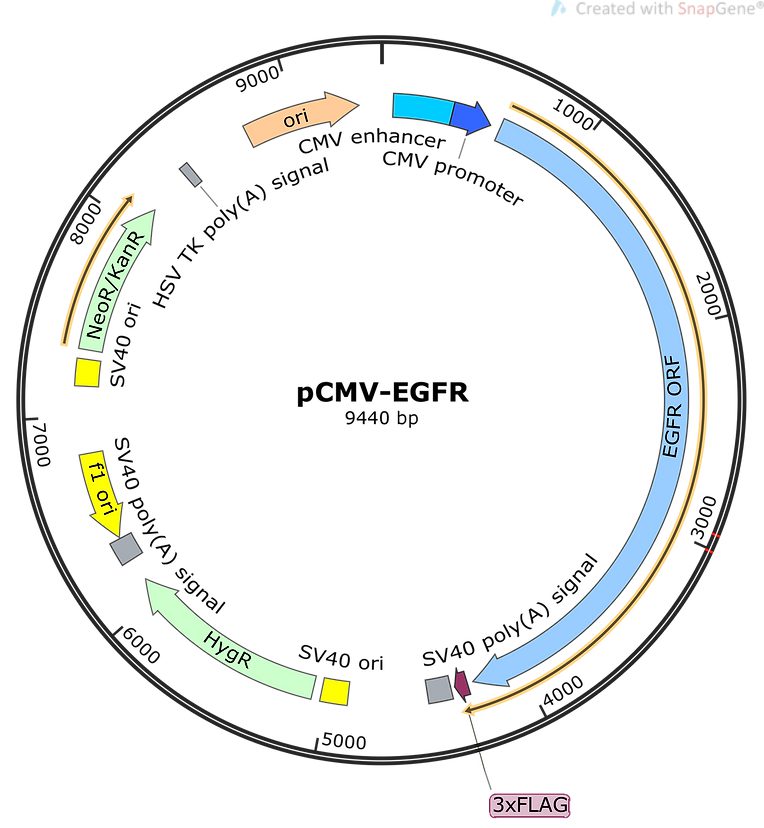
The panel includes more than 12 ready-to-use EGFR mutants.

DYKDDDDK-Tag for Easy Detection

This tag is engineered to downstream of EGFR gene to facilitate Western blot.

Perfect for Stable Cell Line Establishment

The hygromycin-resistant gene is embedded in the vector for stable cell line establishment.

Custom Service Available

We also offer the custom service with the same list price. If you have any mutants in your mind, please let us know.

pLenti-MSCV-EGFR-GFP Vectors

Epidermal growth factor receptor (EGFR) is a transmembrane tyrosine kinase receptor that plays a critical role in regulating cell division and death. Mutations in EGFR gene that lead to overexpression of the protein have been associated with a number of different cancers. The EGFR gene is present on chromosome 7p11.2 and has 28 coding exons in which exons 5–7 and 13–16 code for the ligand-binding domain and exons 18–24 code for the TK domain. Downstream effects of EGFR activation include modulation of three major pathways of RAS-RAF-MAPK, PI3K/AKT, and JAK/STAT pathways which can lead to cell survival, proliferation, and migration of cancer cells.

​We offer a collection of unique cloning and expression cDNA vectors contain WT and mutated EGFR gene. These mutations are clinically approved and are under further investigation by many researchers. Our high-quality vectors are fully sequenced and are ready to use. We also offer custom cloning services for your specific construct needs.

pCMV-EGFR Vectors Research Application

  • ​Overexpressing EGFR in cells to study the effects of EGFR on cell proliferation, differentiation, and survival.
  • Generating stable cell lines expressing EGFR for long-term studies of EGFR signaling pathways and downstream effects.
  • Investigating the mechanisms of resistance to EGFR inhibitors or combination therapies in cells that overexpress EGFR.
  • Developing animal models of EGFR-driven cancers by transfecting cells with pCMV-EGFR vectors and injecting them into animals.
  • Evaluating the efficacy of various inhibitors or combination therapies targeting EGFR in vitro and in vivo.

pLenti-MSCV-EGFR-GFP Vectors

Benefits

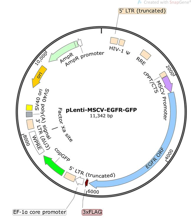
Infect both dividing and nondividing cells DYKDDDDK-tag engineered to downstream of EGFR gene for Western blot

Super bright CopGFP under EF-1 promoter as a selection marker

Contain HIV-1 LTRs and the lentiviral packaging signal (Ψ)

Specific elements (WPRE, cPPT, RRE) included for improving transgene expression

pLenti-MSCV-EGFR-GFP Vectors

Epidermal growth factor receptor (EGFR) is a transmembrane tyrosine kinase receptor that plays a critical role in regulating cell division and death. Mutations in EGFR gene that lead to overexpression of the protein have been associated with a number of different cancers. The EGFR gene is present on chromosome 7p11.2 and has 28 coding exons in which exons 5–7 and 13–16 code for the ligand-binding domain and exons 18–24 code for the TK domain. Downstream effects of EGFR activation include modulation of three major pathways of RAS-RAF-MAPK, PI3K/AKT, and JAK/STAT pathways which can lead to cell survival, proliferation, and migration of cancer cells.

We offer a collection of unique cloning and expression cDNA vectors contain WT and mutated EGFR gene. These mutations are clinically approved and are under further investigation by many researchers. Our high-quality vectors are fully sequenced and are ready to use. We also offer custom cloning services for your specific construct needs.

pLenti-MSCV-EGFR-GFP Vectors Research Application

  • ​Generating stable cell lines expressing EGFR for long-term studies of EGFR signaling pathways and downstream effects.
  • Investigating the effects of EGFR mutations on downstream signaling pathways and cellular processes, such as cell proliferation, differentiation, and survival.
  • Identifying novel therapeutic strategies for targeting EGFR in cancer by testing the efficacy of various inhibitors or combination therapies.
  • Screening for small molecule compounds or antibodies that selectively target EGFR mutants over wild-type EGFR.
  • Developing animal models of EGFR-driven cancers by transfecting cells with pLenti-MSCV-EGFR-GFP vectors and injecting them into animals.
  • Evaluating the potential of EGFR and its mutants as diagnostic and prognostic biomarkers for specific types of cancer.

Benefits

EGFR under MSCV promoter for high expression in hematopoietic or stem cells

Super bright CopGFP under EF-1 promoter as a selection marker

DYKDDDDK-tag engineered to downstream of EGFR gene for Western blot

Description Catalog No.
pLenti-EGFR (L718Q, L858R) ELM-0006
pLenti-EGFR (D770_N771insSVD, C797S) ELM-0004
pLenti-EGFR (H773_V774iinsNPH) ELM-0003
pLenti-EGFR (L718Q, T790M, L858R) ELM-0008
pLenti-EGFR (L792H, L858R) ELM-0007
pLenti-EGFR (Viii) ELM-0010
pLenti-EGFR (H773_V774insH, C797S) ELM-0005
pLenti-EGFR (A767_dupASV, C797S) ELM-0002
pLenti-EGFR (T790M, L792H, L858R) ELM-0009
pLenti-EGFR (WT) ELM-0001
pCMV-EGFR (G719S) EV-0002
pCMV-EGFR (T790M C797S Exon 19 Deletion) EV-0014
pCMV-EGFR (L858R) EV-0007
pCMV-EGFR ( L718Q L858R) EV-0017
pCMV-EGFR (T790M L792H L858R) EV-0020
pCMV-EGFR (L792H L858R ) EV-0019
pCMV-EGFR Exon 19 Insertion EV-0010
pCMV-EGFR (L858Q) EV-0006
pCMV-EGFR (G719C) EV-0004
pCMV-EGFR (D770_N771insSVD, C797) EV-0015
pCMV-EGFR Exon 20 Insertion EV-0011
pCMV-EGFR (G719A) EV-0003
pCMV-EGFR (L861Q) EV-0009
pCMV-EGFR Exon 19 Deletion EV-0008
pCMV-EGFR (T790M) EV-0005
pCMV-EGFR (H773insNPH) EV-0016
pCMV-EGFR ( L718Q T790M L858R) EV-0018
pCMV-EGFR (T790M C797S) EV-0013
pCMV-EGFR (C797S) EV-0012
pCMV-EGFR (WT) EV-0001

Tyrosine Kinases
JAK2 Mutants Stable Cell Lines

Gene function analysis, target discovery and validation, assay development, and compound screening often need cell-based assays. Stable cell lines that are engineered to express a gene of interest via transgene integrations into the host genome provide an efficient approach to conduct such analysis.

At Signosis, we help researchers save time and labor in generating and validating stable cell lines so scientists can spend more effort on solving the big questions. We offer a wide selection of validated luciferase reporter cell lines that measure transcription factor activity as a read-out for various signaling pathways.

Principle

The cell line was generated with expression vector in combined with hygromycin resistant and or puromycin resistant as well as GFP expression into BaF3 cells, and the single clones have been selected with hygromycin treatment, GFP expression. The positive clone has been confirmed with PCR-sequencing and Western blot with anti-JAK antibody.

Benefits

Non Viral Involvement

Our engineered expression vector has been delivered into the cells with electroporation.

Double Selection

The single clones have been selected with GFP and hygromycin.

Solid Validation

The selected clones have been validated with PCR-sequencing and Western blot analysis.

Functional Analysis

​The cell lines have been conducted the proliferation test with the corresponding inhibitors.

Products

Description Catalog No.
Jak2 V617F + EPOR expressing Ba/F3 cell line SL-0202 (G418 resistance)  SL-0202
Jak2 V617F Expressing Ba/F3 Cell Line SL-0201
Jak2WT Expressing Ba/F3 Cell Line  SL-0200

Parental Cell Lines

Signosis offers some of the parental (Host) cells used for generating its stable cell lines. These cell lines can be used as a matched control for their related stable cell lines. All cell lines tested negative for mycoplasma.

Principle

A parental cell line is a group of cells that are derived from the same source or organism and are used as a starting point for creating different cell lines. Parental cell lines are often used in biomedical research and drug development to establish a baseline or control group for comparison purposes.

​Parental cell lines can also be used to produce biologics, such as monoclonal antibodies or recombinant proteins, which are used as therapeutics. In this case, the parental cell line serves as the foundation for the production of these products.

Benefits

Time Saving

Cell line can be used for experiments right away to study different signaling pathways

Consistent

Exogenous gene is stably integrated into the genome to avoid experimental/cell-to-cell variations

Cell Lines

Research Application

HeLa cells are a commonly used cell line in cancer research, derived from cervical cancer cells. They are an immortal cell line, meaning they can divide indefinitely. HeLa cells have been extensively studied and used in many different research applications due to their rapid growth, adaptability, and ability to easily transfect with exogenous DNA.

Research applications:

  • Studying the mechanisms of cancer development and progression.
  • Screening for novel cancer therapies.
  • Evaluating the efficacy of various cancer treatments, such as chemotherapy, radiation, and immunotherapy.
  • Investigating the effects of viral infections, such as HPV, on cancer development.
  • Studying the regulation of cell cycle and apoptosis.

HEK293 cells are a widely used human embryonic kidney cell line. They are also an immortal cell line and can be easily transfected with exogenous DNA. HEK293 cells have been extensively used in research for protein expression and biochemical studies due to their high transfection efficiency and robust protein expression.

​Research applications:

  • Protein expression and purification for biochemical and structural studies.
  • Studying signaling pathways and protein-protein interactions.
  • Developing recombinant viral vectors for gene therapy.
  • Developing and optimizing cell-based assays for drug screening.
  • Studying the effects of gene mutations on protein function.

A549 cells are a human lung carcinoma cell line commonly used in respiratory research. They were originally isolated from a patient with adenocarcinoma and have been extensively studied for their ability to differentiate into respiratory epithelial cells.

Research applications:

  • Studying respiratory diseases, such as asthma, chronic obstructive pulmonary disease (COPD), and lung cancer.
  • Investigating the effects of environmental toxins, such as cigarette smoke, on lung function.
  • Developing and optimizing cell-based assays for drug screening.
  • Studying the regulation of inflammation and immune response in the respiratory system.
  • Studying the mechanisms of viral infections, such as influenza and respiratory syncytial virus (RSV).

NIH/3T3 cells are a mouse embryonic fibroblast cell line. They are widely used in research for their ability to rapidly divide and their ease of transfection with exogenous DNA.

​Research applications:

  • Studying the regulation of cell cycle and apoptosis.
  • Studying the effects of gene mutations on protein function.
  • Developing and optimizing cell-based assays for drug screening.
  • Studying the mechanisms of cancer development and progression.
  • Studying the regulation of cellular metabolism and energy production.

Jurkat cells are a human T lymphocyte cell line commonly used in immunology research. They were originally isolated from a patient with acute T cell leukemia and have been extensively studied for their role in immune function.

​Research applications:

  • Studying the regulation of immune function, including T cell activation, differentiation, and apoptosis.
  • Studying the mechanisms of immune-related diseases, such as autoimmune disorders and cancer.
  • Developing and optimizing cell-based assays for drug screening.
  • Studying the effects of gene mutations on immune function.
  • Studying the regulation of cellular metabolism in immune cells.

K562 cells are a human erythroleukemia cell line commonly used in cancer research. They were originally isolated from a patient with chronic myelogenous leukemia and have been extensively studied for their role in erythropoiesis and differentiation.

​Research applications:

  • Studying the regulation of erythropoiesis and differentiation.
  • Studying the mechanisms of cancer development and progression.
  • Developing and optimizing cell-based assays for drug screening.
  • Studying the effects of gene mutations on protein function.
  • Studying the regulation of cellular

COS-7 is a cell line derived from the African green monkey kidney cells, commonly used in molecular biology and biotechnology research. The cells are highly transfectable, allowing for the introduction of exogenous genes and expression of recombinant proteins.

Research applications:

  1. Protein expression and purification: COS-7 cells are commonly used for the production of recombinant proteins due to their high transfection efficiency and ability to express high levels of proteins.
  2. Gene expression studies: COS-7 cells are often used to study the function and regulation of genes, as they can be easily transfected with plasmids containing reporter genes and other genetic constructs.
  3. Viral vector production: The high transfection efficiency of COS-7 cells makes them useful for the production of viral vectors for gene therapy and other applications.
  4. Drug discovery: COS-7 cells can be used in high-throughput screening assays to identify potential drug candidates for various diseases.

MCF7 is a breast cancer cell line derived from a human patient with metastatic breast adenocarcinoma. The cells are widely used in breast cancer research as a model system for studying the molecular mechanisms underlying the disease.

​Research applications:

  1. Breast cancer biology: MCF7 cells are used to study various aspects of breast cancer biology, including the mechanisms of tumor progression, drug resistance, and metastasis.
  2. Drug discovery: MCF7 cells are used in drug discovery research to screen for potential therapeutic compounds that may be effective in treating breast cancer.
  3. Hormone receptor signaling: MCF7 cells express estrogen receptors and are used as a model system to study the mechanisms of hormone receptor signaling in breast cancer.
  4. Gene expression studies: MCF7 cells are commonly used to study the expression and regulation of genes associated with breast cancer.

Associated Products

Benefits

High signal and responsive

Produces strong and stable light signal.

Simple, easy to use One-step solutions

Ready to use without any prior preparation or mixing needed

Compatible

Works with all Signosis luciferase products and third-party firefly luciferase systems

Cost Effective

Less $ spent per reaction compared to our competitors

Description Catalog No.
Gaussia Luciferase Substrate (10mL for 200 reactions) GLUC010
Gaussia Luciferase Substrate (50mL for 1000 reactions) GLUC050
Firefly Luciferase Substrate (100mL) for 2000 reactions LUC100
Firefly Luciferase Substrate (15mL) for 300 reactions LUC015
Firefly Luciferase Lysis Buffer 5X (10mL) LS-001

EGFR Vectors

Principle

Mutations in the epidermal growth factor receptor (EGFR) gene are commonly observed in many type of cancers including lung, colorectal, head and neck and breast cancer. These mutations play an important role in particular in non-small-cell lung cancer as they can be not only therapeutic target but also a predictive tool for response to EGFR tyrosine kinase inhibitors such as gefitinib and erlotinib. Cell lines have played crucial roles in the identification and characterization of these driver mutations.

Signosis has developed expression vectors for the most common EGFR mutations harboring FLAG marker with high expression suitable for your cancer research. We also accept any other customized EGFR mutation for your research need, contact us for more information.

Benefits

A panel of EGFR Mutant Vectors

The panel includes more than 12 ready-to-use EGFR mutants.

DYKDDDDK-Tag for Easy Detection

This tag is engineered to downstream of EGFR gene to facilitate Western blot

Perfect for Stable Cell Line Establishment

The hygromycin-resistant gene is embedded in the vector for stable cell line establishment.

Custom Service Available

​We also offer the custom service with the same list price. If you have any mutants in your mind, please let us know.

Cell Vialbility Assay (CVC)

Principle

Cell Viability/Cytotoxicity (CVC) Reagent is a colorimetric WST-8 based assay that provides a simple and accurate method to measure cell proliferation. This is a tetrazolium reduction assays based on the cleavage of the tetrazolium salt WST-8 to water-soluble, orange formazan dye by cellular mitochondrial dehydrogenases (Fig.1). Intensity of dye is a direct proportion of increased activity of the mitochondrial dehydrogenases and therefore number of viable cells which can be quantified by measuring the absorbance at 450 nm.

Ready to use CVC Reagent allow for rapid, sensitive and reliable method for measuring cell viability/cytotoxicity directly from a 96-well plate without any additional processing.

Description Catalog No.
Gaussia Luciferase Substrate (10mL for 200 reactions) GLUC010
Gaussia Luciferase Substrate (50mL for 1000 reactions) GLUC050
pCMV-EGFR (G719S) EV-0002
pCMV-EGFR (G719A) EV-0003
pCMV-EGFR (G719C) EV-0004
pCMV-EGFR (T790M) EV-0005
pCMV-EGFR (L858Q) EV-0006
pCMV-EGFR (L858R) EV-0007
pCMV-EGFR Exon 19 Deletion EV-0008
pCMV-EGFR (L861Q) EV-0009
pCMV-EGFR Exon 19 Insertion EV-0010
pCMV-EGFR Exon 20 Insertion EV-0011
pCMV-EGFR (C797S) EV-0012
pCMV-EGFR (T790M C797S) EV-0013
pCMV-EGFR (T790M C797S Exon 19 Deletion) EV-0014
pCMV-EGFR (D770_N771insSVD, C797) EV-0015
pCMV-EGFR (H773insNPH) EV-0016
pCMV-EGFR ( L718Q L858R) EV-0017
pCMV-EGFR ( L718Q T790M L858R) EV-0018
pCMV-EGFR (L792H L858R ) EV-0019
pCMV-EGFR (T790M L792H L858R) EV-0020
pLenti-EGFR (A767_dupASV, C797S) ELM-0002
pLenti-EGFR (H773_V774iinsNPH) ELM-0003
pLenti-EGFR (D770_N771insSVD, C797S) ELM-0004
pLenti-EGFR (H773_V774insH, C797S) ELM-0005
pLenti-EGFR (L718Q, L858R) ELM-0006
pLenti-EGFR (L792H, L858R) ELM-0007
pLenti-EGFR (L718Q, T790M, L858R) ELM-0008
pLenti-EGFR (T790M, L792H, L858R) ELM-0009
pLenti-EGFR (Viii) ELM-0010
pLenti-EGFR (WT) ELM-0001
pCMV-EGFR (WT) EV-0001
Cell Viability (CVC) Reagent 1ml CVC001
Cell Viability (CVC) Reagent 5ml CVC005
Firefly Luciferase Lysis Buffer 5X (10mL) LS-001
Firefly Luciferase Substrate (15mL) for 300 reactions LUC015
Firefly Luciferase Substrate (100mL) for 2000 reactions LUC100

Transcription Factor Assays

Transcription factors (TFs) are DNA binding proteins that play essential roles in regulating gene expression. They act as sensors to monitor changes of cells and convert the signals into changes of gene expression. Often, a specific cellular signal pathway can activate multiple TFs and the expression of a specific gene is under the control of multiple TFs.

Signosis provides a variety of tools to meet a variety of needs and facilitate the analysis of transcription factors. The TF Activation Profiling Plate Arrays, Transcriptional Interaction TF Plate Arrays, and Promoter-Binding TF Profiling Array are for the analysis of multiple TFs, and TF Filter Plate Assays, EMSA Kits, Luciferase Reporter Vectors and Vector Sets, Luciferase Stable Cell Lines, TF ELISA, and TF Western Blot Detection Assay Kits can monitor individual TFs.

TF Activation Plate Arrays

Profile the activation for up to 192 TFs at one time

Principle

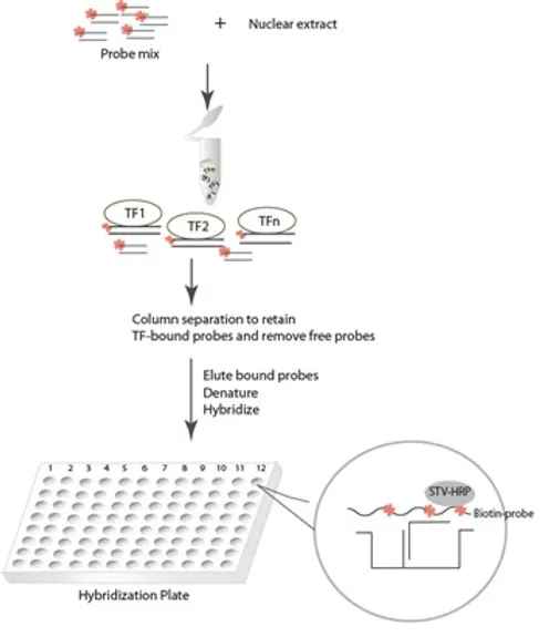
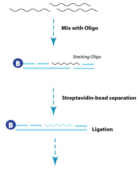
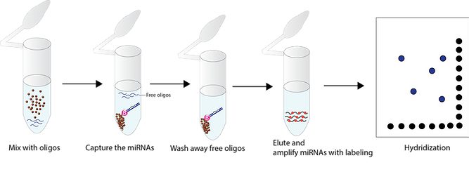
Signosis’ TF Activation Profiling Arrays are used for monitoring the activities of multiple TFs simultaneously. In the technology, a series of biotin-labeled probes are made based on the consensus sequences of TF DNA-binding sites. When the probe mixtures incubate with nuclear extracts, individual probes will find their corresponding TFs and form TF/probe complexes, which can be easily separated from free probes through a simple spin column purification.

The bound probes are then detached from the complexes and analyzed through hybridization with a plate, which each well is specifically pre-coated with complementary sequences of the probes.

Benefits

Multiplex

​One assay allows the measurement of the activities for various transcription factors.

Quantitative Comparison
Differences between samples can be quantitatively compared and analyzed
Simple Procedures

Use simple procedures similar to ELISA & use standard laboratory instrument, Lumniex is not needed

Works with Mammalian Nuclear Extracts

Samples can be taken from human, rat, and mouse

TF Profiling Plate Array

Profile the activation for up to 192 TFs at one time.

Combo TF Activation Profiling Plate Array-192 Catalog No.
Combo TF Activation Profiling Plate Array-192 FA-1016
TF Activation Profiling Plate Array I FA-1001
TF Activation Profiling Plate Array II FA-1002
TF Activation Profiling Plate Array III-96 FA-1011

FewCell TF Plate Arrays

Profile the activation of TFs with limited amount of cells.

Combo TF Activation Profiling Plate Array-192 Catalog No.
FewCell Stem Cell TF Activation Profiling Plate Array FA-1103
FewCell ER Stress (UPR) TF Activation Profiling Plate Array FA-1106

Functional Plate Arrays

Profile the activation of TFs for a specific pathway.

Combo TF Activation Profiling Plate Array-192 Catalog No.
Stem Cell TF Activation Profiling Plate Array I FA-1003
Cancer Stem Cell TF Activation Profiling Plate Array FA-1004
Oxidative Stress TF Activation Profiling Plate Array FA-1005
ER Stress (UPR) TF Activation Profiling Plate Array FA-1006
Wnt/β-Catenin TF Activation Profiling Plate Array FA-1007
Cholesterol Metabolism TF Activation Profiling Plate Array FA-1008
Metal Toxicity TF Activation Profiling Plate Array FA-1009
Mitochondrial UPR TF Activation Profiling Plate Array FA-1010
Lysosomal Stress TF Activation Profiling Plate Array FA-1012
Neurodegenerative Disease TF Activation Profiling Plate Array FA-1013
Insulin Resistance TF Activation Profiling Plate Array FA-1014
T-Cell TF Activation Profiling Plate Array FA-1015

Promoter-Binding Profiling Plate Arrays

Profile the activation for up to 192 TFs at one time

Principle

Promoter-binding TF profiling plate arrays are a competition assay of Signosis’ TF activation plate arrays. In the TF activation plate arrays, if all of 48 or 96 targeted transcription factors exist in the assayed samples, they will form 48 or 96 types of complexes, each TF with its corresponding biotin-labeled oligo (just like the complex in the gel shift assay). After a simple spin separation of the complexes from unbound free biotin-labeled oligos with a membrane-based column, TF-bound oligos eluted from the column and used for plate hybridization in which a complementary DNA of biotin-labeled oligo. The captured oligo is then detected with streptavidin-HRP and a chemiluminescent substrate. If any TF is not present, it will not form a complex, leading to no detection of TF in the following plate assay of biotin-labeled oligo. In promoter-binding TF profiling arrays, PCR fragment containing the promoter of your interest is mixed with a set of 48 or 96 biotin-labeled oligos corresponding to 48 or 96 TFs along with an assayed sample. If unlabeled promoter DNA fragment contains a TF binding sequence, it will compete with the biotin-labeled oligo to bind to the TF in the sample, leading to no or less biotin labeled TF/DNA complex formation and no or lower detection. Through comparison in the presence and absence of the competitor promoter DNA fragment, promoter-bound TFs can be identified.

Benefits

Multiplex

​A single assay allows the characterization of binding of 48 or 96 TFs to a specific promoter

Simple Procedures

Probe incubation, spin column separation, plate hybridization, and HRP detection

Promoter-Binding TF Profiling Array II Catalog No.
Promoter-Binding TF Profiling Array II FA-2002
FA-2002-NE
Promoter-Binding TF Profiling Array I FA-2001
FA-2001-NE

TF Interaction Plate Arrays

Monitor TF/TF or TF/coregulator interactions among 48 or 96 different TFs simultaneously in relation to one TF or coregulator of interest.

Principle

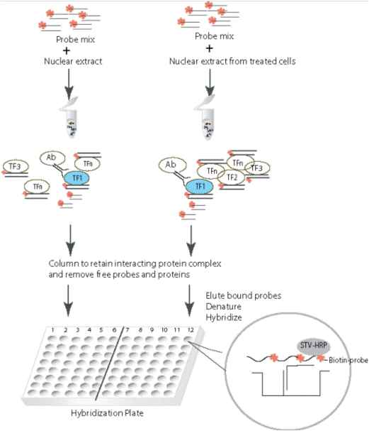
Signosis’ TF-TF Interaction Plate Array can simultaneously profile the transcriptional interaction of multiple TFs with a TF or co-regulator of interest. In this assay, a series of unique biotin-labeled probes are provided that correspond with the consensus sequences of individual TF DNA-binding sites. Therefore, each probe represents an individual TF.


When the probe mix is incubated with nuclear extract, individual probes bind their corresponding TF. The TF or co-regulator of interest is then immunoprecipitated, along with transcriptionally interacting TFs, using a corresponding antibody and protein G or A agarose beads in a tube. Unbound probes and proteins are washed away. The bound probes are then detached from the complex and are subsequently denatured. The biotin-labeled DNA strands are hybridized on a pre-coat plate and detected with streptavidin-HRP and substrate. The detected signals reflect the interacting TFs with the particular TF or co-regulator. Luminescence is reported as relative light units (RLUs) on a microplate luminometer.

Benefits

Description Catalog No.
TF-TF Interaction Plate Array II FA-3002
TF-Coregulator CBP Interaction Plate Array II FA-5032
TF-Coregulator SRC-1 Interaction Plate Array I FA-4021
TF-Coregulator SRC-1 Interaction Plate Array II FA-5022
TF-Coregulator PGC-1a Interaction Plate Array II FA-5042
TF-Coregulator P300 Interaction Plate Array II FA-5012
TF-Coregulator CBP Interaction Plate Array I FA-4031
TF-Coregulator Interaction Plate Array II FA-4002
TF-Coregulator PGC-1a Interaction Plate Array I FA-4041
TF-Coregulator Interaction Plate Array I FA-4001
TF-Coregulator P300 Interaction Plate Array I FA-4011
TF-TF Interaction Plate Array I FA-3001
Multiplex

​A single assay allows the characterization of binding of 48 or 96 TFs to a specific promoter

Quantitative Comparison

​Differences between samples can be quantitatively compared and analyzed.

No Special Equipment Required

Compatible with common chemiluminescence plate readers

Simple Procedures

Co-IP of TF complexes with magnetic beads is quick, easy, and effective.

EMSA

100+ non-radioactive EMSA Kits are available. The probes are pre-labeled with biotin, and all reagents are included in the kit.

Principle

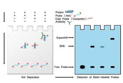
When one TF is activated, it binds to a biotin-labeled probe, which is designed based on the consensus binding sequence of the TF. When the formed complex is subjected to electrophoresis, it runs slower than the free probe, and can be easily distinguished. Two or more samples can be compared and differences can be determined by the density of the shifted band.

Benefits

Isotope Free

It is a chemiluminescent-based detection.

No Probe Preparation

The probes included are pre-labeled and optimized.

Highly Sensitive

With optimized conditions, the kit sensitivity is compatible with that from radioactive-based detection method.

Simple Procedures

All of the reagents are included in the kit.

Description Catalog No. Description Catalog No.
Combination EMSA Kit with Four Probes GS-1001 Myc-Max EMSA Kit GS-0026
EMSA Kits without probes GS-0000 NF-1 EMSA Kit GS-0027
GAS/ISRE EMSA Kit GS-0017 NFAT EMSA Kit GS-0028
NFkB EMSA Kit GS-0030 NF-E2 EMSA Kit GS-0029
Stat1 EMSA Kit GS-0043 NRF2(ARE) EMSA Kit GS-0031
SMAD (MADH) EMSA Kit GS-0039 OCT4 EMSA Kit GS-0032
Sp1 EMSA Kit GS-0040 p53 EMSA Kit GS-0033
SRF EMSA Kit GS-0041 Pax-5 EMSA Kit GS-0034
SATB1 EMSA Kit GS-0042 Pbx1 EMSA Kit GS-0035
Stat3 EMSA Kit GS-0044 Pit EMSA Kit GS-0036
Stat4 EMSA Kit GS-0045 PPAR EMSA Kit GS-0037
Stat6 EMSA Kit GS-0047 PXR EMSA Kit GS-0038
Stat5 EMSA Kit GS-0046 TR EMSA Kit GS-0050
TCF/LEF EMSA Kit GS-0048 YY1 EMSA Kit GS-0051
TFIID EMSA Kit GS-0049 USF-1 EMSA Kit GS-0052
RUNX EMSA Kit GS-0080 VDR EMSA Kit GS-0053
RXR EMSA Kit GS-0081 HSF EMSA Kit GS-0054
AP3 EMSA Kit GS-0082 FOXD3 EMSA Kit GS-0055
AP4 EMSA Kit GS-0083 Prox1 EMSA Kit GS-0056
COUP-TF EMSA Kit GS-0084 SOX18 EMSA Kit GS-0057
HOX4C EMSA Kit GS-0085 SOX5 EMSA Kit GS-0058
MZF EMSA Kit GS-0086 SOX9 EMSA Kit GS-0059
TFE3 EMSA Kit GS-0087 SOX2 EMSA Kit GS-0060
Oct-1 EMSA Kit GS-0088 HOXA-5 EMSA Kit GS-0061
Snail EMSA Kit GS-0089 FoxC1 EMSA Kit GS-0062
AP1 EMSA Kit GS-0001 FOXA1 EMSA Kit GS-0063
AP2 EMSA Kit GS-0002 MyoD EMSA Kit GS-0064
AR EMSA Kit GS-0003 Nkx2-5 EMSA Kit GS-0065
ATF2 EMSA Kit GS-0004 Nkx3-2 EMSA Kit GS-0066
Brn-3 EMSA Kit GS-0005 Pax2 EMSA Kit GS-0067
C/EBP EMSA Kit GS-0006 PIT1 EMSA Kit GS-0068
CAR EMSA Kit GS-0007 Pax3 EMSA Kit GS-0069
CBF EMSA Kit GS-0008 Pax8 EMSA Kit GS-0070
CDP EMSA Kit GS-0009 WT1 EMSA Kit GS-0071
CREB EMSA Kit GS-0010 Gli EMSA Kit GS-0072
E2F-1 EMSA Kit GS-0011 RB site EMSA Kit GS-0073
EGR EMSA Kit GS-0012 SRY EMSA Kit GS-0074
ELK EMSA Kit GS-0013 FOXO1 (FKHR) EMSA Kit GS-0075
ER EMSA Kit GS-0014 FOXG1 EMSA Kit GS-0076
Ets EMSA Kit GS-0015 Gfi-1 EMSA Kit GS-0077
FAST-1(FOXH1) EMSA Kit GS-0016 SMUC EMSA Kit GS-0078
GATA EMSA Kit GS-0018 PLAG1 EMSA Kit GS-0079
GR/PR EMSA Kit GS-0019 KLF4 EMSA Kit GS-0090
PR EMSA Kit GS-0020 NRF1 EMSA Kit GS-0091
HIF EMSA Kit GS-0021 ROR(RZR) EMSA Kit GS-0092
HNF-4 EMSA Kit GS-0022 HNF-1 EMSA Kit GS-0093
IRF EMSA Kit GS-0023 HEN(NSCL-1) EMSA Kit GS-0094
MEF2 EMSA Kit GS-0024 FREAC-2 (FOXF2) EMSA Kit GS-0095
Myb EMSA Kit GS-0025 AHR EMSA Kit GS-0096

TF Filter Plate Arrays

This “EMSA on a plate” assay allows you to monitor the activation of a TF in vitro in up to 96 samples at one time.

Principle

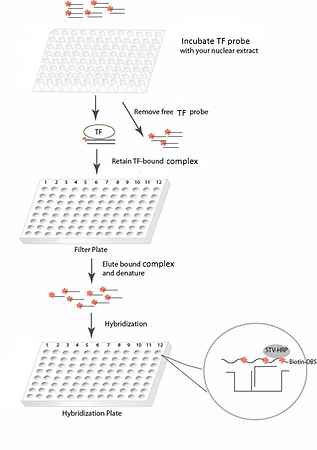
In Signosis’ TF filter plate assays, a specific biotin-labeled TF DNA binding sequence is mixed with nuclear extracts to allow formation of TF-DNA complexes. A filter plate is used to retain bound DNA probe and remove free probe. The bound pre-labeled DNA probe is then eluted from the filter and collected for quantitative analysis through DNA plate hybridization. The captured DNA probe is further detected with streptavidin-HRP. Luminescence is reported as relative light units (RLUs) on a microplate luminometer.

Benefits

High Throughput Screen

A specific TF in multiple samples can be analyzed.

Flexible

Can analyze 1 to 96 samples simultaneously. Easy to reseal and store for later use.

Quantitative Comparison

TF activity differences between two or more samples can be quantitatively analyze

Wide Selection

100+ popular TFs Kits are available from Signosis.

Description Catalog No.
STAT3 Filter Plate Assay FA-0005
YY1 Filter Plate Assay FA-0006
NRF2 Filter Plate Assay FA-0007
TF Filter Plate Assay without Probe (Use with PP-xxxx) FA-0010
PP-0001
HIF Filter Plate Assay FA-0002
AP1 Filter Plate Assay FA-0004
ER Filter Plate Assay FA-0003
NF?B Filter Plate Assay FA-0001

TF ELISA Kits

TF ELISA Kit allows High-throughput profiling TF-DNA binding activity.

Principle

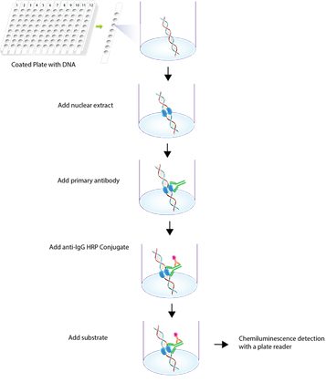
TF ELISA Kit is high sensitive and specific assay with a simple and optimized procedure. The 96-well (8×12 strip) clear plate is pre-immobilized with the TF consensus sequencing oligo. The activated TF in nuclear extract or the while cell lysate is added in the well and binds to the oligo. The activated TF is detected with a specific antibody against the subunit and a HRP conjugated secondary antibody. The assay utilizes colorimetric detection method, which can be easily measured by spectrophotometry.

Benefits

Positive Protein Control Included

The kit contains an active recombinant protein for a positive control.

Highly Effective

Distinguishing two pathways in one ELISA assay.

Highly Specific

Specifically monitor TF activation both the DNA binding sequence and gene specific antibody against the specific gene.

High-Throughput

With Whole Cell Lysis Buffer provided by Signosis, cell lysate can be prepared and high-through TF ELISA can be performed with limited number of cells from 96-well tissue plate. 

Description Catalog No.
ATF4 ELISA Kit (Colorimetric) TE-0039
SATB1 ELISA Kit (Colorimetric) TE-0008
ATF6 ELISA Kit (Colorimetric) TE-0041
STAT3 ELISA Kit (Colorimetric) TE-0020
STAT1 ELISA Kit (Colorimetric) TE-0023
STAT1/STAT3 ELISA Kit (Colorimetric) TE-0025
TFEB ELISA Kit (Colorimetric) TE-0029
SOX2 ELISA Kit (Colorimetric) TE-0033
OCT4 ELISA Kit (Colorimetric) TE-0031
OCT4/SOX2 ELISA Kit (Colorimetric) TE-0035
PPARγ ELISA Kit (Colorimetric) TE-0009
Smad1/5/8 ELISA Kit (Colorimetric) TE-0013
Smad2/3 ELISA Kit (Colorimetric) TE-0011
Smad2/3 and 1/5/8 ELISA Kit (Colorimetric) TE-0015
NFκB p65 ELISA Kit (Colorimetric) TE-0001
IRF3/IRF7 ELISA Kit (Colorimetric) TE-0007
NFκB p50 ELISA Kit (Colorimetric) TE-0002
IRF3 ELISA Kit (Colorimetric) TE-0005
IRF7 ELISA Kit (Colorimetric) TE-0006
ATF ELISA Kit ( ATF4 or ATF6)(Colorimetric) TE-0037

Snapshot Magnetic Beads ChIP Assays

Analyze the interaction of transcription factors and associated coactivitors and corepressors on target promoters in 10^6 cells.

Principle

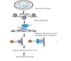
The magnetic beads ChIP assay typically comprises four steps: (1) cross-linking proteins to DNA; (2) chromatin fragmentation; (3) protein precipitation; and (4) target identification and quantitation. The initial cross-linking is to insure that protein-DNA complexes remain associated through the subsequent steps. Formaldehyde is the most commonly used reagent for the cross-linking of proteins that are directly bound to DNA, such as transcription factors and histones, but not for coactivators and corepressors indirectly associated with DNA due to short spacer arm.


Several improvements in Signosis’ ChIP assay are highlighted as follows: firstly, the dual cross-linking, formaldehyde (short space arm) and second cross-linking reagent (long space arm), is introduced to ensure the detection of both TFs and associated cofactors. Secondly, DNA is broken down to mononucleosomes and 500bp in size by MNase digestion, which enhances greatly the sensitivity and reproducibility. The cross-linked protein-DNA complexes are subsequently pulled down by a specific antibody and ChIP grade protein A/G magnetic beads. The last improvement is to release DNA from cross-linked proteins with one-step chelex-100 without tedious proteinase K digestion and further purification, significantly increasing the assay efficiency.

Benefits

High Reproducibility

Controllable MNase digestion for DNA fragmentation instead of sonication.

Highly Specificity

The cross-linked protein-DNA complexes are subsequently pulled down by a specific antibody and ChIP grade protein A/G magnetic beads.

Simple Procedure

Use one step Chelex-100 treatment to release cross-linked DNA instead of tedious proteinase K digestion and column purification.

High-Throughput

Kit comes with everything you will need to perform the assay, including crosslinkers and enzyme. Dual cross linking to analyze both transcription factors and the associated factors.

Description Catalog No.
Snapshot Magnetic Beads ChIP Assay CA-0001
OCT3/4 Snapshot Magnetic Beads ChIP Assay CA-0101
NRF2 Snapshot Magnetic Beads ChIP Assay CA-0102
HDAC1 Snapshot Magnetic Beads ChIP Assay CA-0103
SMAD1/5/8 Snapshot Magnetic Beads CHIP Assay CA-0104
SMAD2/3 Snashot Magnetic Beads CHIP Assay CA-0111
STAT3 Snapshot Magnetic Beads ChIP Assay CA-0105
NFkB p65 Snapshot Magnetic Beads ChIP Assay CA-0106
NFkB p50 Snapshot Magnetic Beads ChIP Assay CA-0112
HIF-1a Snapshot Magnetic Beads ChIP assay CA-0107
P300 Snapshot Magnetic Beads ChIP Assay CA-0109
P53 Snapshot Magnetic Beads ChIP Assay CA-0110
Akt1/2/3 Snapshot Magnetic Beads ChIP assay CA-0113
ERK1/2 Snapshot Magnetic Beads ChIP assay CA-0134
EGFR Snapshot Magnetic Beads ChIP assay CA-0114
Stat1 Snapshot Magnetic Beads ChIP assay CA-0116
C/EBPa Snapshot Magnetic Beads ChIP assay CA-0117
C/EBPb Snapshot Magnetic Beads ChIP assay CA-0118
RelB Snapshot Magnetic Beads ChIP assay CA-0119
caspase-1 Snapshot Magnetic Beads ChIP assay CA-0120
caspase-3 Snapshot Magnetic Beads ChIP assay CA-0121
caspase-9 Snapshot Magnetic Beads ChIP assay CA-0122
c-Rel Snapshot Magnetic Beads ChIP assay CA-0123
Annexin V Snapshot Magnetic Beads ChIP assay CA-0124
Sox-2 Snapshot Magnetic Beads ChIP assay CA-0125
PPARr Snapshot Magnetic Beads ChIP assay CA-0126
TFEB Snapshot Magnetic Beads ChIP assay CA-0127
c-Myc Snapshot Magnetic Beads ChIP assay CA-0128
Bad Snapshot Magnetic Beads ChIP assay CA-0129
twist Snapshot Magnetic Beads ChIP assay CA-0130
Histone H3 Snapshot Magnetic Beads ChIP assay CA-0131
RXRa Snapshot Magnetic Beads ChIP assay CA-0132
b-TrCP/HOS Snapshot Magnetic Beads ChIP assay

TF Luciferase Vectors

Measure TF activation in cells. 100+ vectors are available. We also provide custom service to create a vector for any unlisted TFs.

pTF-Luc reporter vectors are a series of firefly luciferase-based reporter constructs for quantitative measurement of the activities of transcription factors (TFs) in cells. Each vector contains a cis-element (DNA binding sequence), a minimal promoter, and firefly luciferase gene. When a TF is activated, it binds to cis-element and results in induction of luciferase gene expression. Therefore, luciferase activity represents the activity of the TF.

Principle

The Luciferase Reporter Vectors have been specially constructed to report the binding activity of a single TF. Multiple copies (at least four) of the cis-acting enhancer element have been inserted into each vector upstream of a minimal TA promoter. Upon vector transfection and cell stimulation, the activated TF is bound to the enhancer element and initiate the transcription of downstream luciferase reporter gene. The chemiluminescent measurement of luciferase activity is proportional to the amount of expressed enzyme and thus the binding activity of the targeted TF.

Benefits

Functional Assay

Monitor the transactivation of transcription factors in cells.

Quantitative Analysis

Determine TF activation by quantitative measurement ofluciferase activity.

Stable Cell Lines

​The vectors can be used for establishing stable cells expressing the reporters.

Firefly Luciferase Substrate

Firefly Luciferase Substrate for use with the reporter vectors.

Description Catalog No. Description Catalog No.
pFOXC1-Luc LR-2097 pFOXD1-Luc LR-2098
pETS-Luc LR-2035 pFOXF2-Luc LR-2099
pFAST-1-Luc LR-2036 pFOXI1-Luc LR-2100
pAP1-Luc LR-2006 pFOXO3-Luc LR-2101
TA-Luc Control LR-2200 pSMAD-Luc LR-2017
pCBF-Luc LR-2023 pMyc-Luc LR-2018
pHNF1a-Luc LR-2039 pYY1-Luc LR-2019
pHOX4C-Luc LR-2010 pIRF-Luc LR-2020
pNF?B-Luc LR-2001 pBRN3-Luc LR-2021
pPit1-Luc LR-2043 pC/EBP-Luc LR-2022
pSP1-Luc LR-2007 pCDP-Luc LR-2024
pUSF1-Luc LR-2074 pCDXA/NKX2-Luc LR-2025
pHIF-Luc LR-2002 pMyb-Luc LR-2028
pHNF3-Luc LR-2040 pCoup-TF-Luc LR-2029
pHOXA5-Luc LR-2104 pE2F-Luc LR-2031
pSTAT1-Luc LR-2003 pE47-Luc LR-2032
pSTAT3-Luc LR-2004 pEGR-Luc LR-2033
pSTAT4-Luc LR-2005 pEKLF-Luc LR-2034
pCREB-Luc LR-2008 pFKHR-Luc LR-2037
pHSF-Luc LR-2009 pFREAC-Luc LR-2038
pER-Luc LR-2011 pP53-Luc LR-2041
pNFAT-Luc LR-2015 pPbx1-Luc LR-2042
pGAS/ISRE-Luc LR-2016 pMEF3-Luc LR-2056
pGR-Luc LR-2012 pNkx2-5-Luc LR-2057
pPur-1-Luc LR-2044 pNkx3-2-Luc LR-2058
pRAR-Luc LR-2013 pNF-1-Luc LR-2059
pPR-Luc LR-2014 pNFE2-Luc LR-2060
pSRY-Luc LR-2045 pELK1-Luc LR-2061
pTAX/CRE-Luc LR-2046 pNRF1-Luc LR-2062
pTCF/LEF-Luc LR-2047 pOct1-Luc LR-2063
pGATA-Luc LR-2103 pPax2-Luc LR-2064
pAR-Luc LR-2105 pPax3-Luc LR-2065
pNRF2/ARE-Luc LR-2106 pPax4-Luc LR-2066
pTFE3-Luc LR-2048 pPax5-Luc LR-2067
pOCT4-Luc LR-2049 pPax6-Luc LR-2068
pNANOG-Luc LR-2050 pPax8-Luc LR-2069
pLEF1-Luc LR-2053 pRXR-Luc LR-2070
pMEF1-Luc LR-2054 pSRF-Luc LR-2071
pMEF2-Luc LR-2055 pStat5-Luc LR-2072
pWT1-Luc LR-2077 pTR-Luc LR-2073
pXBP1-Luc LR-2078 pVDR-Luc LR-2075
pPLAG1-Luc LR-2081 pV-MAF-Luc LR-2076
pProx1-Luc LR-2082 pSOX5-Luc LR-2090
pRREB1-Luc LR-2084 pSOX9-Luc LR-2091
pRUNX1-Luc LR-2085 pTAL1/TCF3-Luc LR-2092
pSOX10-Luc LR-2086 pTEAD1-Luc LR-2093
pSOX17-Luc LR-2087 pTFAP2A-Luc LR-2094
pSOX18-Luc LR-2088 pGli1-Luc LR-2095
pSOX2-Luc LR-2089 pFOXA1-Luc LR-2096
Description Catalog No.
Nuclear Extraction Kit SK-0001
TF Binding Buffer Mix (FA-100X) (30�l) TBB001
TF Plate Hybridization Buffer (10ml) TPHB001
TF Probe Mix I (FA-X001) (10�l) TPM001-1
TF Probe Mix II (FA-X002) (20�l) TPM001-2
5X Detection Wash Buffer (40ml) CDWB001-40
Substrate A (1ml) SAP001-T
Substrate B (1ml) SBP001-T
Substrate B for Membranes (1.2ml) SBP001-E
5X Binding Buffer (EMSA) (120�l) GSBB001
10X Loading Buffer (EMSA) (100�l) GSLB001
Polyd (I-C) (EMSA) (30�l) GSPD001

Cytokine ELISA

Cytokines and growth factors are signaling molecules that have critical roles in many biological processes such as cellular growth, differentiation, gene expression, migration, immunity and inflammation. Disruption in these signals can lead to a variety of diseases, including arthritis, liver disease, inflammatory bowel disease, cardiac-related diseases, and cancers.

Signosis provides a number of tools to profile one specific cytokine or growth factor, or to monitor multiple simultaneously.

Cytokine ELISA Plate Arrays

Cytokines and growth factors are signaling molecules that have critical roles in many biological processes such as cellular growth, differentiation, gene expression, migration, immunity and inflammation. Disruption in these signals can lead to a variety of diseases, including arthritis, liver disease, inflammatory bowel disease, cardiac-related diseases, and cancers.

Signosis provides a number of tools to profile one specific cytokine or growth factor, or to monitor multiple simultaneously. See our ELISA biomarker kit.

Principle

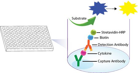
In this assay, the test sample initially reacts with the solid phase capture antibody, resulting in the cytokine being bound to the well. The wells are then washed to remove unbound proteins, and biotin-linked antibodies are added to bind to the cytokines. After washing away the unbound antibodies, Streptavidin-HRP conjugate is added to form a complex with the antibody-bound cytokines.

​For colorimetric kits, the HRP substrate, TMB, is added and forms a blue color when the HRP-linked antibodies are detected. The reaction is then terminated with Stop Solution, which changes the color from blue to yellow. The cytokine concentration in each well is directly proportional to its color intensity and can be quantified by measuring its optical density at 450 nm (OD450) in a microplate reader.

​For chemiluminescence kits, the plate is detected with HRP luminescent substrate. Luminescence is reported as relative light units (RLUs) on a microplate luminometer. The level of expression for each specific cytokine is directly proportional to the luminescent intensity.

Benefits

High Sensitivity & Responsiveness

Requires minimal amount of sample. Compatible with common plate readers. Simple procedure with reagents included.

Multiplex

​One pre-coated plate can measure 48 cytokines for 2 human samples, 32 cytokines for 3 human samples; 32 cytokines for 3 mouse samples, 24 cytokines for 4 mouse samples; or 16 cytokines for 6 rat samples.

Quantitative Profiling

Allows quantitative comparison of expression among samples

Wide Sample Type Selection

The assay is suitable for any biological liquid material and cell lysate, such as sera, cerebrospinal fluid (CSF), urine and tissue homogenate.

Rat Cytokine ELISA Plate Array

Profile an array of rat cytokines using our ELISA kit

Description Catalog No.
Rat Cytokine ELISA Plate Array (Chemiluminescence) EA-4004
Rat Cytokine ELISA Plate Array (Colorimetric) EA-4006

Mouse Cytokine ELISA Plate Array

Profile an array of mouse cytokines using our ELISA kit

Description Catalog No.
Mouse Cytokine ELISA Plate Array IV (Colorimetric) EA-4014
Mouse Cytokine ELISA Plate Array II (Chemiluminescence) EA-4021
Mouse Cytokine ELISA Plate Array III (Chemiluminescence) EA-4022
Mouse Cytokine ELISA Plate Array III (Colorimetric) EA-4013
Mouse Cytokine ELISA Plate Array I (Colorimetric) EA-4005
Mouse Cytokine ELISA Plate Array II (Colorimetric) EA-4008
Mouse Cytokine ELISA Plate Array I (Chemiluminescence) EA-4003

Human Cytokine ELISA Plate Array

Profile an array of human cytokines using our ELISA kit

Description Catalog No.
Human Cytokine ELISA Plate Array II (Chemiluminescence) EA-4019
Human Cytokine ELISA Plate Array I (Colorimetric) EA-4002
Human Cytokine ELISA Plate Array IV (Chemiluminescence) EA-4020
Human Cytokine ELISA Plate Array IV (Colorimetric) EA-4015
Human Cancer Cytokine ELISA Plate Array (Colorimetric) EA-4016
Human Cytokine ELISA Plate Array II (Colorimetric) EA-4010
Human Angiogenesis ELISA Plate Array (Colorimetric) EA-4012
Human IL Family ELISA Plate Array (Colorimetric) EA-4011
Human TGF-beta Family ELISA Plate Array (Colorimetric) EA-1791
Human TGF-beta 1 and 3 ELISA Plate Array (Colorimetric) EA-1781

Research Discovery Objectives

Drug Screening and Discovery

Stable cell lines expressing a specific receptor or target protein can be used to screen for potential drug candidates and evaluate their efficacy and toxicity.

Protein Production

Stable cell lines can be used to produce recombinant proteins for various applications, including biopharmaceutical production, biochemical assays, and structural biology studies.

Gene Editing and Gene Therapy

Stable cell lines can be used to study gene function and develop gene editing or gene therapy approaches for genetic diseases.

Disease Modeling

Stable cell lines can be used to model genetic diseases and study their pathogenesis and potential therapeutic targets.

Vaccine Development

Stable cell lines can be used to produce recombinant proteins for vaccine development and production.

Cell-Based Assays

Stable cell lines can be used in various cell-based assays, such as reporter gene assays, cell proliferation assays, and apoptosis assays, to study cellular processes and signaling pathways.

HEK293

Widely used for protein expression and gene editing studies

U2OS

For studying DNA damage response and genome stability

Jurkat

T-cell leukemia line used for immunology and HIV research

PC-3

Prostate cancer line used for drug discovery and metastasis studies

MDAMB-231

Triple negative breast cancer line used for drug discovery and metastasis studies

A431

Epidermoid carcinoma line used for skin and cancer research

Caco-2

Colon adenocarcinoma line used for drug absorption and intestinal transport studies

CHO

For production of therapeutic proteins and monoclonal antibodies

A549

Lung cancer cell line used for respiratory virus research and drug screening

HeLa

Cervical cancer line used for virology and cancer research

THP-1

Monocyte line used for immunology and infectious disease research

HL-60

Leukemia line used for hematopoietic research and drug development

MDA-MB-468

Breast cancer line used for hormone receptor and drug resistance studies

SW480

Colorectal cancer line used for drug discovery and cancer research

HCT116

Colorectal cancer cell line used for drug discovery and cancer research

MCF-7

Breast cancer cell line used for hormonal and drug resistance studies

SH-SY5Y

Neuroblastoma line used for neuroscience and drug development

MDCK

Canine kidney line used for influenza virus production and vaccine development

SK-N-SH

Neuroblastoma line used for neuroscience and drug development

293T

Derivative of HEK293 line used for lentiviral production and gene editing

Cytokine ELISA Strips

Cytokines and growth factors are signaling molecules that have critical roles in many biological processes such as cellular growth, differentiation, gene expression, migration, immunity and inflammation. Disruption in these signals can lead to a variety of diseases, including arthritis, liver disease, inflammatory bowel disease, cardiac-related diseases, and cancers. Signosis provides a number of tools to profile one specific cytokine or growth factor, or to monitor multiple simultaneously. See our ELISA biomarker kit.

Principle

In this assay, the test sample initially reacts with the solid phase capture antibody, resulting in the cytokine being bound to the well. The wells are then washed to remove unbound proteins, and biotin-linked antibodies are added to bind to the cytokines. After washing away the unbound antibodies, Streptavidin-HRP conjugate is added to form a complex with the antibody-bound cytokines.

​For colorimetric kits, the HRP substrate, TMB, is added and forms a blue color when the HRP-linked antibodies are detected. The reaction is then terminated with Stop Solution, which changes the color from blue to yellow. The cytokine concentration in each well is directly proportional to its color intensity and can be quantified by measuring its optical density at 450 nm (OD450) in a microplate reader.

​For chemiluminescence kits, the plate is detected with HRP luminescent substrate. Luminescence is reported as relative light units (RLUs) on a microplate luminometer. The level of expression for each specific cytokine is directly proportional to the luminescent intensity.

Benefits

Multiplex

​One ELISA strip allows the measurement of 8 cytokines.

Flexible

​Comparisons can be made between 2 to 12 samples.

Quantitative Profiling & Analysis

Allows quantitative comparison of expression among samples. Protein standard is available for purchase separately.

Wide Sample Selection

​The assay is suitable for any biological liquid material and cell lysate, such as sera, cerebrospinal fluid (CSF), urine and tissue homogenate.

Rat Cytokine ELISA Strips

Profile 8 different rat cytokines using our ELISA strip kit.

Description Catalog No.
Rat Obesity ELISA Strip (Chemiluminescence) EA-1821
Rat Oxidative Stress ELISA Strip (Chemiluminescence) EA-1831
Rat Inflammation ELISA Strip (Colorimetric) EA-1201
Rat Obesity ELISA Strip (Colorimetric) EA-1221
Rat Angiogenesis ELISA Strip (Colorimetric) EA-1211
Rat Inflammation ELISA Strip (Chemiluminescence) EA-1801
Rat Angiogenesis ELISA Strip (Chemiluminescence) EA-1811
Rat Oxidative Stress ELISA Strip (Colorimetric) EA-1501

Mouse Cytokine ELISA Strips

Profile 8 different mouse cytokines using our ELISA strip kit

Description Catalog No.
Mouse Angiogenesis ELISA Strip (Chemiluminescence) EA-1701
Mouse Inflammation ELISA Strip (Colorimetric) EA-1051
Mouse Obesity ELISA Strip II (Colorimetric) EA-1421
Mouse Cytokine ELISA Strip (Colorimetric) EA-1091
Mouse Interleukin ELISA Strip (Chemiluminescence) EA-1751
Mouse ER Stress ELISA Strip (Chemiluminescence) EA-1761
Mouse Oxidative Stress ELISA Strip (Chemiluminescence) EA-1741
Mouse Cytokine ELISA Strip (Chemiluminescence) EA-1731
Mouse Oxidative Stress ELISA Strip (Colorimetric) EA-1401
Mouse ER Stress ELISA Strip (Colorimetric) EA-1131
Mouse Interleukin ELISA Strip (Colorimetric) EA-1411
Mouse Obesity ELISA Strip I (Colorimetric) EA-1071
Mouse Inflammation ELISA Strip (Chemiluminescence) EA-1711
Mouse Obesity ELISA Strip (Chemiluminescence) EA-1721
Mouse Angiogenesis ELISA Strip (Colorimetric) EA-1021

Human Cytokine ELISA Strips

Profile 8 different human cytokines using our ELISA strip kit

Description Catalog No.
Human Adipokine ELISA Strip (Chemiluminescence) EA-1695
Human Oxidative Stress ELISA Strip (Chemiluminescence) EA-1671
Human Cytokine ELISA Strip (Chemiluminescence) EA-1651
Human Interleukin ELISA Strip (Colorimetric) EA-1311
Human ER Stress ELISA Strip (Chemiluminescence) EA-1694
Human Angiogenesis ELISA Strip I (Colorimetric) EA-1011
Human Angiogenesis ELISA Strip II (Colorimetric) EA-1041
Human Neuroinflammation ELISA Strip (Colorimetric) EA-1121
Human Cytokine ELISA Strip (Colorimetric) EA-1081
Human Obesity ELISA Strip (Colorimetric) EA-1001
Human Oxidative Stress ELISA Strip (Colorimetric) EA-1301
Human Angiogenesis ELISA Strip I (Chemiluminescence) EA-1611
Human Inflammation ELISA Strip (Chemiluminescence) EA-1621
Human Growth Factor ELISA Strip I (Chemiluminescence) EA-1641
Human Interleukin ELISA Strip (Chemiluminescence) EA-1681
Human Angiogenesis ELISA Strip II (Chemiluminescence) EA-1631
Human Inflammation ELISA Strip (Colorimetric) EA-1031
Human Adipokine ELISA Strip (Colorimetric) EA-1151
Human Neuroinflammation ELISA Strip (Chemiluminescence) EA-1691
Human Growth Factor ELISA Strip II (Colorimetric) EA-1101
Human ER Stress ELISA Strip (Colorimetric) EA-1141

Protein Standards

Description Catalog No.
Rat Obesity ELISA Strip Protein Standards EA-1222
Human Growth Factor ELISA Strip II Protein Standards EA-1102
Mouse Obesity ELISA Strip II Protein Standards EA-1422
Human Inflammation ELISA Strip Protein Standards EA-1032
Mouse Oxidative Stress ELISA Strip Protein Standards EA-1402
Rat Angiogenesis ELISA Strip Protein Standards EA-1212
Human Growth Factor ELISA Strip I Protein Standards EA-1062
Human Neuroinflammation ELISA Strip Protein Standards EA-1122
Human Angiogenesis ELISA Strip I Protein Standards EA-1012
Mouse Interleukin ELISA Strip Protein Standards EA-1412
Mouse Cytokine ELISA Strip Protein Standards EA-1092
Mouse Angiogenesis ELISA Strip Protein Standards EA-1022
Mouse Inflammation ELISA Strip Protein Standards EA-1052
Mouse Obesity ELISA Strip I Protein Standards EA-1072
Mouse ER Stress ELISA Strip Protein Standards EA-1132
Human Angiogenesis ELISA Strip II Protein Standards EA-1042
Human Adipokine ELISA Strip Protein Standards EA-1152
Rat Inflammation ELISA Strip Protein Standards EA-1202
Human Cytokine ELISA Strip I Protein Standards EA-1082
Human Oxidative Stress ELISA Strip Protein Standards EA-1302
Human Obesity ELISA Strip Protein Standards EA-1002
Rat Oxidative Stress ELISA Strip Protein Standards EA-1502
Human Interleukin ELISA Strip Protein Standards EA-1312

Cytokine ELISA Kits

Cytokines and growth factors are signaling molecules that have critical roles in many biological processes such as cellular growth, differentiation, gene expression, migration, immunity and inflammation. Disruption in these signals can lead to a variety of diseases, including arthritis, liver disease, inflammatory bowel disease, cardiac-related diseases, and cancers. Signosis provides a number of tools to profile one specific cytokine or growth factor, or to monitor multiple simultaneously. See our ELISA biomarker kit.

Principle

In this assay, the test sample initially reacts with the solid phase capture antibody, resulting in the cytokine being bound to the well. The wells are then washed to remove unbound proteins, and biotin-linked antibodies are added to bind to the cytokines. After washing away the unbound antibodies, Streptavidin-HRP conjugate is added to form a complex with the antibody-bound cytokines.

​For colorimetric kits, the HRP substrate, TMB, is added and forms a blue color when the HRP-linked antibodies are detected. The reaction is then terminated with Stop Solution, which changes the color from blue to yellow. The cytokine concentration in each well is directly proportional to its color intensity and can be quantified by measuring its optical density at 450 nm (OD450) in a microplate reader.

​For chemiluminescence kits, the plate is detected with HRP luminescent substrate. Luminescence is reported as relative light units (RLUs) on a microplate luminometer. The level of expression for each specific cytokine is directly proportional to the luminescent intensity.

Colorimetric ELISA Diagram

Benefits

Cost-Effective Analysis

​High Quality with the most competitive price.

Efficient & Flexible

​Multiple samples can be analyzed simultaneously.

Specific & Simple Procedure

A pair of highly selective antibodies against specific cytokine. All in one system with a pre-coated 96-well plate.

Wide Sample Selection

​The assay is suitable for any biological liquid material and cell lysate, such as sera, cerebrospinal fluid (CSF), urine and tissue homogenate.

Rat Cytokine ELISA Kits

Analyze a specific rat cytokine using our ELISA kit

Description Catalog No.
Rat IL-1α ELISA EA-3004
Rat MCP-1 ELISA EA-3006
Rat IL-1β ELISA EA-3005
Rat IL-5 ELISA EA-3010
Rat SCF ELISA EA-3014
Rat IP-10 ELISA EA-3012
Rat Rantes ELISA EA-3007
Rat IL-15 ELISA EA-3011
Rat VEGF ELISA EA-3015
Rat Leptin ELISA EA-3013
Rat IFNγ ELISA EA-3003
Rat MIP-1α ELISA EA-3008
Rat FGFb ELISA EA-3009
Rat IL-6 ELISA EA-3002
Rat TGFβ ELISA EA-3016
Rat TNFα ELISA EA-3001

Mouse Cytokine ELISA Kits

Analyze a specific mouse cytokine using our ELISA kit

Description Catalog No.
Mouse GM-CSF ELISA EA-2502
Mouse IL-23 ELISA EA-2517
Mouse IGF-1 ELISA EA-2204
Mouse IL-15 ELISA EA-2515
Mouse VEGF ELISA EA-2401
Mouse IL-17a ELISA EA-2516
Mouse Resistin ELISA EA-2520
Mouse Insulin ELISA EA-2522
Mouse SCF ELISA EA-2407
Mouse FGFb ELISA EA-2402
Mouse MIP-1α ELISA EA-2409
Mouse IL-12 ELISA EA-2514
Mouse β-NGF ELISA EA-2518
Mouse TNFα ELISA EA-2203
Mouse IL-4 ELISA EA-2510
Mouse EGF ELISA EA-2403
ouse MCP-1 ELISA EA-2408
Mouse IL-5 ELISA EA-2511
Mouse IFNγ ELISA EA-2404
Mouse Leptin ELISA EA-2202
Mouse IL-6 ELISA EA-2206
Mouse PDGF-BB ELISA EA-2519
Mouse IL-1β ELISA EA-2508
Mouse IL-2 ELISA EA-2509
Mouse G-CSF ELISA EA-2501
Mouse TGFβ ELISA EA-2521
Mouse Rantes ELISA EA-2506
Mouse IL-1α ELISA EA-2503
Mouse IL-10 ELISA EA-2513

Human Cytokine ELISA Kits

Analyze a specific human cytokine using our ELISA kit

Description Catalog No.
Human Leptin ELISA EA-0202
Human β-NGF ELISA EA-0406
Human IGF-1 ELISA EA-0204
Human PDGF-BB ELISA EA-0404
Human IFNγ ELISA EA-0507
Human IL-17a ELISA EA-0514
Human IL-2 ELISA EA-0508
Human IL-1RA ELISA EA-0517
Human MCP-1 ELISA EA-0408
Human VEGF ELISA EA-0401
Human PAI-1 ELISA EA-0207
Human IL-1? ELISA EA-0531
Human IL-13 ELISA EA-0511
Human TGF-β3 ELISA EA-0209
Human Adiponectin ELISA EA-0201
Human G-CSF ELISA EA-0501
Human ICAM-1 ELISA EA-0518
Human IL-15 ELISA EA-0516
Human IL-12 ELISA EA-0510
Human IL-4 ELISA EA-0509
Human EGF ELISA EA-0403
Human FGFb ELISA EA-0402
Human IL-10 ELISA EA-0513
Human TGF-β1/β3 ELISA EA-0210
Human IL-1α ELISA EA-0503
Human IL-6 ELISA EA-0206
Human TGF-β1 ELISA EA-0208
Human Rantes ELISA EA-0506
Human Eotaxin-3 ELISA EA-0512
Human PIGF-1 ELISA EA-0405
Human SCF ELISA EA-0407
Human VCAM-1 ELISA EA-0519
Human IP-10 ELISA EA-0505
Human TNFα ELISA EA-0203
Human GM-CSF ELISA EA-0502
Human Resistin ELISA EA-0205
Human MIP-1α ELISA EA-0409

Associated Kits and Components

Signosis offers an extra supply of reagents alongside associated kits needed for certain products.

Benefits

Cost-Effective Analysis

​High Quality with the most competitive price.

Efficient & Flexible

​Multiple samples can be analyzed simultaneously.

Specific & Simple Procedure

A pair of highly selective antibodies against specific cytokine. All in one system with a pre-coated 96-well plate.

Wide Sample Selection

​The assay is suitable for any biological liquid material and cell lysate, such as sera, cerebrospinal fluid (CSF), urine and tissue homogenate.

Description Catalog No.
2X Cell Lysis Buffer for ELISA EA-0001
1x Diluent Buffer EADB01
5x Assay Wash Buffer EAWB01
Streptavidin Conjugated to HRP EASH001
Substrate EAS01
Stop Solution EASP01
Substrate A PASA001
Substrate B PBSB001
Substrate Dilution Buffer PASDB001

Cytokine ELISA Mix and Match

Cytokines and growth factors are signaling molecules that have critical roles in many biological processes such as cellular growth, differentiation, gene expression, migration, immunity and inflammation. Disruption in these signals can lead to a variety of diseases, including arthritis, liver disease, inflammatory bowel disease, cardiac-related diseases, and cancers. Signosis’ Customized Mix and Match ELISA kits can monitor multiple different cytokines of your choice simultaneously. You can choose any cytokine from our wide selection of human, mouse, and rat cytokines.

Cytokine Panel

Principle

In this assay, the test sample initially reacts with the solid phase capture antibody, resulting in the cytokine being bound to the well. The wells are then washed to remove unbound proteins, and biotin-linked antibodies are added to bind to the cytokines. After washing away the unbound antibodies, Streptavidin-HRP conjugate is added to form a complex with the antibody-bound cytokines.

​For colorimetric kits, the HRP substrate, TMB, is added and forms a blue color when the HRP-linked antibodies are detected. The reaction is then terminated with Stop Solution, which changes the color from blue to yellow. The cytokine concentration in each well is directly proportional to its color intensity and can be quantified by measuring its optical density at 450 nm (OD450) in a microplate reader.

Colorimetric ELISA Diagram

Benefits

Multiplex

​One ELISA strip allows the measurement of 8 cytokines.

Flexible

​Comparisons can be made between 2 to 12 samples.

Quantitative Profiling & Analysis

Allows quantitative comparison of expression among samples. Protein standard is available for purchase separately.

Wide Sample Selection

​The assay is suitable for any biological liquid material and cell lysate, such as sera, cerebrospinal fluid (CSF), urine and tissue homogenate.

Description Catalog No.
Customized Mix and Match ELISA Strip for Human, Mouse, or Rat Cytokines Cus-EA-1001

Gene Expression&miRNA

miRNA Research

miRNAs are small non-coding RNA molecules that regulate up to 30% of mammalian gene expression. They serve as post-transcriptional regulators and bind to complementary sequences on target messenger RNA transcripts (mRNAs), resulting in translational repression or target degradation. So far, more than 400 miRNAs have been identified in the human genome and many of them are only different in one or a few nucleotides. Dysregulation of miRNA can be associated with many diseases such as cancer; therefore, the research of miRNAs is very important.

Signosis provides a variety of innovative tools for the research of miRNAs including miRNA arrays for profiling multiple miRNAs simultaneously or monitoring one specific miRNAs.

miRNA Real Time PCR

With the miRNA Real-Time PCR kits, miRNA can be analyzed from total RNA or from cell lysates directly without cDNA conversion. This kit can be used for 12 samples and 50 PCR reactions.

Principle

In the assay, the target miRNA molecule is hybridized with two oligos to form a RNA/DNA duplex. When the sequences are perfectly matched, they are aligned with the miRNA and the joint can be ligated with DNA ligase. A single nucleotide difference among miRNAs will block either the hybridization or the ligation. After the pair of oligos is ligated, the ligated molecules are subjected to real-time PCR analysis.

Benefits

Convenient

All reagents from cell lysis to PCR, are included in one kit

No RNA Preparation

Cell lysates can be used directly in reverse transcription, although total RNA is perfectly fine.

Real-Time

Analysis can be monitored in real time.

Description Catalog No.
Let-7 Isoform Real-Time PCR Assay Kit CL-0006
miRNA Real-Time PCR Assay Kit CL-0004
PO-0001
miR-17-92 Cluster Real-Time PCR Assay Kit CL-0005

miRNA Northern Blot Assays

The assay kits are non-radioactive with all reagents included, and the probes are biotin pre-labeled. Signosis also provides the Highly Sensitive Northern Blot Assay kits, which are 100 times more sensitive than conventional chemiluminescent detection.

Principle

RNA samples are separated through gel electrophoresis and transferred onto a membrane. Expression of a specific miRNA is detected with a biotin-labeled probe. This regular Northern blot assay is suitable to most moderate to high expressors of miRNA. High sensitive Northern blot (NB-1001 and HP-XXXX) is needed to analyze low expressors of miRNA. The biotin-labeled probes contain two moieties – complementary sequence of the miRNA and a tag sequence. The tag sequence is then detected by an amplifier enriched with biotin molecules.

Benefits

No Isotope required

Safe method of detection using chemiluminescence.

No Probe Preparation

Pre-labeled biotin probes included in each kit.

All Components in One Kit

Everything needed to perform the assay is included.

Simple Procedure

No miRNA isolation required, total RNA can be used directly.

Description Catalog No.
miRNA Northern Blot Assay Kit without gels NB-0002
Highly sensitive miRNA Northern Blot Assay Kit without gels NB-1002

miRNA Key Components

Signosis offers an extra supply of reagents needed for certain products.

Description Catalog No.
Substrate Dilution Buffer (8ml) SDB002
Detection Sheets (EMSA) GSDS001
Membranes for EMSA NBM01
Streptavidin HRP conjugate (60�l) PASH001
5X Plate Assay Hybridization Wash Buffer (FA-100X) (40ml) PAHWB001
Blocking Buffer (60ml) PABB001
1X NB Hybridization Buffer for NB Assay (35ml) NBHB01
5X NB Hyb Wash Buffer for NB Assay (40ml) NBHW01
5X Detection Wash Buffer (50ml) PADWB001-50
Substrate A for Membranes (1.2ml) CAS001
Substrate B for Membranes (1.2ml) CBS001
NB Gel Loading Buffer (90�l) NBLB001
Molecular Markers (20nt & 60nt & 100nt) (30�l) NBMM001
1X Hybridization Buffer for cDNA Plate Assay (30ml) CHB01
Reverse Trascriptase Buffer Mix (40�l) AP002

miRNA Luciferase Reporter Vectors

miRNAs have been shown to regulate many developmental and physiological processes, and the deregulation of miRNAs has been linked to a number of disease processes, including cancer. miRNAs play important roles in fine-tuning gene expression regulators referred to as “dimmer switches” because of their ability to repress gene expression without completely silencing it. The precise monitoring of miRNA expression particularly in cell-based assay, can help to reveal the potential miRNA impacts on cancer etiology, differential stages and therapy development. Signosis developed the miRNA reporter vectors. Each reporter vector was constructed by inserting miRNA exactly complementary sequence (~20nt) as corresponding miRNA binding sites on the downstream of luciferase reporter gene. When miRNA in cells binds to the complementary site at 3’UTR downstream of reporter luciferase gene, it leads to repression of the expression of luciferase and results in reduction of luciferase enzyme activity. The miRNA expression level quantitatively corresponds with the luciferase activities.

100+ luciferase reporter vectors are available for monitoring different miRNAs in vivo. Signosis also provides custom services for any unlisted miRNA that you might be interested in.

Principle

The specific miRNA complementary sequence has been engineered at 3′ untranslated region of luciferase gene to make a miRNA luciferase reporter vector. When miRNA targets the binding site at 3’UTR downstream of reporter luciferase gene, it leads to repression of the expression of luciferase and results in reduction of luciferase enzyme activity. Through comparison of luciferase activity in two samples, up- or down-regulated miRNA can be quantitatively measured.

Benefits

Functional Assay

Monitor miRNA expression in cells

Quantitative Analysis

Expression and activation of miRNAs are measured through suppression of firefly luciferase gene expression.

through suppression of firefly luciferase gene expression.

Description Catalog No. Description Catalog No.
pLet7b-Luc LR-0001 pMiR-143-3p-Luc LR-0047
pMiR-7-5p-Luc LR-0002 pMiR-145-5p-Luc LR-0048
pMiR-7b-5p-Luc LR-0003 pMiR-15b-5p-Luc LR-0049
pMiR-15a-5p-Luc LR-0004 pMiR-181b-5p-Luc LR-0050
pMiR-16-5p-Luc LR-0005 pMiR-18a-5p-Luc LR-0051
pMiR-17-5p-Luc LR-0006 pMiR-19b-3p-Luc LR-0053
pMiR-17-3p-Luc LR-0007 pMiR-9-5p-Luc LR-0054
pMiR-19a-3p-Luc LR-0008 pMiR-205-5p-Luc LR-0055
pMiR-20a-5p-Luc LR-0009 pMiR-208a-3p-Luc LR-0056
pMiR-21-5p-Luc LR-0010 pMiR-34b-5p-Luc LR-0057
pMiR-24-3p-Luc LR-0011 pMiR-34c-5p-Luc LR-0058
pMiR-26a-5p-Luc LR-0012 pMiR-210-3p-Luc LR-0059
pMiR-29a-3p-Luc LR-0013 pMiR-451a-Luc LR-0060
pMiR-34a-5p-Luc LR-0014 pMiR-106b-3p-Luc LR-0061
pMiR-101-3p-Luc LR-0015 pMiR-18b-5p-Luc LR-0062
pMiR-106a-5p-Luc LR-0016 pMiR-29b-3p-Luc LR-0063
pMiR-107-Luc LR-0017 pMiR-153-3p LR-0064
pMiR-122a-5p-Luc LR-0018 pMiR-223-3p LR-0066
pMiR-125a-5p-Luc LR-0019 (mum)pMiR-380-5p-Luc LR-0067
pMiR-125b-5p-Luc LR-0020 pMiR-496a-3p LR-0068
pMiR-127-5p-Luc LR-0021 pMiR-448 LR-0069
pMiR-132-3p-Luc LR-0022 pMiR-885-5p LR-0070
pMiR-144-3p-Luc LR-0023 pMiR-455-5p LR-0071
pMiR-146a-5p-Luc LR-0024 pMiR-208b-3p LR-0072
pMiR-155-5p-Luc LR-0025 pMiR-200c-3p LR-0073
pMiR-181a-5p-Luc LR-0026 pMiR-29c-3p LR-0074
pMiR-194-5p-Luc LR-0027 pMiR-28-Luc LR-0075
pMiR-196a-5p-Luc LR-0028 pMiR-122-Luc LR-0076
pMiR-199a-5p-Luc LR-0029 pMiR-141-Luc LR-0077
pMiR-206-Luc LR-0030 pMiR-198-Luc LR-0078
pMiR-214-3p-Luc LR-0031 pMiR-200-Luc LR-0079
pMiR-221-3p-Luc LR-0032 pMiR-320-Luc LR-0080
pMiR-222-3p-Luc LR-0033 pMiR-349-Luc LR-0081
pMiR-372-3p-Luc LR-0035 pMiR-451-Luc LR-0082
pMiR-373-5p-Luc LR-0036 pMiR-655-Luc LR-0083
pLet7a-Luc LR-0037 pMiR-181-Luc LR-0084
pLet7c-Luc LR-0038 pMiR-101a-Luc LR-0085
pLet7d-Luc LR-0039 pMiR-200a-Luc LR-0086
pLet7e-Luc LR-0040 pMiR-19-5p-Luc LR-0087
pLet7f-Luc LR-0041 pMiR-27a-Luc LR-0088
pLet7g-Luc LR-0042 pMiR-30b-Luc LR-0089
pLet7i-Luc LR-0043 pMiR-380-Luc LR-0090
pMiR-1-Luc LR-0044 pMiR-885-Luc LR-0091
pMiR-10a-5p-Luc LR-0045 pMiR-99a-Luc LR-0092
pMiR-10b-5p-Luc LR-0046 pMiR-Luc-Control LR-1000

miRNA Arrays

The miRNA Arrays are used for simultaneously profiling up to 132 miRNAs.

Principle

miRNA is different from large messenger RNA in three aspects; (1) miRNAs are small size molecules with quite a big difference in abundance, (2) mature miRNAs co-exist with their precursor pre-miRNA and pri-miRNA, which only differ in length, and (3) many miRNAs are very closely related in sequences, such as isoforms, only differing by one or a few nucleotides. Therefore, the conventional mircoarray technologies cannot directly be applied to analyzing these molecules. A number of miRNA microarray products are commercially available, but they are either tedious in requiring pre-isolation of microRNA, lack the discriminative power to differentiate between isoforms, or are not sensitive enough to monitor low abundant miRNAs.

In our array assay, each miRNA molecule is targeted by two oligos that hybridize with the target miRNA to form a RNA/DNA duplex. When the sequences are perfectly matched, the oligos are aligned with the miRNA and the joint can be ligated by DNA ligase (figure 1). A single nucleotide difference among miRNAs will block either the hybridization or the ligation; Thus, miRNA isoforms can be differentiated. Due to the small size of miRNA, the hybrid might not be stable; Therefore, we introduce the stacking sequences. By extending these two oligos along with their complementary oligos, the stability is increased. Once the pair of oligos is ligated, the ligated molecules are subjected to linear amplification via T7 transcription into RNA in the presence of biotin-UTP, which are used as probes for array hybridization. To differentiate each isoform, we assigned unique tag sequences to the ligation oligos, so that single nucleotide differences are converted into unique tag sequences. In this way, each isoform can be easily distinguished by array hybridization.

The assay procedure includes three steps: (1) mix the miRNA with provided oligos to form miRNA/oligo hybrids; (2) select the hybrids and remove free oligos, and ligate miRNA-directed pairing of oligos to become a single DNA; and (3) amplify the ligated DNA with T7 transcription.

Benefits

Highly discriminative power

​It is able to differentiate all miRNA isoforms. All let7 isoforms can be clearly distinguished in the assay.

Linear amplification

Captured miRNAs are subjected to T7 amplification without introduction of bias in PCR.

No pre-isolation of miRNA
Total RNA can be directly used for the assay without pre-isolation of miRNA.
Simple procedure

​The assay is simple and straightforward.

Description Catalog No.
Human miRNA Array IV AP-0009
Human miRNA Array I AP-0001
Human miRNA Array II AP-0002
Cancer miRNA Array AP-0003
Apoptosis-Associated miRNA Array AP-0004
Mouse miRNA Array AP-0005
Rat miRNA Array AP-0006
Stem cell-associated miRNA array AP-0007
Human miRNA Array III AP-0008

miRNA Plate Assays

With the Plate Assay kits, the conversion of miRNA into cDNA is not needed. It is 100 times more sensitive than Northern Blot Assay, and the discrimination power of distinguishing the isomers of miRNAs is also higher.

Principle

In the proprietary miRNA plate assay, one miRNA molecule is flanked by a capture oligo and a biotinated detection oligo through two bridges oligos. One of the bridge oligos is partially hybridized with the miRNA molecule and the capture oligo and another with the miRNA and the detection oligo. The hybrid is immobilized onto plate through hybridization with an immobilized oligo and detected by a streptavidin-HRP conjugate and chemiluminecscent substrate. This hybrid structure is sensitive to the sequence of the miRNA molecule. One nucleotide difference will prevent the formation of the hybrid and therefore miRNA isoform can be differentiated, which normally is hard to tackle with Northern blot. In addition, the sensitivity of the assay is higher than miRNA Northern blot assay.

Benefits

Simple

​Do miRNA detection as doing ELISA. It does not need enzymatic conversion from miRNA into cDNA. The entire assay procedure is “incubation and wash”.

Higher Sensitivity & Increased Discrimination Power

The detection is approximately 100 times more sensitive than miRNA Northern blot. In addition, it has higher discrimination power than miRNA Northern blot in differentiating miRNA isoforms.

Quantitative Comparison

​The difference of two or more samples can be quantitatively analyzed.

Description Catalog No.
miR-21 Plate Assay MA-0104
miR-17-92 Cluster Plate Assay MA-0102
Description Choose probe of interest Catalog No.
miRNA Plate Assay Kit (Use with MO-XXXX) let-7a-5p

MA-0101

MO-0001

miR-1

MA-0101

MO-0002

miR-7-5p

MA-0101

MO-0003

miR-9-5p

MA-0101

MO-0004

miR-30a-5p

MA-0101

MO-0005

miR-10a-5p

MA-0101

MO-0006

miR-15a-5p

MA-0101

MO-0007

miR-16-5p

MA-0101

MO-0008

miR-17-3p

MA-0101

MO-0009

miR-17-5p

MA-0101

MO-0010

miR-124a-3p

MA-0101

MO-0037

miR-125a-5p

MA-0101

MO-0038

miR-125b-5p

MA-0101

MO-0039

miR-126-3p

MA-0101

MO-0040

miR-128a-3p

MA-0101

MO-0041

miR-128b-3p

MA-0101

MO-0042

miR-131

MA-0101

MO-0043

miR-132-3p

MA-0101

MO-0044

miR-133a-3p

MA-0101

MO-0045

miR-133b

MA-0101

MO-0046

miR-134-5p

MA-0101

MO-0047

miR-135b-5p

MA-0101

MO-0048

miR-137

MA-0101

MO-0049

miR-140-5p

MA-0101

MO-0050

miR-141-3p

MA-0101

MO-0051

miR-142-3p

MA-0101

MO-0052

miR-136-5p

MA-0101

MO-0053

miR-143-3p

MA-0101

MO-0054

miR-145-5p

MA-0101

MO-0055

miR-146a-5p

MA-0101

MO-0056

miR-148a-3p

MA-0101

MO-0057

miR-198

MA-0101

MO-0079

miR-199a-5p

MA-0101

MO-0081

miR-199b-5p

MA-0101

MO-0082

miR-200a-3p

MA-0101

MO-0083

miR-202-5p

MA-0101

MO-0084

miR-203a

MA-0101

MO-0085

miR-204-5p

MA-0101

MO-0086

miR-205-5p

MA-0101

MO-0087

miR-206

MA-0101

MO-0088

miR-210-3p

MA-0101

MO-0089

miR-214-3p

MA-0101

MO-0090

miR-215-5p

MA-0101

MO-0091

miR-216-5p

MA-0101

MO-0092

miR-218-5p

MA-0101

MO-0093

miR-219

MA-0101

MO-0094

miR-221-3p

MA-0101

MO-0095

miR-222-3p

MA-0101

MO-0096

miR-146b-5p

MA-0101

MO-0097

miR-224-5p

MA-0101

MO-0098

miR-296-5p

MA-0101

MO-0099

miR-329

MA-0101

MO-0100

miR-342-3p

MA-0101

MO-0101

miR-372-3p

MA-0101

MO-0102

miR-373-5p

MA-0101

MO-0103

miR-375

MA-0101

MO-0104

miR-488-5p

MA-0101

MO-0105

miR-213 (miR-181a)

MA-0101

MO-0106

miR-497-5p

MA-0101

MO-0107

miR-499-5p

MA-0101

MO-0200

miR-507

MA-0101

MO-0500

miR-369-3p

MA-0101

MO-0501

miR-369-5p

MA-0101

MO-0502

miR-720

MA-0101

MO-0503

miR-223-3p

MA-0101

MO-0504

miR-609

MA-0101

MO-0505

miR-760

MA-0101

MO-0506

sno234

MA-0101

MO-0507

RNU48

MA-0101

MO-0508

U6

MA-0101

MO-0509

let-7b-5p

MA-0101

MO-0510

let-7e-5p

MA-0101

MO-0511

let-7c-5p

MA-0101

MO-0512

let-7d-5p

MA-0101

MO-0513

let-7g-5p

MA-0101

MO-0515

let-7i-5p

MA-0101

MO-0516

miR-29b-3p

MA-0101

MO-0517

miR-29c-3p

MA-0101

MO-0518

miR-181b-5p

MA-0101

MO-0519

miR-196b-5p

MA-0101

MO-0520

miR-200c-3p

MA-0101

MO-0521

mir-376c-3p (miR-368)

MA-0101

MO-0522

miR-100-5p

MA-0101

MO-0523

miR-127-5p

MA-0101

MO-0524

miR-142-5p

MA-0101

MO-0525

miR-31-5p

MA-0101

MO-0526

miR-144-3p

MA-0101

MO-0527

miR-152-3p

MA-0101

MO-0528

miR-184

MA-0101

MO-0529

miR-337-3p

MA-0101

MO-0530

miR-338

MA-0101

MO-0531

miR-345-5p

MA-0101

MO-0532

miR-371a-3p

MA-0101

MO-0533

miR-96-5p

MA-0101

MO-0534

miR-193a-3p

MA-0101

MO-0535

miR-193a-5p

MA-0101

MO-0536

Gene Expression Analysis

Gene Expression is the process by which information from a gene is used to direct the synthesis of a functional gene product. These products are often proteins. Gene expression analysis is critical to understand the functions of genes and its protein products, as well as to get a clearer picture of the complex regulatory networks that effect fundamental biological processes.

Signosis has a number of assay products for the gene expression study, including the cDNA Plate Arrays for monitoring the expression of genes that are up or down regulated by TFs, and the single cell PCR kits for analyzing gene expression, which allows direct analysis of cell lysates without RNA isolation prior assay.

Gene Amplification Reagents

Introduction

Signosis has launched a series of enzymes and buffers for facilitating gene amplification including qPCR, RT-qPCR , reverse transcription and RNA synthesis. Particularly we offer one step RT-qPCR buffer which optimizes reagents for not only conducting reverse transcription and qPCR in one ready-to-use buffer, but also can be used with multiple probes for multiplex detection.

Benefits

High Quality

All of provided reagents are well-validated

Competitive Price

Guarantee the lowest price on the market.

Fully Optimized

Our one-Step buffer is ready to use for RT- qPCR with a separate enzyme mix to achieve the maximal efficiency

Size from Small to Large

Reagent size is flexible for your convenience.

DescriptionCatalog No.
Hi-T7 RNA Polymerase
Heat-labile Uracil-DNA Glycosylase
HS-Taq DNA Polymerase
One Step RT-qPCR Assay Kit(Enzyme Included)
qPCR SYBR Green Master Mix
ROX Reference Dye (High)
ROX Reference Dye (Low)
M-MLV Reverse Transcriptase (RNase H-) (Enzyme Included)
Bsu DNA Polymerase, Large fragment (Enzyme Included)

Cell Lysis Buffer

Principle

The study of gene expression often needs RNA preparation followed by cDNA synthesis and PCR, but most of the time, you don’t want to waste a large amount of cells for RNA preparation. This is a particular problem for researchers using laser dissected samples, FACS sorted cells, cultured cells in 96-wells, and liquid biopsy.

Signosis’ Direct cDNA cell lysis buffer allows researchers to prepare cell lysate from small samples, which can be used for direct reverse transcription without RNA preparation. Cell lysate made from even a few cells is good enough for reverse transcription of RNA to cDNA, which you can then use for PCR analysis or cDNA plate array assays.

​Signosis Direct cDNA cell lysis buffer. A: The indicated cells were lysed with Direct cDNA cell lysis buffer from Signosis and competitor respectively, and subjected to RT-PCR for ß-actin with 30 cycles. B: Testing for genomic DNA contamination. Lane1. ß-actin was amplified with 36 PCR cycles directly from cell lysate without reverse transcription (RT). Lane2. ß-actin was amplified with 36 PCR cycles directly from cDNA transcribed from cell lysate.

​​Comparison of PCR products amplified from lysates made with Signosis Direct cDNA cell lysis buffer or TRIzol without DNAse treatment. By using intron-spanning primers, DNA amplified from genomic DNA and reverse-transcribed mRNA can be distinguished. PCR amplification from RNA prepared with TRIzol contains a band that corresponds with the predicted length of the genomic DNA, as well as the predicted length of the mRNA. However, PCR amplification from cell lysate prepared with Signosis Direct cDNA cell lysis buffer only contains a band that corresponds with the predicted length of the mRNA and not the genomic DNA contamination.

Benefits

Simplified Procedures

Reverse transcription can be performed in cell lysate without RNA preparation

Limited Cell Numbers Required

A few cells are good enough to make cDNA for PCR

Description Catalog No.
Direct cDNA Cell Lysis Buffer CL-0001
Single Cell RT-PCR Assay Kit CL-0002
Single Cell Real-Time RT-PCR Assay Kit CL-0003
Real-Time PCR Assay for Monitoring NFkB Regulated Genes CL-1001

cDNA Plate Arrays

Principle

NFkB is a ubiquitous transcription factor that plays a key role in cellular responses to stimuli such as stress, cytokines, free radicals, ultraviolet irradiation, oxidized LDL, and bacterial or viral antigens. Incorrect regulation of NFkB has been linked to cancer, inflammatory and autoimmune diseases, septic shock, viral infection, and improper immune development. When NFkB is activated, it is dissociated from its inhibitor IkB and moves from the cytoplasm to the nucleus, where it binds to target DNA elements and positively regulates the transcription of genes involved in immune and inflammatory responses, cell growth control, and apoptosis. To get a better overview of gene expression of NFkB regulated genes, Signosis has developed a cDNA Plate Array which couples its CLTM cDNA synthesis kit which allows reverse transcription directly from cell lysates without RNA preparation. This plate-based assays allows profiling the expression of 20+ NFkB-regulated genes directly in cell lysates. This assay provides quantitative data, allowing researchers to screen multiple samples, as well as high sensitivity, allowing researchers to use less sample.

​Unlike conventional reverse transcription which needs total RNA, Signosis has developed a CL(TM) cDNA synthesis-based plate array. It can synthesize cDNA probes through reverse transcription directly in cell lysates without RNA preparation. The synthesized cDNA probes are then applied for a plate array hybridization. Targeted genes are specifically captured onto individual wells on a plate through a pre-coated gene-specific oligonucleotide. The captured cDNAs are further detected with streptavidin-HRP. Luminescence is reported as relative light units (RLUs) on a microplate luminometer. The expression level of genes is directly proportional to the luminescent intensity.

​​Human NFkB-regulated cDNA plate array analysis. HeLa cells were treated with (red bars) or without (light blue) 10ng/ml TNF for 30 min. Cells were collected for profiling 23 genes with the cDNA plate array assay (top). Time course of IL-6 induction to TNFa treatment in HeLa cells was monitored with the cDNA plate array assay.

Description Catalog No.
Human NFkB-Regulated cDNA Profiling in Cell Lysates AP-3101
Human HIF-Regulated cDNA Profiling in Cell Lysates AP-3102

Benefits

Simple

DNA can be synthesized directly in cell lysates without RNA preparation. In addition, the detection is as simple as ELISA

Highly Sensitive Detection

Detected mRNA molecules are converted into multiple biotin-labeled cDNAs for sensitive detection

Quantitative Comparison

The difference of two or more samples in gene expression can be quantitatively analyzed

Biomarkers

Biomarker & Key Proteins

The presense or altered level of a specific molecule or biomarker can help to confirm a particular disease state. The ability to detect and quantifiably compare levels of disease markers can yield great insight for the study of diseases and the development of treatments.

Signosis provides a variety of ELISA kits to detect biomarkers for autoimmune, cardiac, and tumor diseases. With the tools, certain diseases can be detected more efficiently.

Mouse Autoimmune ELISA Kits

The presense or altered level of a specific molecule or biomarker can help to confirm a particular disease state. The ability to detect and quantifiably compare levels of disease markers can yield great insight for the study of diseases and the development of treatments.

Signosis provides a variety of ELISA kits to detect biomarkers for autoimmune, cardiac, and tumor diseases. With the tools, certain diseases can be detected more efficiently.

New Quantitative Analysis

Antibody standard is now provided for calibration curve with NO extra cost.

Benefits

High Quality

All of provided reagents are well-validated

Simple Procedure

All in one system with a pre-coated plate with 12×8-well strips.

Efficient & Flexible

Multiple samples can be analyzed simultaneously with a flexible number of assay wells.

Cost-Effective Analysis

High quality with an affordable price.

Description Catalog No.
Mouse Anti-GAD65 ELISA Kit EA-5211
Mouse Anti-Jo-1 ELISA Kit EA-5207
Mouse Anti-Scl-70 ELISA Kit EA-5205
sulin ELISA Kit EA-5210
Mouse Anti-SSA (Ro-52) ELISA Kit EA-5203
Mouse Anti-Histone ELISA Kit EA-5208
Mouse Anti-SmD1 ELISA Kit EA-5209
Mouse Anti-CENP-B ELISA Kit EA-5206
Mouse Anti-SSA (Ro-60) ELISA Kit EA-5202
Mouse Anti-SSB (La) ELISA Kit EA-5204
Mouse Anti-dsDNA ELISA Kit EA-5201

Mouse Tumor Marker ELISA Kits

Tumor markers are substances found in the body that are produced by cancer cells or healthy cells in response to cancer. These markers provide important information because they can be used to help detect the presence of a cancer during diagnosis and determine the best type of treatment for the cancer. In addition, cancer biomarkers are used by physicians to monitor how a cancer is progressing and responding to a certain treatment. Signosis provides a series of tumor marker ELISA kits, including CA125, CTSD, CEA, E-selectin, and galectin-3.

Principle

Autoimmune ELISA kits measure autoimmune antibodies in the serum. It is based on the principle of a solid phase enzyme-linked immunosorbent assay. The assay utilizes a specific antigen for immobilization on the microtiter wells and anti-mouse IgG antibody conjugated to horseradish peroxidase (HRP) for detection. The test sample is allowed to react simultaneously with the two components, resulting in autoimmune antibodies being sandwiched between the solid phase antigen and the enzyme-linked antibodies. After incubation, the wells are washed to remove unbound antibodies. Then an HRP substrate is added to develop a blue color. The color development is stopped with the addition of Stop Solution, changing the color to yellow. The concentration of autoimmune antibodies is directly proportional to the color intensity of the test sample. Absorbance is measured spectrophotometrically at 450 nm.

Benefits

Specific

A pair of highly selective antibodies are specific against the marker.

Simple Procedure

All in one system with a pre-coated plate with 12×8-well strips.

Efficient & Flexible

Multiple samples can be analyzed simultaneously with a flexible number of assay wells.

Cost-Effective Analysis

High quality with an affordable price.

Description Catalog No.
Mouse AFP ELISA Kit EA-9003
Mouse E-selectin ELISA Kit EA-9008
Mouse Galectin-3 ELISA Kit EA-9009

Human Autoimmune ELISA Kits

The presense or altered level of a specific molecule or biomarker can help to confirm a particular disease state. The ability to detect and quantifiably compare levels of disease markers can yield great insight for the study of diseases and the development of treatments.

Signosis provides a variety of ELISA kits to detect biomarkers for autoimmune, cardiac, and tumor diseases. With the tools, certain diseases can be detected more efficiently.

Principle

Autoimmune ELISA kits measure autoimmune antibodies in the serum. It is based on the principle of a solid phase enzyme-linked immunosorbent assay. The assay utilizes a specific antigen for immobilization on the microtiter wells and anti-human IgG antibody conjugated to horseradish peroxidase (HRP) for detection. The test sample is allowed to react simultaneously with the two components, resulting in autoimmune antibodies being sandwiched between the solid phase and enzyme-linked antibodies. After incubation, the wells are washed to remove unbound-labeled antibodies. A HRP substrate, TMB, is added to result in the development of a blue color. The color development is then stopped with the addition of Stop Solution changing the color to yellow. The concentration of autoimmune antibodies is directly proportional to the color intensity of the test sample. Absorbance is measured spectrophotometrically at 450 nm.

Benefits

Simple Procedure

All in one system with a pre-coated plate with 12×8-well strips.

Efficient & Flexible
Multiple samples can be analyzed simultaneously with a flexible number of assay wells.
Cost-Effective Analysis

High quality with an affordable price.

Description Catalog No.
Human Anti-SSA (Ro-60) ELISA Kit EA-5003
Human Anti-SmD1 ELISA Kit EA-5011
Human Anti-SSA (Ro-52) ELISA Kit EA-5004
Human Anti-Insulin ELISA Kit EA-5014
Human Anti-SSB (La) ELISA Kit EA-5005
Human Anti-Jo-1 ELISA Kit EA-5009
Human Eight-ANA ELISA Screen Assay EA-5101
Human Anti-U1-snRNP (68 kDa) ELISA Kit EA-5006
Human Anti-CENP-B ELISA Kit EA-5008
Human Anti-Scl-70 ELISA Kit EA-5007
Human ANA ELISA Kit EA-5013
Human Anti-Histone ELISA Kit EA-5010
Human Anti-GAD65 ELISA Kit EA-5015
Human Anti-dsDNA ELISA Kit EA-5002

Reactive oxygen species (ROS) can be easily detected in live cells using fluorescent dye-based tests like DCFDA because they are temporary. However, measuring ROS in tissues and fluids is challenging. When the cell struggles to get rid of ROS, it can lead to oxidative stress, potentially damaging crucial parts of the cell like proteins, lipids, and DNA. This stress is linked to various human diseases like cancer, Parkinson’s, and Alzheimer’s. Signosis has two specialized kits to gauge oxidative stress by measuring ROS production, especially hydrogen peroxide. These kits are versatile, able to check ROS levels both inside and outside the cell.

Principle

Amplex Red H2O2 Assay

The Hydrogen Peroxide (H2O2) Assay kit utilizes the Amplex Red reagent and horseradish peroxidase (HRP) to detect H2O2 in biological samples, such as cell media. Because cells secrete H2O2 into their surrounding environment, cells under oxidative stress can be analyzed by measuring their extracellular H2O2 levels. The H2O2 levels in samples is quantified with the Amplex Red reagent, which reacts with HRP and H2O2 to produce a red fluorescent signal.

DCFDA ROS Assay

The DCFDA (H2DCFDA) Assay kit utilizes 2’,7’ –dichlorofluorescin diacetate (DCFDA), a cell permeable reagent, to detect reactive oxygen species in live cells. DCFDA is capable of measuring hydroxyl, peroxyl, and other reactive oxygen species activity within the cell. When DCFDA enters the cell, it is deacetylated by cellular esterases into a non-fluorescent compound. Then, it is oxidized by reactive oxygen species into 2’, 7’ –dichlorofluorescein (DCF), which is highly fluorescent and can be detected by fluorescence spectroscopy.

Benefits

Quantitative Measurement

The kit provides a quantitative assessment of reactive oxygen species (ROS), specifically hydrogen peroxide, allowing for precise measurement.

Cellular Insights

By detecting and quantifying ROS levels, the kit offers valuable insights into the oxidative stress status of cells, contributing to a better understanding of cellular health.

Versatility

The kit is versatile, capable of evaluating ROS levels both intracellularly and extracellularly, providing a comprehensive view of oxidative stress in different cellular environments.

Early Detection

It enables early detection of oxidative stress, allowing researchers to identify potential cellular damage before it progresses to more severe conditions.

Research Applications

The kit is applicable across various research areas, including studies related to cancer, neurodegenerative diseases (e.g., Parkinson’s and Alzheimer’s), and other conditions associated with oxidative stress.

Products

Description Catalog No.
DCFDA ROS Assay Kit EA-7001
Amplex Red H2O2 Assay Kit EA-7000

Custom Services

Our Services

Personalize your research kit with our customized solutions

By choosing this service you will benefit from;

  1. Quick and Reliable stable cell line generation with minimum turn around time of 8 weeks.
  2. Experienced Technical Expertise: Our highly experienced scientific team has established 60+ TF luciferase reporter and gene overexpression stable cell lines.
  3. Exceptional Client Service: The experienced scientists will consult, discuss and conduct your projects effectively. The complete functional test is also available upon your request.
  4. Mycoplasma Testing: We guarantee the absence of mycoplasma in generated cell lines.

Signosis provides custom services for making stable cell lines with vectors of your choice or vectors available in the Signosis catalog. You can also provide your own cell lines or choose from our in-house collection. The schematic for generating a stable cell line is pictured on the right and the process takes about 5-8 weeks on average, depending on the complexity of the project.

Signosis’ products and assays can be customized to suit your project. Probes, plate layout, and more can all be customized to your research specifications. The following products that can be customized include:

  • TF Profiling Arrays
  • EMSA
  • TF Filter Assays
  • Cytokine ELISA Plate Arrays
  • Cytokine ELISA Strips
  • miRNA Arrays
  • miRNA Northern Blot Assay Kits
  • miRNA Real-Time PCR
  • Co-IP Kits

Signosis provides cloning services for all of our vectors, whether they be our TF Reporter Vectors, miRNA vectors, or any other customized DNA cloning service that may suit your needs.

Signosis provides protein expression services for expression of recombinant proteins such as SH2 domains in E. Coli.


Citations

Contact UsView All Products Stable Cell LineTranscription Factor AssaysCytokines ELISAmiRNA ResearchGene Expression AnalysisBiomarkers and Key ProteinsOthers Stable Cell Line Luciferase Read More...

Cytokines and Hormones

Cytokines and growth factors are signaling molecules that have critical roles in many biological processes such as cellular growth, differentiation, Read More…

Biomarkers & Key Proteins

The presense or altered level of a specific molecule or biomarker can help to confirm a particular disease state. The Read More…

miRNA Research

miRNAs are small non-coding RNA molecules that regulate up to 30% of mammalian gene expression. They serve as post-transcriptional regulators, Read More…

TF ELISA Kits

  Benefits Highly specific – Specifically detect activation by binding sequence and with  an antibody against the specific gene. High-throughput – With Whole Cell Read More…

Gene Expression Analysis

Gene Expression is the process by which information from a gene is used to direct the synthesis of a functional gene Read More…

miRNA Real Time PCR

Principle In the assay, the target miRNA molecule is hybridized with two oligos to form a RNA/DNA duplex. When the Read More…

Transcription Factor Assays

Transcription factors (TFs) are DNA binding proteins that play essential roles in regulating gene expression. They act as a sensor to Read More…

Resisting Cell Death

In order to survive and proliferate, cancer cells must overcome and avoid the diverse range and number of cell death Read More…

Oligos and Amplification

Overview PCR is a mainstay Molecular Biology technique and over the years has been refined, developed and advanced to become Read More…

Analysis of PCR

Overview Analysing and visualising your results is often the culmination of weeks if not months of work and can be Read More…

Genetic Engineering – Master the Essentials

Overview In Molecular Biology, the ability to successfully manipulate DNA is essential.  Stratech offers cutting edge cloning systems, genome editing Read More…

Visualisation

  Overview There are many ways of visualising your results; some of them more visual than others! Stratech supply sensitive Read More…