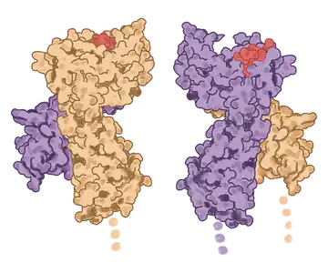
For novel drug development.

Design and manufacturing of high-quality and bioactive recombinant proteins.

We are a quality-focused biotechnology company dedicated to enhancing the latest therapeutic technologies, including cell therapies, gene editing, immunotechnology, and vaccines.

Innovative drug discovery requires innovative reagents.
Our customers develop novel therapies and so they need customized or first-to-market proteins and enzymes.

We carefully select our catalog products based on unmet needs in the market.

Our focus is customization and tailored solutions so that your innovation potential is limitless.


Products

Product Literature

Why work with KACTUS

We are an innovative biotechnology company dedicated to providing high-quality proteins & enzymes that meet novel customer demands.

Design Expertise

Our expertise in protein design and enzymatic studies, along with our SAMS™ platform, will help you create a protein configuration that will be functional for your application while also optimizing purity & yield.

Tailored Expression System

After generating 3D models of your protein/enzyme, we test the configurations in various expression systems to optimize protein folding, maximize bioactivity, and enhance stability.

Quality Bulk Supply

We’ll take your protein configuration from bench to large-scale production. Our quality control systems ensure batch-to-batch consistency and our facilities are prepared for consistent high-quantity production.

Reliable and bioactive commercial proteins are fundamental to successful and timely drug development.

SAMS

Structure Aided Design & Multiplex Screening

Researchers face common challenges when using recombinant proteins including:

  • Membrane protein antigen production
  • Recombinant antigen immunogenicity
  • Antibody affinity
  • Batch-to-batch consistency
  • Production time & production cos

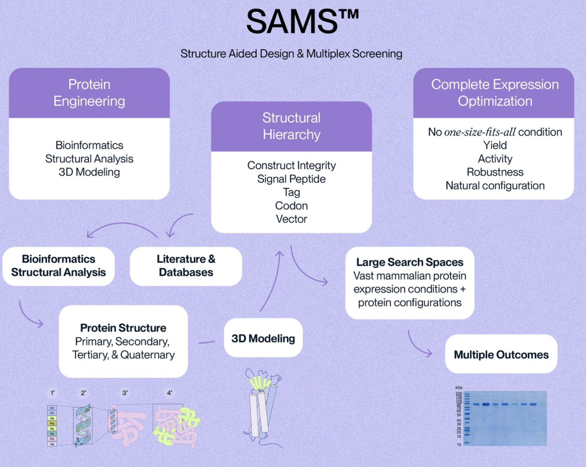
SAMS (Structure Aided Design and Multiplex Screening) is our high-throughput protein expression and purification screening system.

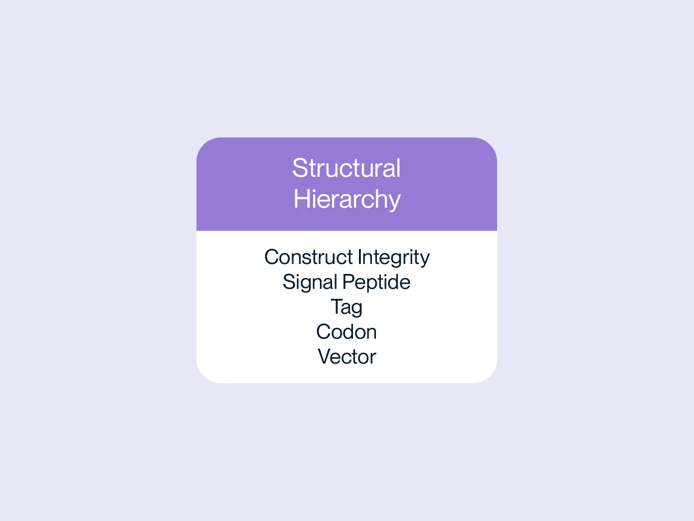
The structural hierarchy is the overarching list of variables that will affect protein structure and function.

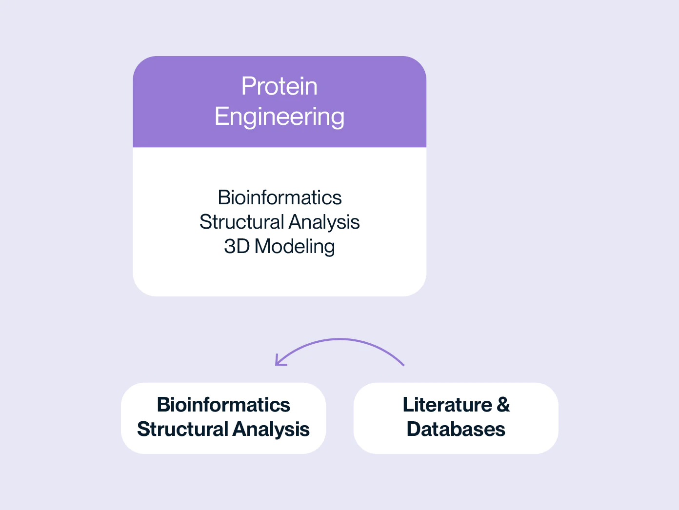
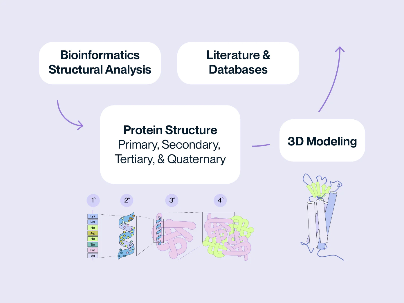
Unmet needs for commercial proteins are identified via literature and database searches. We then use bioinformatics and structural analysis to design a variety of protein sequences/structures based on variables from the structural hierarchy.

Integrity, signal peptide, codons, etc. are analyzed using structural informatics and 3D modeling to determine how they will influence all four levels of protein structure.

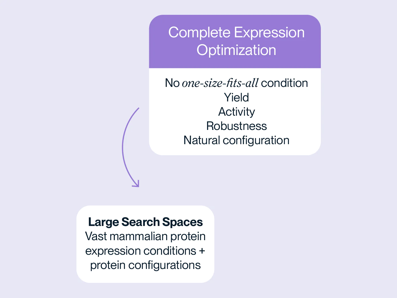
The optimal protein configuration is identified from large search spaces of protein configurations and expression conditions. We prioritize natural protein configuration, yield, and bioactivity.

Protein designs are tested for bioactivity in multiple cell types/settings. Because we ensure protein function in multiple settings, you can be confident that the protein you purchase will work for your application.

Advantages of SAMS Platform

Success with SAMS

Our products are used worldwide for diverse applications, including basic research, drug discovery, diagnostic study, antibody production, quality control testing, and more. Through the SAMS platform, we have already produced multiple challenging proteins, including the first ever full-length Claudin 18.2 protein.

Drug development is full of unknowns. Don’t let your reagents be one of them.

Quality Control & Assurance

At KACTUS, we focus on quality at every step of the process, from protein engineering to manufacturing, so you can be confident in the products you receive.

KACTUS recombinant proteins are designed and manufactured in-house, giving a high level of control over every step from protein engineering to product validation. We test each prodcut for bioactivity throughout the development process to ensure function in multiple in vitro settings. Each batch undergoes strict quality control testing including purity, activity, and endotoxin. Our high level of control results in superior batch to batch consistency. Moreover, the majority of our products are expressed using mammalian cell lines, allowing for post-translational modification, native protein conformation, and low endotoxin levels.

Vertical integration of protein engineering & manufacturing

Mammalian cell line expression systems

Comprehensive quality control testing

Quality Control

Quality control is a critical aspect of recombinant protein production that ensures the purity, potency, and consistency of the final product. Our quality control systems are designed to monitor and assess the quality of the production process and final product.


Process Validation

We monitor the entire production process to ensure expression conditions are optimal for that specific protein. This includes monitoring the growth of the host cells, expression of the target gene, and purification of the recombinant protein. The expression process is optimized to minimize the presence of contaminants that can affect the purity or activity of the final product.

Analytical Testing

PURITY The purity of each recombinant protein is ensured using HPLC and by quantifying the level of impurities, such as host cell proteins, endotoxin, and host cell DNA in the final product.

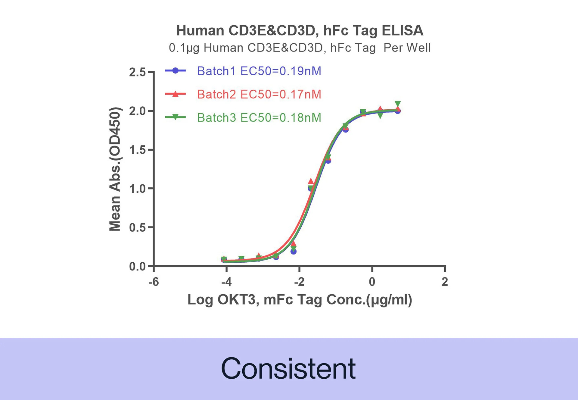
ACTIVITY The potency of our recombinant proteins is analyzed by measuring its in vitro activity, such as its ability to bind to its target molecule via ELISA or SPR assay.

CONSISTENCY Our products are tested for batch-to-batch consistency to ensure stable bioactivity across lots. Additionally, we assess stability of our products by measuring degradation over time and resistance to environmental stress factors, such as temperature and pH changes.

Batch to Batch Consistency

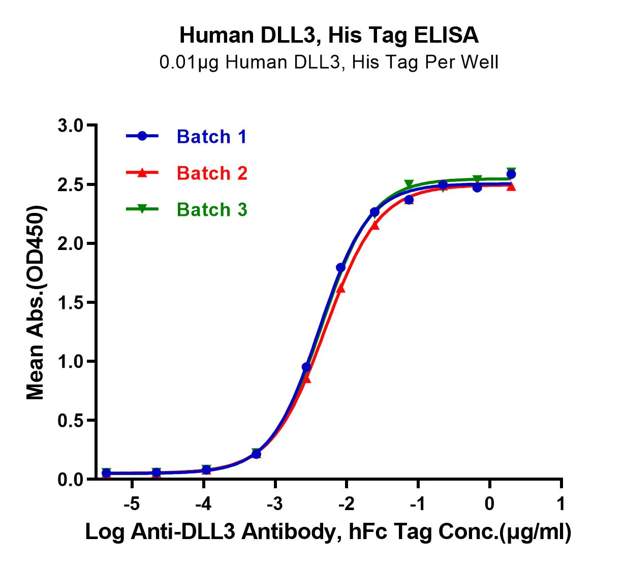
Fig 1. Imobilized Human DDL3, His Tag at 0.1µg/mL (100µl/well) on the plate. Dose response curve for Anti-DDL3 Antibody, hFc Tag with an EC50 of 5.0, 4.5 and 5.0 ng/mL respectively determined by ELISA

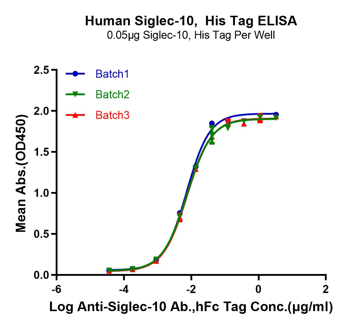
Fig 2. Immobilized Human Siglec-10, His Tag at 0.5µg/mL (100µl/well) Dose response curve for Anti-Siglec-10 Ab., hFc Tag with an EC50 OF 7.3, 7.7 and 7.4ng/mL, respectively asgetermined by ELISA

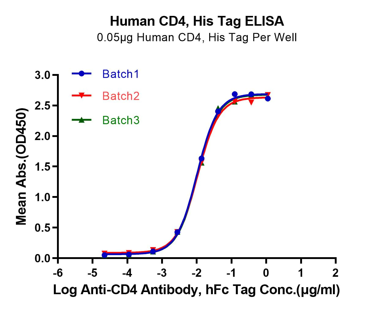
Fig 3. Immobilized Human CD4 at 0.5µg/mL (100µl/well) on the plate. Dose response curve for Anti-CD4 Antibody, hFc Tag with an EC50 OF 10.2, 10.6 and 10.6 ng/mL respectively as determined by ELISA

Quality Assurance

Our quality management system monitors the entire production process to ensure each batch is manufactured under the same optimal conditions for that protein. This includes monitoring the growth of the host cells, expression of the target gene, and purification of the recombinant protein. Moreover, the protein expression process is optimized during development to minimize the presence of contaminants that can affect the purity or activity of the final product.

  • R&D production headquarters of over 50,000 square feet
  • GMP-production facility of over 100,000 square feet
  • Scientific team of 200 researchers and production engineers
  • Structure Aided Design and Multiplex Screening (SAMS)
  • Optimizes protein configuration and expression conditions
  • Bioactive proteins in multiple cell lines.
  • High-purity and bioactive proteins
  • Extensive quality control testing, including activity testing
  • Optional GMP-grade material for commercial manufacturing

Custom Production

Scientific support for structure optimization specific to your application

Bulk material supply for research and process development

GMP-Grade material for clinical manufacturing

Equipment certification, batch records, etc. for regulatory filing

High-quality, custom design & manufacturing of recombinant proteins

Antigens

Enzymes

Transmembrane Proteins

Cytokines

KACTUS offers CRO services and large-scale contract manufacturing for protein engineering, expression, and production of custom recombinant proteins. Our recombinant protein production experience along with our proprietary SAMS™ platform will ensure your product has the optimal configuration and expression conditions for bioactivity and purity.

Structure Optimization

Optimized protein configuration to enhance specificity, sensitivity, and yield

Purity & Bioactivity

Analyzed for purity and bioactivity to ensure successful application.

Large Batch

Large-scale production batches with batch-to-batch consistency.

GMP-Grade Production

ISO certified and batch auditing records available for optional GMP-level production.

SAMS™ Platform

Our proprietary structure aided design and multiplex screening technology uses bioinformatics and 3D modeling combined with in vitro expression condition optimization to optimize protein configuration.

Quality Control

KACTUS performs quality control testing criteria including endotoxin, residual host cell protein, etc. Proteins are synthesized in an antibiotic-free and animal-free environment.

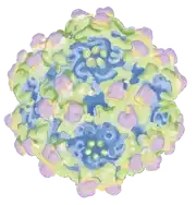
Transmembrane proteins are difficult to express in their natural 3D configuration. We use virus-like-particles (VLPs) derived from self-assembling viral capsid proteins to express transmembrane proteins in their natural conformation. VLPs are non-infectious and have high immunogenicity. Our line of VLP-displayed antigens can be used for antibody drug discovery of otherwise difficult targets.

Products

GMP-Grade & GMP-Ready

Template Preparation, Transcription, & Modification

T7 RNA Polymerase

Our GMP-grade T7 RNA polymerase is engineered to provide high mRNA yield and accurate transcription of mRNA from DNA templates. It is designed to minimze dsRNA contaminzation in transcripts and is free of animal-free of antiobiotics and animal-derived products. Our T7 polymerase has been successfully submitted to the FDA Drug Master Files (#037660).

Restriction Enzymes

Restriction enzymes are used in mRNA synthesis to cleave DNA templates at sequence-specific sites. The enzymes introduce specific and well-defined breaks in the DNA, which then allow for the transcription of RNA with defined lengths and sequences.

KACTUS offers various GMP-Grade and GMP-Ready restriction enzymes. Learn more about a specific enzyme below.

Capping Enzymes

Vaccinia capping enzyme is a type of RNA capping enzyme that adds a 7-methylguanosine cap (m7G or m7Gppp) to the 5′-end of an mRNA molecule. This creates the Cap0 structure which protects the mRNA from degradation and is important for efficient translation and export of the mRNA from the nucleus to the cytoplasm.

mRNA cap 2′-O-methyltransferase adds a methyl group to the first nucleotide at the 2′-O position of ribose, resulting in the creation of the Cap1 strcuture (m7GpppNm). This 2′-O-methylation plays a role in authorizing foreign RNA as “self RNA” and hence in the non-self discrimination of the innate immune response against the mRNA.

KACTUS provides GMP-Grade Vaccinia Capping Enzyme and mRNA Cap 2′-O-Methyltransferase designed for use in a one-step capping reaction system to generate the Cap1 structure. Our capping enzymes demonstrate greater than 95% capping efficiency.

E. Coli Poly(A) Polymerase Tailing Enzyme

Poly(A) polymerase plays a crucial role in mRNA polyadenylation. The enzyme catalyzes the addition of adenosine monophosphates (AMPs) to the 3′ end of the mRNA molecule, creating what is known as the poly(A) tail. The poly(A) tail protects RNA from degradation by nucleases, assists in the transport of RNA out of the nucleus, and stimulates translation of the RNA into protein.

Cyclization Enzymes

Cyclization enzymes are responsible for forming a unique 5′-5′ phosphodiester bond between the first and last nucleotides of the mRNA molecule, creating what is known as a circular mRNA. This circularization process is important for increasing the stability and translational efficiency of the mRNA molecule, as well as protecting it from degradation by cellular exonucleases.

RNase R digests linear RNA.

RNase R leaves circular RNA intact.

KACTUS offers GMP-Grade CRISPR Cas9 enzymes suitable for manufacturing of cell & gene therapeutics.

FDA Drug Master Files #036578

What makes our Cas9 different?

Using our proprietary protein engineering platform, SAMS™, we have successfully developed a highly active Cas9 protein. Our Cas9 has undergone nuclear localization signal (NLS) design. Additionally, through a series of steps such as protein structure analysis, expression system screening, and formulation optimization, our Cas9 has been optimized for purity and editing activity.

High editing efficacy in multiple cell types

Gene knockout efficiency analyzed in nucleofected 293T, Jurkat, and T cells using TIDE analysis. Results show greater than 95% editing efficacy across all three cell types, comparable to leading suppliers.

GMP-Grade

Our CRISPR Cas9 enzyme has been submitted to the FDA Drug Master Files #036578. Additionally, we provide necessary regulatory documentation including but not limited to COA, COO, MSDS, and compliance statements.

Recombinant Proteins

Virus-like Particle (VLP)-Displayed Antigens

Research and development for antibody drugs has increasingly fierce competition. The discovery of antibodies specific to difficult targets can help you maintain competitiveness in the market.

What are VLP-displayed antigens?

Nanoparticles (20-200nm) derived from outer capsid protein of virus

Formed by the automatic assembly of one or more viral capsid proteins

Surface display provides multiple copies of antigen per VLP to enhance immunogenicity

Why use VLP-displayed antigens?

Do not contain viral infectious genomes, making them safe to use in manufacturing operations

Efficiently activate the body’s humoral and cellular immune responses

Natural configuration of transmembrane proteins for discovery of differentiated antibodies

APPLICATIONS

ELISA

SPR Analysis

Antibody Discovery

Antibody Screening

Immunization

PD/PK

ADVANTAGES

Transmembrane proteins

High activity

High immunogenicity

Soluble expression of different proteins

Clear structure

Not pathogenic

Validation Data

Fig 1. Flurescent Human Claudin 18.2 VLP used to detect the positive rate of anti-Claudin 18.2 CAR 293T cells

Fig 2. Immobilized Human GPRC5D VLP at 5μg/ml (100μl/Well) on the plate. Dose response curve for Anti-GPRC5D Antibody, hFc Tag with the EC50 of 11.8ng/ml determined by ELISA.

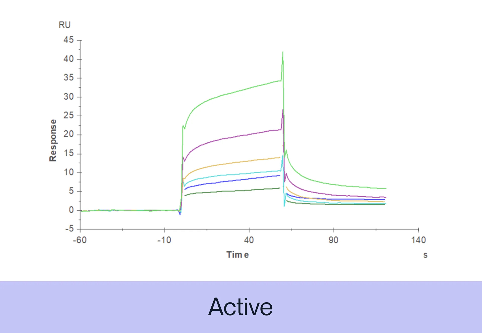
Fig 3. Biotinylated  Human Claudin 6 VLP captured on CM5 Chip via Streptavidin can bind Anti-Claudin6 Antibody with an affinity constant of 0.65 nM as determined in SPR assay (Biacore T200)

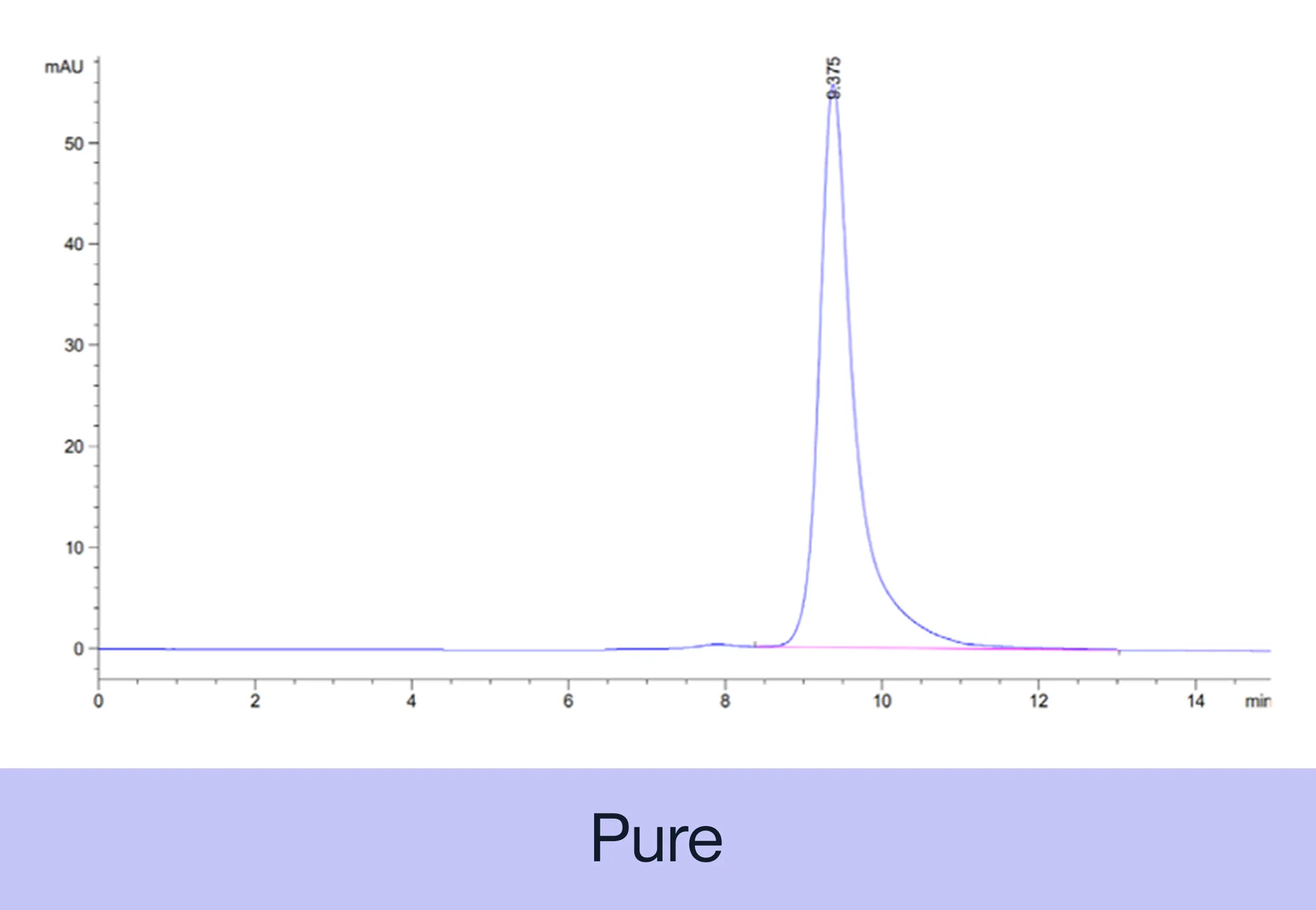
Fig 4. The Purity of Human SSTR2 VLP is greater than 95% as determined by SEC-HPLC

Custom Projects

Let us know if you’re looking for a VLP or modification not listed on our site. KACTUS can provide customization of VLP-displayed antigens.

The coronavirus disease 2019 (COVID-19) pandemic, due to the novel severe acute respiratory syndrome coronavirus 2 (SARS-CoV-2), has caused a worldwide health crisis. To support COVID-19 research, KACTUS has developed a series of SARS-CoV-2 and SARS antigens including domains of spike (S) proteins and host cell receptor ACE2.

We are also offering personalized service that customers demand.

Product Features

  • Low-endotoxin(<1EU per μg by the LAL method) and high-purity (>95%)
  • High avidity
  • High degree of batch-to-batch consistency
  • Featured protein expression screening system for high-yield product (up to several grams)
  • Offering biotinylated proteins, with highest bioactivity and sensitivity.

Validation Data

Fig. 1 SDS-PAGE of SARS-CoV-2 Spike S1 and bioactivity assay by ELISA.

Fig. 2 Human ACE2 captured on Protein A chip, can bind SARS-COV-2 Spike RBD, His Tag with an affinity constant of 13.8nM as determined in a SPR assay(Biacore T200).

Fig. 3 Human ACE2 captured on Protein A chip, can bind SARS-COV-2 Spike RBD(N501Y), His Tag with an affinity constant of 1.74nM as determined in a SPR assay(Biacore T200).

Immune blockade therapy is becoming a new weapon in oncology due to the development of immune checkpoint proteins targeting in cancer, especially after the success of antibody drugs targeting programmed cell death-1 (PD-1), programmed cell death ligand-1 (PD-L1) and cytotoxic T-lymphocyte antigen-4 (CTLA-4).
Although immune checkpoint blockade therapy often leads to a more durable response than chemo or targeted therapies in some cases, clinical data shows a limitation that in most cancers, the response rate is low (10-30%). Therefore, more associated studies focus on the mechanisms of non-responsiveness, as well as developing new targeting strategies using other immune checkpoint proteins.
Utilizing our featured SAMSTM platform, KACTUS has developed a series of immune checkpoint proteins with multiple pre-labels.

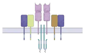
Highly active CD3 proteins including monomers, homodimers, heterodimers, and biotinylated

Product Features

High Protein Affinity

We verified the activity of our heterodimers, such as CD3E/G-His Tag and CD3E/D-His Tag, via ELISA and SPR.

.

Equal Expression of Subunits

We adopted a specialized design that ensures the two subunits of the heterodimers are expressed in equal proportions with greater than 95% purity.

Stability

We demonstrated via ELISA high consistency across batches and long-term stability of our products.

Applications

Antibody Discovery

Immunization

Antibody Screening

Functional characterization

Affinity Determination (ELISA, SPR, BLI)

Featured Products

Validation Data

Fig 1. Immobilized Human CD3E/CD3G, hFc Tag, at 1μg/ml (100μl/well). Dose-response curve for anti-CD3E/CD3G antibody (OKT3, mFc Tag). The EC50s are 20.7, 19.5, 20.0 and 20.7ng/ml respectively, as determined by ELISA

Fig 2. Human  CD3E/CD3D His Tag, captured on CM5 chip via anti-His antibody, binds OKT3 mFc Tag with an affiinty constant of 0.36nM as determined in SPR assay (Biacore T200).

Fig 3. Immobilized Human CD3E/CD3G, hFc Tag, at 1μg/ml (100μl/well). Dose-response curve for anti-CD3E/CD3G antibody (OKT3, mFc Tag) The EC50s are 20.7,21.0 and 20.0ng/mL respectively as determined by ELISA

Fig 4. Human CD3E/CD3G His Tag, captured on C5 Chip via anti-His antibody, can bind OKT M mFc tag with an affinity constant of 0.22nM as determined in SPR assay (Biacore T200)

Fig 5. The purity of Cynomolgus CD3E is greater than 95% as determined by SEC-HPLC

Fig 6. Cynomolgus CD3E/CD3D on Tris-Bis PAGE under reduced conditions. The purity is greater than 95%

Looking for something else?

Contact us to request a custom CD3 protein.

CELL THERAPY

Peptide-MHC I Complexes

Catalog & customized Peptide-MHC complexes

Custom Peptide-MHC in 6-8 Weeks

→ Fluorophore Labeling (PE, Cy5, FITC, etc.)

→ Biotinylation

→ Single Chain Peptide-MHC Expression

→ Mammalian & E. coli

→ Tetramerization

→ Custom Peptide Sequences

→ Allelles from Multiple Species (human, mouse, cyno)

About MHC-I Complexes

MHC Class I molecules play a crucial role in the immune system by presenting peptide antigens to cytotoxic T cells. These heterotrimers consist of a transmembrane heavy chain, a light chain known as β2-microglobulin (β2m), and an 8-10 peptide antigen. The heavy chain contains two peptide binding domains (α1 and α2), an immunoglobulin-like domain (α3), and a transmembrane region. The folding of the α1 and α2 domains forms a groove where peptide antigens bind to the MHC-I molecule. β2m stabilizes the peptide binding groove and MHC I presentation.

A single nucleated cell expresses 105 copies of each MHC I molecule, presenting a variety of peptides simultaneously on the cell surface to CTLs. Accumulating genomic mutations in cancers result in the production of tumor-specific antigens or neoantigens, which can be presented by MHC I molecules of tumor cells to CTLs.


Our MHC Engineering Platform

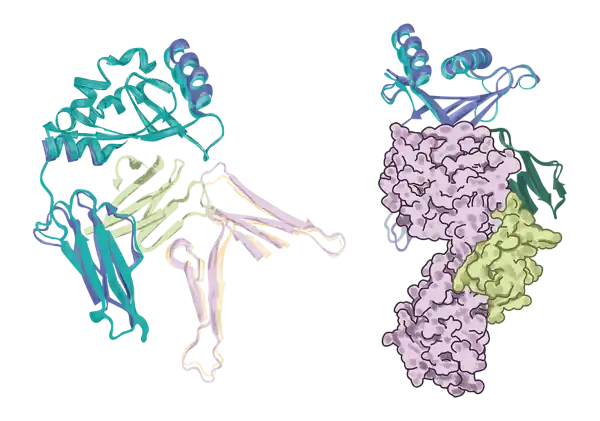
Heterotrimeric MHC I molecules could be engineered as a single chain protein that sequentially incorporates all three subunits on one construct. Here in KACTUS we offer engineered MHC I monomer, MHC I tetramer, MHC I multimer and peptide-ready MHC I using our protein engineering platform based on advanced bioinformatics, structural analysis, and 3D modeling. Our analysis of >200 TCR-pMHCI/II complex crystal structures shows that the linker connecting the C-terminal peptide does not interfere with the TCR and pMHC binding interface at all (Figure 1), which justifies our single chain design of peptide-MHC. The single chain design enhances the stability of MHC I molecules and reduces the chance of displacement by other peptides, allowing for more accurate and reliable detection of antigen-specific T cells. It also leads to extended cell-surface half-life and resistance to displacement by high-affinity competitor peptides.

Figure 1. Crystallographic alignment of TCR-MCH I/II complexes clears out single-chain MHC design concerns.

Our Products

Our MHC product collection features neoantigens from various tumor targets, including KRAS, PRAME, p53, MART-1, NY-ESO-1, GP100, etc. We currently have 105 catalog MHC products and we offer custom MHC services, provided with your MHC allele and neoantigen sequences. Our MHC protein engineering platform guarantees stable and active products with 100% peptide loading efficiency. We also offer custom MHC services with a lead time of 6 to 8 weeks.

Immobilized Anti-P53 R175H (HLA-A*02:01) Antibody, hFc Tag at 5μg/ml (100μl/well) on the plate. Dose response curve for Biotinylated Human P53 R175H (HLA-A*02:01) , His Tag with the EC50 of 1.6μg/ml determined by ELISA. See testing image for detail.

Human GP100 (HLA-A*02:01) Tetramer Protein (MHC-HM402T)

The purity of Human GP100 (HLA-A*02:01) Tetramer was greater than 95% as determined by SEC-HPLC

Human NY-ESO-1 (HLA-A*02:01) Tetramer Protein (MHC-HM405T)

Anti-NY-ESO-1 (HLA-A*02:01) Antibody, hFc Tag captured on CM5 Chip via Protein A can bind Human NY-ESO-1 (HLA-A*02:01) Tetramer, His Tag with an affinity constant of 0.09 nM as determined in SPR assay (Biacore T200).

Human KRAS G12V (HLA-A*03:01) Protein (MHC-HM418)

Immobilized Human NY-ESO-1 (HLA-A*02:01) Tetramer, His Tag at 5μg/ml (100μl/Well) on the plate. Dose response curve for Anti-NY-ESO-1 (HLA-A*02:01) Antibody, hFc Tag with the EC50 of 6.0ng/ml determined by ELISA (QC Test).

Monomers/Tetramers

We offer single-chain peptide-MHC monomers with the C-terminus His-Avi tag. The Avi tag is a 15-amino acid sequence, which has a high affinity for biotin and can be specifically biotinylated by BirA. Our biotinylated MHC monomers are achieved by the covalent bonding between the lysine residue within the Avi tag and biotin.

Streptavidin is a homotetrameric protein that has a very high affinity for the small molecule biotin. The streptavidin-biotin interaction is one of the strongest non-covalent interactions in nature, with a dissociation constant (Kd) of 10-14M. Each streptavidin subunit is about 13.3 kDa in size and contains a binding site for biotin. The high affinity of streptavidin for biotin and its small size makes it a powerful tool in making MHC tetramers. MHC tetramers are complexes of four MHC biotinylated monomers bound to streptavidin molecules. The enhanced avidity of MHC tetramers and TCR interactions can have a significant impact for detecting antigen-specific T cells.

In Vitro and In Vivo Fluorescently-Labeled MHC Tetramer

We offer MHC I tetramers that are assembled in vitro via the interaction between fluorescently labeled streptavidin and biotin on MHC I monomers.

Additionally, we provide the option for in vivo assembly of fluorescently labeled MHC I tetramers through Expi293 expression by fusing the fluorescent protein to the MHC I molecule. The folding nature of fluorescent protein generates fluorescently labeled MHC I tetramers in vivo by single step expression and purification, without extra steps of biotinylation, tetramerization or labeling. This cost-effective design produces uniform populations of fluorescently labeled MHC-I tetramers for downstream applications.

Fluorescent MHC tetramers can be used to identify T cells that recognize a particular peptide-MHC complex. By labeling T cells with MHC tetramers, the frequency and distribution of antigen-specific T cells can be determined and sorted by FACS in a cell population. MHC tetramers can also be used to monitor immune responses to vaccines, infections and diseases by measuring the frequency of antigen-specific T cells over time to track the efficacy of a treatment.

We validated that our in vitro and in vivo fluorescently labeled MHC I tetramers show equivalent bind to 293 cell line expressing LILRB2.

MHC Multimers: Virus-Like Particle (VLP)-Displayed Fluorescent MHC I

In combination with our Virus-Like Particles (VLP) technology platform, we are introducing multivalent fluorescent MHC I.  These MHC I-VLP complexes are about 750 Å in diameter, compared to the MHC I monomer size of 70 Å (see Figure 2). Each VLP contains approximately 250 copies of MHC I, resulting in boosted fluorescence quantum yield for enhanced detection of TCR binding. We currently offer FITC- and APC-equivalent options for MHC I-VLP fluorescence labeling.

Figure 2. Multimeric MHC I-VLP. The structure of MHC I-VLP is illustrated, revealing multiple copies of  MHC I molecules displayed on the surface of the VLP (A). The individual MHC I monomer has a diameter of approximately 70 Å (B), whereas the entire MHC I-VLP complex measures approximately 750 Å in diameter in an electron microscopy image (C).

Other MHC Protein Engineering

Chimeric MHC

Our protein engineering team has also designed a chimeric MHC by replacing the human α3 immunoglobulin-like domain with the mouse α3 domain (Figure 3). This modification enhances antigen specificity for antibody discovery.

Figure 3. Structural simulation of chimeric MHC. (Left: front view. Right: side view)

Peptide-Ready MHC Complex

While neoantigens offer a distinct advantage in their unique tumor-specific and normal tissue-absent feature, presenting ideal targets for effective and personalized tumor-specific immunotherapy, we developed peptide-ready MHC, which is only composed of the α heavy chain and β2-microglobulin (β2m) light chain for neoantigen peptide fishing. Peptide-ready MHC I and full-length tumor gene are co-expressed in the target cell line and then peptide loaded MHCs were enriched and purified, followed by mass spectrometry analysis of the peptide sequences. As a proof of concept, we confirmed “ALLPAVPSL” and “VLDFAPPGA” for WT1 (Wilms tumor protein 1) in our analysis from published papers [1, 2].

References

1. Kurosawa et al., Development of a T‐cell receptor mimic antibody targeting a novel Wilms tumor 1‐derived peptide and analysis of its specificity. Cancer Sci. 2020 Oct; 111(10): 3516–3526.

2. Doubrovina et al., Mapping of novel peptides of WT-1 and presenting HLA alleles that induce epitope-specific HLA-restricted T cells with cytotoxic activity against WT-1(+) leukemias. Blood. 2012 Aug 23;120(8):1633-46

Research-Grade and Preclinical-Grade Laminin 521

Matrix protein for maintenance of pluripotent stem cells.

Features

→ High purity (≥ 95%)

→ Low internal toxicity (< 10EU/mg)

→ Applicable to a wide range of cells, from human iPSCs and MSCs to most anchorage-dependent progenitor cell types

→ Compatible with a variety of feeder-free stem cell culture media on the market

→ Can maintain the homogeneous growth and karyotype stability of stem cells

KACTUS has successfully developed a high-quality Laminin 521 product that can be applied to scientific research (research-grade) and preclinical development (preclinical-grade). It can effectively support the normal growth and passage of PSCs, maintain good stemness, and ensure the homogeneous growth of cells with genetic stability. Laminin 521 can be applied to the cultivation of stem cells and other primary cells and the development of related stem cell products.

Recombinant Laminin for Stem Cell Culture

Due to their unique ability for self-renewal and directional differentiation, stem cells can be applied in many fields such as embryonic development research, drug screening modeling, and cell therapy. At the same time, stem cell culture methods continue to be optimized. Laminin 521 is an important matrix adhesion protein, which can recreate a suitable growth environment for primary cells such as stem cells, and is currently one of the most commonly used trophoblast-free culture substrates.

Laminin 521 can maintain the pluripotency of stem cells, promote cell proliferation, support the clonal expansion of pluripotent human stem cells and the growth of skin keratinocytes. It is also involved in the regulation of important physiological processes such as cell adhesion, migration, differentiation, phenotype maintenance, and matrix-mediated signal transduction [3]. Compared with Laminin 511, Laminin 521 can support the monolayer maintenance and expansion of stem cells in the absence of apoptosis inhibitors (such as ROCK inhibitors). It can be an excellent alternative to Laminin 511 and to some extent, is more suitable for human pluripotency stem cell culture than Laminin 511.

Figure 1. The main components of globular basement membrane (GBM) [2].


Product Validation

Figure 2. Laminin 521 can effectively maintain the growth of human iPSCs.

Figure 3. Normal expression of stem cell markers OCT3/4, SOX2, SSEA4, TRA-1-60, etc.

Applications

Stem Cell Expansion Culture

Laminin 521 can support the low-density growth and self-renewal of stem cells in a chemically defined, animal-free environment. It is suitable for the culture of hSC, iSC and cloned stem cells, such as hPSC [4], MSC, hematopoietic stem cells, etc.

Directed Differentiation of Stem Cells

Laminin 521 promotes the differentiation of hPSCs into different cell types such as hepatocytes, cardiomyocytes, retinal pigment epithelial (RPE) cells [5] and endothelial cells [6]. hESCs grown on Laminin 521 can exhibit molecular phenotypic characteristics of hepatocytes and undergo cell self-organization [7].

Organoid Culture

Indeterminate media replacing Matrigel is a major target for organoid culture, and full-length laminin (rather than laminin-derived peptides) has been shown to be a key component for the correct formation of organoids in gel models. Lucendo-Villarin and collaborators developed an economical automated platform for the generation of human hepatic spheroids from pluripotent stem cell-derived hepatic progenitors, endothelial cells, and hepatic stellate cells with the help of laminin 521, which can be used in disease modeling and drug screening research [8].

Growth Maintenance of Differentiated Primary Cells

Laminin 521 can support the growth and proliferation of differentiated primary cells, such as cardiomyocytes, retinal pigment epithelial (RPE) cells, nerve cells, endothelial cells, islet cells, etc. For example, the basement membrane damage of the isolated islets will cause islet function decline and cell death, but when cultured on a matrix containing Laminin 521, it can significantly promote the survival and function maintenance of islet cells [9], and has now been successfully applied in the original Generation islet cell culture system. Likewise, cardiac progenitor cells differentiated in vitro can proliferate on Laminin 521-containing matrices and transform into mature cardiomyocytes.

Drug Research

Laminin 521 is a new anti-GBM disease pulmonary hemorrhage autoantibody target. Studies have shown that in anti-glomerular basement membrane disease, circulating antibodies can recognize Laminin 521 in addition to type IV collagen, indicating that Laminin 521 is Another major autoantigen against GBM [10], autoantibodies against Laminin 521 may promote lung injury and lead to pulmonary hemorrhage by increasing the total amount of IgG bound to the alveolar basement membrane, which provides theoretical clues for the development of related drugs.

About Laminin 521

Laminin 521 (LN521, LAMB2, LN-11) is a heterotrimeric glycoprotein composed of α5, β2 and γ1 chains, expressed in stem cells and most basement membranes (especially in the nerve sheath, muscle nerve junction and glomerular basement membrane GBM). It is a key matrix protein that supports the growth of stem cells and maintains the stability and performance of the basement membrane.

Laminin 521 can form a huge net-like material environment together with nestin, collagen, fibronectin, glycosaminoglycan, and other extracellular matrix components. It is a key supportor of various tissues such as muscle fiber, epithelium, nerve, capillaries, fat, etc. In addition, the LG1-5 region of Laminin 521 can bind to cell membrane surface integrin α6β1 or α3β1, etc., start the downstream PI3K-Akt signaling pathway, and promote cell proliferation.

Figure 4. Laminin 521 structure [1].

References

[1] Pulido D, Briggs DC, Hua J, Hohenester E. Crystallographic analysis of the laminin β2 short arm reveals how the LF domain is inserted into a regular array of LE domains. Matrix Biol. 2017 Jan;57-58:204- 212.

[2] Naylor RW, Morais MRPT, Lennon R. Complexities of the glomerular basement membrane. Nat Rev Nephrol. 2021 Feb;17(2):112-127.

[3] Yap L, Tay HG, Nguyen MTX, Tjin MS, Tryggvason K. Laminins in Cellular Differentiation. Trends Cell Biol. 2019 Dec;29(12):987-1000.

[4] Dziedzicka D, Markouli C, Barbé L, Spits C, Sermon K, Geens M. A High Proliferation Rate is Critical for Reproducible and Standardized Embryoid Body Formation from Laminin-521-Based Human Pluripotent Stem Cell Cultures. Stem Cell Rev Rep . 2016 Dec;12(6):721-730.

[5] Lynch VJ, Nnamani MC, Kapusta A, Brayer K, Plaza SL, Mazur EC, Emera D, Sheikh SZ, Grützner F, Bauersachs S, Graf A, Young SL, Lieb JD, DeMayo FJ, Feschotte C, Wagner GP . Ancient transposable elements transformed the uterine regulatory landscape and transcriptome during the evolution of mammalian pregnancy. Cell Rep. 2015 Feb 3;10(4):551-61.

[6] Nguyen MTX, Okina E, Chai X, Tan KH, Hovatta O, Ghosh S, Tryggvason K. Differentiation of Human Embryonic Stem Cells to Endothelial Progenitor Cells on Laminins in Defined and Xeno-free Systems. Stem Cell Reports. 2016 Oct 11;7(4):802-816.

[7] Cameron K, Tan R, Schmidt-Heck W, Campos G, Lyall MJ, Wang Y, Lucendo-Villarin B, Szkolnicka D, Bates N, Kimber SJ, Hengstler JG, Godoy P, Forbes SJ, Hay DC. Laminins Drive the Differentiation and Self-Organization of hESC-Derived Hepatocytes. Stem Cell Reports. 2015 Dec 8;5(6):1250-1262.

[8] Meseguer-Ripolles J, Kasarinaite A, Lucendo-Villarin B, Hay DC. Protocol for automated production of human stem cell derived liver spheres. STAR Protoc. 2021 Apr 30;2(2):100502.

[9] Brandhorst D, Brandhorst H, Lee Layland S, Acreman S, Schenke-Layland K, Johnson PRV. Basement membrane proteins improve human islet survival in hypoxia: Implications for islet inflammation. Acta Biomater. 2022 Jan 1;137:92- 102.

[10] Shen CR, Jia XY, Luo W, Olaru F, Cui Z, Zhao MH, Borza DB. Laminin-521 is a Novel Target of Autoantibodies Associated with Lung Hemorrhage in Anti-GBM Disease. J Am Soc Nephrol. 2021 Aug ;32(8):1887-1897.

Mammalian-derived HLA-G and their receptors, LILRB/LILRA in various species and tags for first-in-class drug discovery.

HLA-G, like other immune checkpoints, mediates its function by binding to receptors on immune cells.
The known receptors are from the leukocyte Ig-like receptor (LILR) family and include LILRAs (activating) and LILRBs (suppressing).
When HLA-G binds to LILRBs, tumor cells can escape the surveillance of the immune system.

Owing to their role in activation/supression of immune cells, LILR family proteins have shown broad potential for anti-tumor effects.
LILRBs have become hot targets for drug development, especially for discovery of first-in-class drugs.
The HLA-G and ILT-2/4 signaling pathway is a novel target for monoclonal antibody therapy or cell-based immunotherapy.

Featured Products

Validation Data

We invest considerable time and effort performing quality and activity assays during product development to ensure our proteins are both pure and bioactive.

Fig 1. Human LILRB2, hFc Tag captured onCM5 Chip via Protein A can bind Human HLA-G Tetramer with an affinity constant of 4.62 n/M as determined in SPR assay (Biacore T200)

Fig 2. Serial dilutations of Anti-LILRB2 Antibody were added into Human LILRB2, His Tag: Biotinylated HLA-G Complex Tetramer, His Tag binding reactioins. The half maximal inhibitiory concentration (IC50) is 75.3ng/ml.

Fig 3. The purity of Biotinylated Human APOE3 is greater than 95% as determined by SEC-HPLC

Fig 4. Cynomolgus LILRB2, His Tag immobilized on CM5 Chip can bind Cynomolgus HLA-G Complex Tetramer, His Tag with an affinity constant of 852 nM as determined in SPR assay (Biacore T200)

Custom Products

We offer customization of any of our HLA-G/LILR products or we can work with you to set up an expression system for your novel protein.

GMP-Grade & GMP-Ready CRISPR Nucleases.

What makes our Cas9 different?

Using our proprietary protein engineering platform, SAMS™, we have successfully developed a highly active Cas9 protein. Our Cas9 has undergone nuclear localization signal (NLS) design. Additionally, through a series of steps such as protein structure analysis, expression system screening, and formulation optimization, our Cas9 has been optimized for purity and editing activity.

High editing efficacy in multiple cell types

Figure 1. Gene knockout efficiency of our CRISPR Cas9 analyzed in nucleofected 293T, Jurkat, and T cells using TIDE analysis. Results show greater than 95% editing efficacy across all three cell types, comparable to leading suppliers.

High In Vitro Cleavage Activity for AsCas12a

KACTUS also offers a highly active CRISPR-Cas12a enzyme. Cas12a is known for its ability to recognize and cut DNA at a specific site different from the prototypical Cas9. Additionally, Cas12a has been shown to have a lower off-target activity than Cas9, making it a promising tool for precision genome editing.

Figure 2. In vitro cleavage assay using AsCas12a. More than 85% of substrates can be cleaved by our Cas12a Enzyme.

Gene Therapy Reagents

DNA Amplification and Modification Enzymes

Robust isothermal DNA amplification.

Phi29 DNA Polymerase

Phi29 DNA polymerase is a highly processive enzyme that can efficiently amplify DNA sequences in vitro. It is often used as an alternative to the more commonly used polymerase chain reaction (PCR) method for DNA amplification. Phi29 DNA polymerase amplification has several advantages over PCR, including higher processivity, reduced reaction time, and higher fidelity. It is particularly useful for amplifying DNA from samples with limited starting material, such as ancient DNA or single cells. It is a highly robust enzyme and is suitable for rolling circle amplification (RCA), multiple-displacement amplification (MDA), and whole genome amplification.

Features

Isothermal amplification

High sensitivity

High processivity

High fidelity

Amplification with low starting quantities

Applications

Whole genome amplification

Single-cell genome sequencing

Rolling circle amplification

DNA manufacturing

Replication requiring isothermal or moderate temperature amplification

TelN Protelomerase

TelN Protelomerase cleaves dsRNA at its recognition site and leaves covalently closed ends. It can be used for DNA vaccine development as well as for non-viral therapy vectors. TelN Protelomerase is isolated from phage N15.

Exonuclease III

Exonuclease III is a highly processive enzyme that hydrolyzes the phosphodiester bonds in DNA in the 3′ to 5′ direction, resulting in stepwise removal of nucleotides. Exonuclease III catalyzes the removal of nucleotides from linear or nicked double stranded DNA. Degradation of Exonuclease III could be initiated from a 3’ blunt end, 3’ recessed end, 3’ overhangs with less than 4 bases and nicked DNA.

AAV9 Titration ELISA Kit

High specificity, sensitivity, and reproducibility

Features

→ Specificity to AAV9

→ Sensitivity as low as 1.0E7 capsids/mL

→ Broad linear range: 1.0E7 – 6.E8 capsids/mL

→ Strong reproducibility

→ Choice of 2 hour rapid procedure for fast turnaround or 6 hour standard procedure for sensitivity

Applications

→ Intact AAV wild type virions

→ AAV recombinant virions

→ Assembled AAV virions

→ Intact empty AAV capsids

How it works

Empty AAV9 capsids lacking the transgene of interest cause an unwanted immune response without providing the therapeutic benefit of the transgene. Alongside DNA and infectious titers, determination of the capsid titer is crucial for validating the efficacy and safety of your AAV gene therapy. Our titration kit detects full and empty AAV9 capsids.

This kit uses a double-antibody sandwich enzyme-linked immunosorbent assay (ELISA) to determine the quantity of AAV9 capsids in the test sample. The AAV monoclonal antibody is pre-coated on a 96-well reaction plate. AAV9 standard or sample is added to the pre-coated plate, which will specifically bind to the reaction plate. The biotinylated detection antibody is then added to bind the AAV9 capsids. Next, the HRP conjugate is added to form an antibody-antigen complex. HRP will then oxidize TMB substrate to produce a color change. The intensity of the color change is proportional to the AAV9 capsid titer in the sample. After reading the absorbance, the AAV9 capsid titer is calculated based on the absorbance values and the known AAV9 titers of the standard.

Standard Curve

Our AAV9 ELISA kit uses an 8-point standard curve of recombinant AAV9 capsids. Monoclonal AAV9 antibodies precoated on the plate bind the AAV9 capsids. Additional biotinylated AAV9 monoclonal antibodies bind as the second label. The antibodies are serotype-specific and bind only intact AAV9 capsids, not partial capsids. The kit offers two versions of the protocol, a standard procedure (6 hours) which provides greater sensitivity, and a rapid procedure (2 hours) for fast turnaround.

GMP-Grade & GMP-Ready CRISPR Nucleases.

What makes our Cas9 different?

Using our proprietary protein engineering platform, SAMS™, we have successfully developed a highly active Cas9 protein. Our Cas9 has undergone nuclear localization signal (NLS) design. Additionally, through a series of steps such as protein structure analysis, expression system screening, and formulation optimization, our Cas9 has been optimized for purity and editing activity.

High editing efficacy in multiple cell types

Figure 1. Gene knockout efficiency of our CRISPR Cas9 analyzed in nucleofected 293T, Jurkat, and T cells using TIDE analysis. Results show greater than 95% editing efficacy across all three cell types, comparable to leading suppliers.

High In Vitro Cleavage Activity for AsCas12a

KACTUS also offers a highly active CRISPR-Cas12a enzyme. Cas12a is known for its ability to recognize and cut DNA at a specific site different from the prototypical Cas9. Additionally, Cas12a has been shown to have a lower off-target activity than Cas9, making it a promising tool for precision genome editing.

Figure 2. In vitro cleavage assay using AsCas12a. More than 85% of substrates can be cleaved by our Cas12a Enzyme.

High In Vitro Cleavage Activity for AsCas12a

KACTUS also offers a highly active CRISPR-Cas12a enzyme. Cas12a is known for its ability to recognize and cut DNA at a specific site different from the prototypical Cas9. Additionally, Cas12a has been shown to have a lower off-target activity than Cas9, making it a promising tool for precision genome editing.

Figure 2. In vitro cleavage assay using AsCas12a. More than 85% of substrates can be cleaved by our Cas12a Enzyme.

GMP-Grade & GMP-Ready

Enzymes for mRNA Production

Template Preparation, Transcription, & Modification

Full Portfolio mRNA Enzymes

KACTUS offers a full selection of mRNA production enzymes. Many of our enzymes are already manufactured according to cGMP guidelines and have been submitted to the FDA Drug Master Files (DMF). Our remaining enzymes are GMP-Ready, meaning that our team will move them to cGMP standards when it suits your needs.

GMP: Currently manufactured according to cGMP guidelines

DMF: Submitted to the FDA Drug Master Files (DMF) for simplified regulatory applications

All of our other mRNA enzymes can be manufactured in our 100,000 sq ft GMP-Grade facility as necessary.

If you don’t see the enzyme you’re looking for here, contact us about our custom enzyme production services

T7 RNA Polymerase

Our GMP-grade T7 RNA polymerase is engineered to provide high mRNA yield and accurate transcription of mRNA from DNA templates. It is designed to minimze dsRNA contaminzation in transcripts and is free of animal-free of antiobiotics and animal-derived products. Our T7 polymerase has been successfully submitted to the FDA Drug Master Files (#037660).

→ High yield

→ Low dsRNA contamination

→ Stability testing & batch-to-batch consistency

Restriction Enzymes

Restriction enzymes are used in mRNA synthesis to cleave DNA templates at sequence-specific sites. The enzymes introduce specific and well-defined breaks in the DNA, which then allow for the transcription of RNA with defined lengths and sequences.

KACTUS offers various GMP-Grade and GMP-Ready restriction enzymes. Learn more about a specific enzyme below:

Capping Enzymes

Vaccinia capping enzyme is a type of RNA capping enzyme that adds a 7-methylguanosine cap (m7G or m7Gppp) to the 5′-end of an mRNA molecule. This creates the Cap0 structure which protects the mRNA from degradation and is important for efficient translation and export of the mRNA from the nucleus to the cytoplasm.

mRNA cap 2′-O-methyltransferase adds a methyl group to the first nucleotide at the 2′-O position of ribose, resulting in the creation of the Cap1 strcuture (m7GpppNm). This 2′-O-methylation plays a role in authorizing foreign RNA as “self RNA” and hence in the non-self discrimination of the innate immune response against the mRNA.

KACTUS provides GMP-Grade Vaccinia Capping Enzyme and mRNA Cap 2′-O-Methyltransferase designed for use in a one-step capping reaction system to generate the Cap1 structure. Our capping enzymes demonstrate greater than 95% capping efficiency.

E. Coli Poly(A) Polymerase Tailing Enzyme

Poly(A) polymerase plays a crucial role in mRNA polyadenylation. The enzyme catalyzes the addition of adenosine monophosphates (AMPs) to the 3′ end of the mRNA molecule, creating what is known as the poly(A) tail. The poly(A) tail protects RNA from degradation by nucleases, assists in the transport of RNA out of the nucleus, and stimulates translation of the RNA into protein.

Cyclization Enzymes

Cyclization enzymes are responsible for forming a unique 5′-5′ phosphodiester bond between the first and last nucleotides of the mRNA molecule, creating what is known as a circular mRNA. This circularization process is important for increasing the stability and translational efficiency of the mRNA molecule, as well as protecting it from degradation by cellular exonucleases.

RNase R digests linear RNA

RNase R leaves circular RNA intact.


Base editing technology leads a new chapter in drug development

View All Products Since its debut in 2016, base editing technology has opened up new perspectives and methods for drug Read More...

MSLN Targeted Drugs Entering the Fast Lane

View All Products Since December 2023, there have been several positive updates about drugs targeting MSLN. On December 19th, RemeGen Read More...

Exploring the Role of MICA in Tumor Immunity and Targeted Therapies

Contact UsView All Products MICA, also known as MHC Class I Polypeptide-Related Sequence A, is a type I MHC molecule Read More...

New advances in CRISPR gene therapy drugs

Contact UsView All Products On April 3, 2023, Vertex Pharmaceuticals and CRISPR Therapeutics announced the completion of the rolling biologics Read More...

Potential Immunotherapy Target TM4SF1

Contact Us View All Products TM4SF1 (Transmembrane 4L six family member 1) is a member of the TM4SF subfamily in Read More...

CDH17: A Safe CAR-T Target for Treatment of Solid Tumors

Contact Us View All Products Chimeric antigen receptor T cell (CAR-T) therapy has been successful in the field of hematological Read More...

GDF15-GFRAL breaks into the new drug track

Contact UsView All Products A variety of metabolic diseases that plague today's society, such as obesity, diabetes, cancer, etc., have Read More...

The culprit of cancer metastasis – uPAR-uPA

Contact UsView All Products Tumor invasion and metastasis are key risk factors for tumor recurrence and patient death. Tumor invasion Read More...