IBA Lifesciences GmbH offers high quality products for life science research in academia and industry based on their proprietary Strep-tag® technology.
The product portfolio offers solutions for the entire production chain of recombinant proteins, from cloning, transfection, protein expression and purification, to detection, immobilization, and assays as well as innovative cell isolation and expansion tools.
This combination of products and services allows IBA’s clients to choose either IBA's proprietary products or alternatively custom services, which are based on the same technologies, to drive their research.


Products

Applications

High performance research tools for

cell & protein isolation

For more than 20 years we have provided life science research products for academia and industry. We develop high performance research tools for protein and cell isolation based on our proprietary Strep-tag® technology. With a worldwide network of distributors, our products are available in more than 40 countries across 5 continents. Learn more about the unique Strep-tag® technology

Popular Products

Brands

Products

Description Size Catalog No.
Anhydrotetracycline 25mg 2-0401-002
Competent E. coli TOP10 20 rxns 5-1600-020
MEXi-293E cells 1.5ml 2-6001-001
MEXi-CM 1000ml 2-6010-010
MEXi-TM 1000ml 2-6011-010
pASG-IBA102 vector 20 µl 5-4102-001
pASG-IBA103 vector 20 µl 5-4103-001
pASG-IBA104 vector 20 µl 5-4104-001
pASG-IBA105 vector 20 µl 5-4105-001
pASG-IBA123 vector 20 µl 5-4123-001
pASG-IBA143 vector 20 µl 5-4143-001
pASG-IBA144 vector 20 µl 5-4144-001
pASG-IBA145 vector 20 µl 5-4145-001
pASG-IBA162 vector 20 µl 5-4162-001
pASG-IBA164 vector 20 µl 5-4164-001
pASG-IBA167 vector 20 µl 5-4167-001
pASG-IBA168 vector 20 µl 5-4168-001
pASG-IBA2 vector 20 µl 5-4002-001
pASG-IBA23 vector 20 µl 5-4023-001
pASG-IBA3 vector 20 µl 5-4003-001
pASG-IBA4 vector 20 µl 5-4004-001
pASG-IBA43 vector 20 µl 5-4043-001
pASG-IBA44 vector 20 µl 5-4044-001
pASG-IBA45 vector 20 µl 5-4045-001
pASG-IBA5 vector 20 µl 5-4005-001
pASG-IBA62 vector 20 µl 5-4062-001
pASG-IBA64 vector 20 µl 5-4064-001
pASG-IBAwt1 vector 20 µl 5-4000-001
pASG-IBAwt2 vector 20 µl 5-4001-001
pASK-IBA2C vector 20 µl 2-1321-000
pASK-IBA3C vector 20 µl 2-1322-000
pASK-IBA4C vector 20 µl 2-1323-000
pASK-IBA5C vector 20 µl 2-1324-000
pASK-IBA6C vector 20 µl 2-1325-000
pASK-IBA7C vector 20 µl 2-1326-000
pCSG-IBA102 vector 20 µl 5-5102-001
pCSG-IBA103 vector 20 µl 5-5103-001
pCSG-IBA104 vector 20 µl 5-5104-001
pCSG-IBA105 vector 20 µl 5-5105-001
pCSG-IBA123 vector 20 µl 5-5123-001
pCSG-IBA142 vector 20 µl 5-5142-001
pCSG-IBA143 vector 20 µl 5-5143-001
pCSG-IBA144 vector 20 µl 5-5144-001
pCSG-IBA145 vector 20 µl 5-5145-001
pCSG-IBA162 vector 20 µl 5-5162-001
pCSG-IBA164 vector 20 µl 5-5164-001
pCSG-IBA167 vector 20 µl 5-5167-001
pCSG-IBA168 vector 20 µl 5-5168-001
pCSG-IBA23 vector 20 µl 5-5023-001
pCSG-IBA3 vector 20 µl 5-5003-001
pCSG-IBA43 vector 20 µl 5-5043-001
pCSG-IBA45 vector 20 µl 5-5045-001
pCSG-IBA5 vector 20 µl 5-5005-001
pCSG-IBA62 vector 20 µl 5-5062-001
pCSG-IBA64 vector 20 µl 5-5064-001
pCSG-IBAwt1 vector 20 µl 5-5000-001
pCSG-IBAwt2 vector 20 µl 5-5001-001
pDSG-IBA102 vector 20 µl 5-5219-001
pDSG-IBA103 vector 20 µl 5-5220-001
pDSG-IBA104 vector 20 µl 5-5221-001
pDSG-IBA105 vector 20 µl 5-5222-001
pDSG-IBA43 vector 20 µl 5-5211-001
pDSG-IBA45 vector 20 µl 5-5214-001
pDSG-IBAwt1 vector 20 µl 5-5200-001
pDSG-IBAwt2 vector 20 µl 5-5201-001
pENTRY-IBA51 vector 20 µl 5-4091-001
pESG-IBA102 vector 20 µl 5-4502-001
pESG-IBA103 vector 20 µl 5-4503-001
pESG-IBA104 vector 20 µl 5-4504-001
pESG-IBA105 vector 20 µl 5-4505-001
pESG-IBA123 vector 20 µl 5-4523-001
pESG-IBA142 vector 20 µl 5-4542-001
pESG-IBA143 vector 20 µl 5-4543-001
pESG-IBA144 vector 20 µl 5-4544-001
pESG-IBA145 vector 20 µl 5-4545-001
pESG-IBA162 vector 20 µl 5-4562-001
pESG-IBA164 vector 20 µl 5-4564-001
pESG-IBA167 vector 20 µl 5-4567-001
pESG-IBA168 vector 20 µl 5-4568-001
pESG-IBA23 vector 20 µl 5-4423-001
pESG-IBA3 vector 20 µl 5-4403-001
pESG-IBA43 vector 20 µl 5-4443-001
pESG-IBA45 vector 20 µl 5-4445-001
pESG-IBA5 vector 20 µl 5-4405-001
pESG-IBA62 vector 20 µl 5-4462-001
pESG-IBA64 vector 20 µl 5-4464-001
pESG-IBAwt1 vector 20 µl 5-4400-001
pESG-IBAwt2 vector 20 µl 5-4401-001
pLSG-IBA102 vector 20 µl 5-4902-001
pLSG-IBA103 vector 20 µl 5-4903-001
pLSG-IBA104 vector 20 µl 5-4904-001
pLSG-IBA105 vector 20 µl 5-4905-001
pLSG-IBA123 vector 20 µl 5-4923-001
pLSG-IBA142 vector 20 µl 5-4942-001
pLSG-IBA143 vector 20 µl 5-4943-001
pLSG-IBA144 vector 20 µl 5-4944-001
pLSG-IBA145 vector 20 µl 5-4945-001
pLSG-IBA162 vector 20 µl 5-4962-001
pLSG-IBA164 vector 20 µl 5-4964-001
pLSG-IBA167 vector 20 µl 5-4967-001
pLSG-IBA168 vector 20 µl 5-4968-001
pLSG-IBA23 vector 20 µl 5-4823-001
pLSG-IBA3 vector 20 µl 5-4803-001
pLSG-IBA43 vector 20 µl 5-4843-001
pLSG-IBA45 vector 20 µl 5-4845-001
pLSG-IBA5 vector 20 µl 5-4805-001
pLSG-IBA62 vector 20 µl 5-4862-001
pLSG-IBA64 vector 20 µl 5-4864-001
pLSG-IBAwt1 vector 20 µl 5-4800-001
pLSG-IBAwt2 vector 20 µl 5-4801-001
pYSG-IBA103 vector 20 µl 5-4703-001
pYSG-IBA105 vector 20 µl 5-4705-001
pYSG-IBA123 vector 20 µl 5-4723-001
pYSG-IBA143 vector 20 µl 5-4743-001
pYSG-IBA145 vector 20 µl 5-4745-001
pYSG-IBA162 vector 20 µl 5-4762-001
pYSG-IBA164 vector 20 µl 5-4764-001
pYSG-IBA167 vector 20 µl 5-4767-001
pYSG-IBA168 vector 20 µl 5-4768-001
pYSG-IBA23 vector 20 µl 5-4623-001
pYSG-IBA3 vector 20 µl 5-4603-001
pYSG-IBA43 vector 20 µl 5-4643-001
pYSG-IBA45 vector 20 µl 5-4645-001
pYSG-IBA5 vector 20 µl 5-4605-001
pYSG-IBA62 vector 20 µl 5-4662-001
pYSG-IBA64 vector 20 µl 5-4664-001
pYSG-IBAwt1 vector 20 µl 5-4600-001
Description Size Catalog No.
Avidin 50mg 2-0204-050
Biotin stock solution 1ml 6-6325-001
10ml 6-6325-010
Buffer CI (10x) 85ml 6-6320-085
CD3 Fab-Strep, human 50 µg 6-8001-150
CD4 Fab-Strep, human 50 µg 6-8002-150
CD8 Fab-Strep, human 50 µg 6-8003-150
CD81 Fab-Strep, human 50 µg 6-8015-150
GFP-Strep-tag®II control protein 500 µl 2-1006-005
GFP-Twin-Strep-tag® control protein 500 µl 2-1007-105
MHC I-Strep H-2 Kb; Ovalbumin (SIINFEKL) 10 µg 6-7015-001
MHC I-Strep HLA-A*0101; CMV pp50 (VTEHDTLLY) 10 µg 6-7024-001
MHC I-Strep HLA-A*0201; CMV pp65 (NLVPMVATV) 10 µg 6-7001-001
MHC I-Strep HLA-A*0201; gp100 (YLEPGPVTA) 10 µg 6-7025-001
MHC I-Strep HLA-A*0201; MART-1 (ELAGIGILTV) 10 µg 6-7007-001
MHC I-Strep HLA-A*0201; NY-ESO-1 (SLLMWITQV) 10 µg 6-7013-001
MHC I-Strep HLA-A*0201; SARS-CoV-2 (YLQPRTFLL) 10 µg 6-7130-001
MHC I-Strep HLA-A*0201; WT1 (VLDFAPPGA) 10 µg 6-7099-001
MHC I-Strep HLA-B*0702; CMV pp65 (TPRVTGGGAM) 10 µg 6-7027-001
Strep-Tactin® AP 0.5ml 2-1503-001
Strep-Tactin® APC 50 µl 6-5010-001
Strep-Tactin® HRP 0.5ml 2-1502-001
Strep-Tactin® PE 50 µl 6-5000-001
Strep-Tactin®XT APC 50 µl 6-5410-001
Strep-Tactin®XT BLI Coupling Kit 1 Kit 2-4380-000
Strep-Tactin®XT DY-488 100 µl 2-1562-050
Strep-Tactin®XT DY-549 100 µl 2-1565-050
Strep-Tactin®XT DY-649 100 µl 2-1568-050
Strep-Tactin®XT PE 50 µl 6-5400-001
Strep-tag®II peptide 1.8 mg 2-1018-002
StrepMAB-Classic 100 µg 2-1507-001
StrepMAB-Classic DY-488 100 µl 2-1563-050
StrepMAB-Classic DY-549 100 µl 2-1566-050
StrepMAB-Classic DY-649 100 µl 2-1569-050
StrepMAB-Classic HRP 75 µl 2-1509-001
StrepMAB-Immo DY-488 100 µl 2-1564-050
StrepMAB-Immo DY-549 100 µl 2-1567-050
StrepMAB-Immo DY-649 100 µl 2-1570-050
Description Size Catalog No.
GFP-Strep-tag®II control protein 500 µl 2-1006-005
Strep-Tactin® 1 mg 2-1204-001
5 mg 2-1204-005
Strep-Tactin® coated microplate 1 plate 2-1501-001
Strep-Tactin®XT BLI Coupling Kit 1 Kit 2-4380-000
Strep-Tactin®XT coated microplate 1 plate 2-5101-001
StrepMAB-Immo 100 µg 2-1517-001
Streptavidin 10 mg 2-0203-010
100 mg 2-0203-100
1 g 2-0203-101
5 g 2-0203-105
Twin-Strep-tag® Capture Kit Classic 2-4370-000
Maxi 2-4370-010
Description Size Catalog No.
Avidin 50 mg 2-0204-050
BioLock 50 ml 2-0205-050
250 ml 2-0205-250
Biotin 2 g 2-1016-002
5 g 2-1016-005
Biotin stock solution 1 ml 6-6325-001
10 ml 6-6325-010
Buffer BXT (10x) 25 ml 2-1042-025
Buffer CI (10x) 85 ml 6-6320-085
Buffer E (10x) 25 ml 2-1000-025
Buffer R (10x) 100 ml 2-1002-100
Buffer W (10x) 100 ml 2-1003-100
Buffer XT-R 250 ml 2-1045-250
CD3 Fab-Strep, human 50 µg 6-8001-150
CD4 Fab-Strep, human 50 µg 6-8002-150
CD8 Fab-Strep, human 50 µg 6-8003-150
CD81 Fab-Strep, human 50 µg 6-8015-150
Desthiobiotin 1 g 2-1000-002
5 g 2-1000-005
Empty Spin Columns 50 columns 2-5150-050
GFP-Strep-tag®II control protein 500 µl 2-1006-005
Magnetic Separator 1 piece 2-1602-000
MagStrep “type3” XT beads 2 ml 2-4090-002
10 ml 2-4090-010
MagStrep® Strep-Tactin® beads 2 ml 2-1613-002
MagStrep® Strep-Tactin®XT beads 2 ml 2-5090-002
10 ml 2-5090-010
MHC I-Strep H-2 Kb; Ovalbumin (SIINFEKL) 10 µg 6-7015-001
MHC I-Strep HLA-A*0101; CMV pp50 (VTEHDTLLY) 10 µg 6-7024-001
MHC I-Strep HLA-A*0201; CMV pp65 (NLVPMVATV) 10 µg 6-7001-001
MHC I-Strep HLA-A*0201; gp100 (YLEPGPVTA) 10 µg 6-7025-001
MHC I-Strep HLA-A*0201; MART-1 (ELAGIGILTV) 10 µg 6-7007-001
MHC I-Strep HLA-A*0201; NY-ESO-1 (SLLMWITQV) 10 µg 6-7013-001
MHC I-Strep HLA-A*0201; SARS-CoV-2 (YLQPRTFLL) 10 µg 6-7130-001
MHC I-Strep HLA-A*0201; WT1 (VLDFAPPGA) 10 µg 6-7099-001
MHC I-Strep HLA-B*0702; CMV pp65 (TPRVTGGGAM) 10 µg 6-7027-001
Strep-Tactin® Buffer Set 1 set 2-1002-001
Strep-Tactin® MacroPrep® resin 20 ml 2-1505-010
Strep-Tactin® Magnetic Microbeads 750 µl 6-5510-050
Strep-Tactin® Sepharose® column 1 ml 2-1202-001
Strep-Tactin® Sepharose® resin 4 ml 2-1201-002
20 ml 2-1201-010
50 ml 2-1201-025
1000 ml 2-1201-500
Strep-Tactin® Superflow® high capacity column 5 x 0.2 ml 2-1209-550
1 ml 2-1209-001
5 ml 2-1209-051
Strep-Tactin® Superflow® high capacity FPLC column 1 ml 2-1239-001
5 ml 2-1240-001
Strep-Tactin® Superflow® high capacity resin 4 ml 2-1208-002
20 ml 2-1208-010
50 ml 2-1208-025
1000 ml 2-1208-500
Strep-Tactin® TACS Agarose 4 ml 6-6350-002
20 ml 6-6350-010
50 ml 6-6350-025
Strep-Tactin® TACS Agarose Column 0.3 ml 6-6310-300
1 ml 6-6310-001
Strep-Tactin®XT 4Flow® column 5 x 0.2 ml 2-5011-005
1 ml 2-5012-001
5 ml 2-5013-001
10 ml 2-5014-001
Strep-Tactin®XT 4Flow® FPLC column 1 ml 2-5023-001
5 ml 2-5024-001
Strep-Tactin®XT 4Flow® high capacity column 5 x 0.2 ml 2-5031-005
1 ml 2-5032-001
5 ml 2-5033-001
10 ml 2-5034-001
Strep-Tactin®XT 4Flow® high capacity FPLC column 1 ml 2-5027-001
5 ml 2-5028-001
Strep-Tactin®XT 4Flow® high capacity resin 4 ml 2-5030-002
20 ml 2-5030-010
50 ml 2-5030-025
1000 ml 2-5030-500
Strep-Tactin®XT 4Flow® high capacity Spin Column Kit 1 Kit 2-5151-000
Strep-Tactin®XT 4Flow® resin 4 ml 2-5010-002
20 ml 2-5010-010
50 ml 2-5010-025
1000 ml 2-5010-500
Strep-Tactin®XT 4Flow® Starter Kit 1 Kit 2-5998-000
Strep-Tactin®XT Buffer Set 1 set 2-1043-000
Strep-tag®II peptide 1.8 mg 2-1018-002
StrepMan Magnet 1 piece 6-5650-065
TACS Column Adapter 0.3 ml 6-3333-001
1 ml 6-6331-001
WET FRED Set 1 ml 2-0911-001
5-10 ml 2-0910-001

Custom Services

With over 20 years of experience and manufacturing expertise our scientists are able meet your needs for customized products.

Our custom protein production service includes cloning of the desired DNA into an expression vector, protein expression of the desired expression construct and protein purification via our proprietary Strep-tag® technology.

Furthermore, we develop and produce MHC I Streptamer® reagents with your specific allele and antigen.

We synthesize the gene of interest (GOI) into an expression vector with the selected features (expression host, affinity tag, signal sequence, etc.). Please download and fill out our questionnaire for custom-specific gene synthesis and cloning, it will help us learn more about your project and requirements.

Requirements for IBA’s custom services:
The customer is requested to provide the complete gene sequence of interest in electronically readable form or its “Genbank“ accession number. Any information concerning the biochemistry of the protein of interest would be helpful for planning a suitable expression strategy.

Alternative procedure – StarGate Cloning

The use of the StarGate cloning system is of advantage for expression projects, where the best expression features (affinity tags, promoters or expression host) for the gene of interest are unknown.

This service comprise the synthesis of the desired gene into StarGate® Entry Vectors and subsequently the transfer into the StarGate Acceptor Vectors (providing a variety of expression features) with optimal control of expression in E. coli and mammalian cells.

We will deliver custom products specifically designed to support unique protein production needs from


  • cloning desired DNA into an expression vector with selected features, to
  • protein expression of the desired construct in E. coli and HEK293 cells, and
  • protein purification via Twin-Strep-tag®
Custom made to your specifications!

The expression procedure is divided into a test and a production phase.

  • Test Phase – expression is performed under standard conditions to evaluate the productivity of the expression system and to determine the culture volume necessary for the production of the amount of protein required by the customer.
  • Production Phase – expression and purification is performed in agreed culture volumes according to the yield of the recombinant protein as evaluated in the test phase.

Custom protein expression in E.coli

Test Phase
  • E. coli test culture (200 ml) and induction of protein expression
  • Strep-Tactin® and/or Strep-Tactin®XT affinity purification, SDS-PAGE
  • Estimation of the culture volume required to produce the requested amount of protein
  • Time required: 1-2 weeks

Production Phase 

  • E. coli culture in the appropriate volume, preparation of protein extract and Strep-Tactin® and/or Strep-Tactin®XT affinity purification
  • Optimization (on request): A set of different E. coli test cultures (200 ml) under various expression conditions and/or different affinity purification attempts can be performed. This set of experiments will be planned in close consultation with the client and depends on the initial results of the E. coli test scale expression.

Custom protein expression in mammalian cells

Test Phase 

  • Transfection of mammalian cells
  • Transient protein production for fast generation of small protein amounts
  • Strep-Tactin® and/or Strep-Tactin®XT affinity purification from cell culture supernatant, SDS-PAGE
  • Estimation of the required culture volume to obtain the requested amount of protein
  • Time required: approximately 2 weeks

Production Phase 

  • Transient transfection of mammalian cells is a powerful technology for the fast production of small amounts of recombinant proteins. Depending on adherent or suspension cells the customer can choose between 1L to 10L cultures.
  • Strep-Tactin® and/or Strep-Tactin®XT affinity purification

Strep-Tactin®XT has a high binding affinity with Twin-Strep-tag® in low picomolar range. Therefore, it is highly suitable for screening for therapeutic proteins and industrial enzymes as well assay development. For assay development we offer unconjugated Strep-Tactin®XT.

For requests, please contact us

MHC I Streptamer® custom production

You cannot find the combination of MHC I allele and antigen you need for your experiment? Or you need an entirely new MHC I allele and/or antigen that you did not see in our current portfolio? We produce customized MHC I-Streps with the alleles and antigens according to your needs. If you already have your own peptide that you would like to use, just mention it in our questionnaire, otherwise we can synthesize the peptide for you.

Please contact us for further information:

Applications

The variety of possible tag-ligand combinations of the Strep-tag® system enables a a wide range of analytical application. Find your application area of interest below.

Protein Affinity Chromatography

Protein purification

The isolation of a target protein from a complex mixture is a precondition for analyzing the protein’s function without interference from other molecules, making protein purification one of the main applications in biotechnology. A target protein can be isolated from other molecules most efficiently by its affinity for another molecule, since affinity is a unique property. It is also possible to use other properties, such as molecular mass, charge, or hydrophobicity, although these properties can be largely similar between different proteins.

To purify a protein with the help of their affinity to another molecule, the interacting partner (ligand), e.g., a protein, a small molecule, or a metal, is immobilized on the stationary phase of the chromatography matrix. The stationary phase mostly consists of agarose or synthetic polymers and is packed into a column in the form of beads. The beads are surrounded and moisturized by a liquid, called the mobile phase. When the target protein-containing sample is applied, it enters the mobile phase and runs through the beads of the stationary phase. Meanwhile, the target protein can bind to the ligand, whereas other molecules remain in the mobile phase and can be removed by washing. For elution of the target protein, the interaction to the ligand is resolved by changing the buffer conditions, for example, the pH value, or by adding a specific competitor that supplants the target protein from the ligand resulting in dissociation of the target, while the ligand remains immobilized on the stationary phase.

Such a direct affinity purification strategy is most commonly used for antibodies based on antigen-antibody interactions. One example is protein A (ligand), which can be utilized to purify immunoglobulin G antibodies (target protein). But how can a protein be purified if an interaction partner is not known so far? In this case, the affinity of known interactions can be utilized for the indirect capture of the target protein. For this purpose, the target protein is tagged with a short peptide of one of the known interactors, which can bind to the other interaction partner immobilized on the stationary phase. The short peptide of the known interactor is called affinity tag and can be, for instance, the His-tag, GST-tag, Strep-tag®II, or Twin-Strep-tag®. They can bind to ligands such as metal ions, glutathione, Strep-Tactin®, or Strep-Tactin®XT, respectively.

Principle of affinity-based protein purification

Comparison of direct and indirect affinity purification strategies

Due to the highly specific selection and the resulting purity of target proteins, affinity-based systems have become the flagship method of purification. However, some of the widely used affinity tags, such as the His-tag, exhibit several drawbacks and limitations, since they can increase the risk of distorting the natural conformation of the target protein or necessitate stringent elution and wash conditions affecting the yield of the target protein. Additionally, many tags are not compatible with varying buffer conditions and must be removed to not impair downstream processing.

The widely used Strep-tag® technology, consisting of two engineered streptavidin variants, Strep-Tactin® and Strep-Tactin®XT, and two affinity tags, Strep-tag®II and Twin-Strep-tag®, is not limited in the use of buffers and its high specificity leads to obtaining highly pure proteins.

Therefore, IBA provides several resins, either coupled with Strep-Tactin® or Strep-Tactin®XT, which are all applicable for Strep-tag®II and Twin-Strep-tag® fusion protein purification. The purification cycle varies between both streptavidin variants, but both resin types serve the same goal: simple, fast, and variable protein purification procedures for highly pure proteins.

The following application notes provide comparison of Strep-Tactin® and Strep-Tactin®XT purification systems: for purification of Latex Clearing Protein , for further proteins (1, 2).

The special differences to other systems, especially between the His-tag system and Strep-tag® technology, are presented in a comprehensive comparison. It summarizes the differences as well as recommends one of the systems depending on the properties of the target protein, expression host, and purification conditions.

The differences between systems in protein purification from Expi supernatants is demonstrated in this application note.

“IBA’s unique Strep-tag® technology is a commonly used tool for the affinity purification of recombinant proteins and is based on one of the strongest non-covalent interactions in nature, which is the interaction of biotin to streptavidin. The system includes two affinity tags: Strep-tag®II and Twin-Strep-tag® (the tandem version of the Strep-tag®II). These peptide sequences exhibit intrinsic affinity towards two specifically engineered streptavidin variants — Strep-Tactin® and Strep-Tactin®XT.”

“The Strep-tag® technology stands for the highest protein purities under physiological conditions and can be used in a variety of different applications: in a field of purification, immobilization of proteins for assay development, and for protein interaction studies.”

Antibody purification

Antibodies are versatile tools in biotechnology. They can be utilized for the direct and indirect detection of antigens in various assays, as capture molecule immobilized on microplates, or as ligand for protein purification. However, before they can be deployed, they have to be purified from cell lysates, cell culture supernatants, or biological probes. The purification can take place without affinity tag via the physicochemical properties, antigen-specific affinity or the antibody class. Physicochemical properties are the molecular weight, charge, or clusters of specific residues. Depending on the origin of the sample, purification with the help of physicochemical properties or antigen-specific affinity can lead to the isolation of further proteins and antibodies with similar properties besides the target antibody. Thus, if a specific antibody with a high purity should be obtained, antibody class-specific affinity chromatography is recommended.

One of the ligands used for antibody class-specific affinity chromatography is Protein A. Originally, Protein A is a surface protein of the Staphylococcus aureus cell wall, which can bind immunoglobulins (antibodies) within the Fc region of their heavy chain without regard to antigen specificity. It is composed of five homologous Ig-binding domains that fold into a three-helix bundle. The IgG binding ability was utilized to produce an affinity tag-free system for direct purification of IgGs from serum, cell supernatants as well as extracts. The Protein A affinity chromatography resin captures IgGs from various mammalian species with different affinities under physiological buffer conditions (pH 7.2–7.4), whereas other molecules and antibodies flow through. Afterwards, the IgGs are released from capture by reduction of the pH to a more acidic value (pH 2.7).

Schematic structure of IgG antibodies

Principle of Protein A affinity chromatography

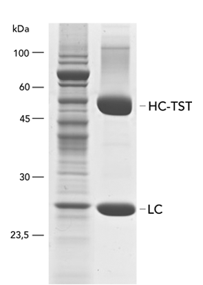
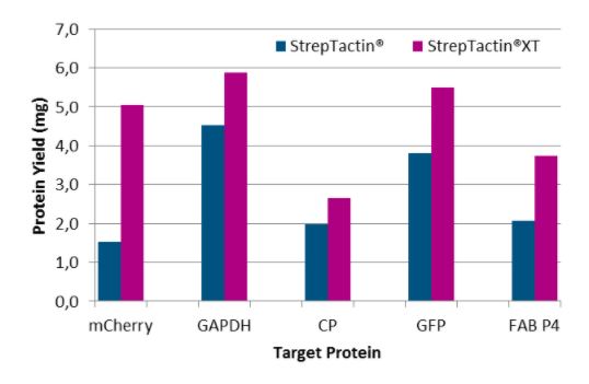
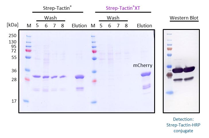
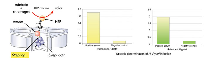
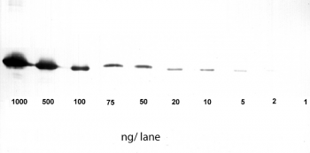
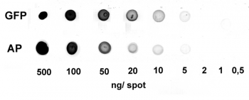
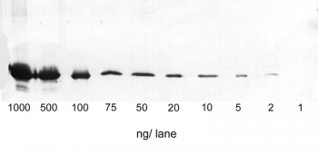
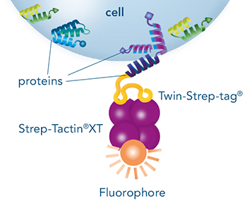
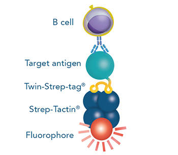
Usually, an affinity tag-free purification method is preferred. However, sometimes an affinity tag-based purification procedure can be beneficial, especially if the antibody is to be implemented in subsequent immobilization or detection. For this purpose, the application of the Strep-tag® technology is highly recommended. The antibody can be simply isolated in high purity with a resin perfectly suitable for large proteins, Strep-Tactin®XT 4Flow®. After removing biotin, the eluent, from the elution fraction via dialysis or size exclusion, the antibody can be immobilized on Strep-Tactin®XT-coated microplates or labeled with a Strep-Tactin®XT conjugate and applied for detection assays.

Versatility of the Strep-tag® technology for antibody purification and assay applications

Biotin blocking

Blocking of free biotin in cell culture supernatants and lysates

Cell culture supernatants often contain high amounts of free biotin. These are unproblematic when working with Strep-Tactin®XT resins, since biotin does neither bind irreversibly to this ligand nor reduces the binding capacity. However, this is the case for the application of resins coupled to Strep-Tactin®. To prevent the impairment of Strep-Tactin®, BioLock, which contains avidin, can be applied to mask free biotin. Avidin is a homolog of streptavidin and can be found in the egg white of birds, reptiles, and amphibians. It consists of four subunits, each of which can bind one molecule of biotin. In contrast to the streptavidin variants, Strep-Tactin® and Strep-Tactin®XT, it is not able to bind Strep-tag®II or Twin-Strep-tag® fusion proteins. Therefore, avidin can be used to selectively mask free biotin in cell lysates or cell culture supernatants, whereas the Strep-tag®II or Twin-Strep-tag® at the target protein remain accessible. An overview of media containing biotin and cell-internal biotin pools is given in the manual for biotin blocking. Blocking of biotin by BioLock is simple and fast. Only a small amount has to be added and, after a short incubation, the sample is ready for protein purification.

Blocking of biotinylated proteins

Besides free biotin, cell lysates also contain small amounts of biotinylated proteins. Normally, these proteins do not influence the purification results with Strep-Tactin® or Strep-Tactin®XT due to their low abundance. However, when it comes to analytic applications with high sensitivity, impurities should be prevented. One option is the treatment of the sample with BioLock prior to protein purification. Otherwise, if the target protein is already purified and should be detected via western blot with Strep-Tactin® or Strep-Tactin®XT conjugates, the application of Biotin Blocking Buffer is possible. Just as BioLock, Biotin Blocking Buffer contains avidin to mask biotin and biotinylated proteins, but the buffer is especially optimized for western blots. Shortly before detection, the membrane is incubated with the diluted buffer for a few minutes. However, if the detection occurs with the aid of StrepMAB-Classic or StrepMAB-Immo, masking of biotinylated proteins is not necessary. In contrast to Strep-Tactin® or Strep-Tactin®XT conjugates, these antibodies do not bind to biotin or biotinylated proteins and therefore do not lead to unspecific signals.

Binding patterns of Strep-tag®II, Twin-Strep-tag® and biotin

Monoclonal antibody development & production

The identification and production of monoclonal antibodies is essential for investigating immune responses and developing therapies. In addition, they can be utilized for the direct and indirect detection of antigens in various assays, as capture molecule immobilized on microplates, or as ligand for protein purification, making them a versatile tool in biotechnology.

Different methods were developed for monoclonal antibody discovery including hybridoma technology, phage display or single B cell sorting.

Their common aim is the identification of high affinity antibodies that recognize a specific antigen of choice. This makes the antigen a central component for antibody development. Fusing a Twin-Strep-tag® to an antigen helps to make this central component applicable for various steps in the development process.

The Twin-Strep-tag® suitable for:

  • Antigen (protein) purification and analysis
  • Immunization
  • Antigen-specific B cell staining and isolation via FACS
  • ELISA and Western blot
  • Biosensor applications
  • Biopanning

This way, the Strep-tag® technology can centralize various steps onto one platform, making antibody development time-saving and cost-efficient.

Figure 1: An antigen fused to a Twin-Strep-tag® as central component for all applications needed in antibody development (e.g. with hybridoma technology, single B cell sorting or phage display)

Antigen expression & purification for antibody development

The production of an antigen is a key process in antibody development. It is needed for several steps in the whole procedure such as immunization, screenings and antibody characterizations.

The advantages of using the Strep-tag® system for the antigen purification process are:

  • Highly pure antigen preparations suitable for immunization and further downstream processes
  • Straightforward isolation procedure without tedious optimization steps
  • Reusability even after CIP (Clean-in-place) procedures

Figure 2: After expressing an antigen with a Twin-Strep-tag®, different formats are available for protein purification. The options range from small to large scale with the possibility to switch between manual and automated systems.

Next to required vectors and an expression system, various purification formats are available, which allow for individual up- or downscaling or automation of the protein production process. In addition to the outstanding target purity, the advantage of fusing an antigen to the Twin-Strep-stag® are the versatile application options for various downstream processes.

The picomolar affinity to its ligand permits the use even for biosensor measurements such as surface plasmon resonance (SPR) or bio-layer interferometry (BLI). This way, switching between different tags depending on the application is unnecessary, which makes the whole procedure quicker and more efficient.

Antigen analysis for antibody development

After the antigen was purified from sources such as cell lysates or cell culture medium, the purity, integrity and concentration should be determined. Coomassie or silver staining as well as analytical size exclusion chromatography are options for determining if unspecific proteins are in a sample. Detecting the produced antigen via its Twin-Strep-tag® in a Western blot is a way to identify if the protein was produced successfully at the expected size and no aggregation or degradation products were formed. To determine the concentration of the yielded antigen, ELISA is a suitable method.

Figure 3: Different Strep-Tactin® conjugates allow for analysis methods such as western blot or ELISA.

Immunization for antibody development

The purification of proteins with the Strep-tag® technology results in a highly pure antigen that is suitable for immunizing a host species. Additional optional processing steps include the removal of biotin, the eluent, from the elution fraction via dialysis or size exclusion and/or TEV cleavage of the Twin-Strep-tag®. However, since the tag has a small size of only 28 amino acids, the immunogenicity is expected to be low.

Figure 4: The Twin-Strep-tag® is a short peptide, therefore the removal prior to immunization is not strictly necessary. Optionally, the tag can be removed via a TEV protease.

Antigen-specific B cell staining & sorting for antibody development

For some antibody development approaches, antigen-specific B cells are isolated from blood or other tissues of seropositive individuals, which encountered the antigen by infection or immunization. Here one can make use of the antigen fused to the Twin-Strep-tag® by incubating it with:

  • Fluorescent Strep-Tactin® or Strep-Tactin®XT conjugates or
  • Strep-Tactin® Magnetic Microbeads or
  • Strep-Tactin® TACS Agarose

This generates selection reagents that are suitable for the staining and sorting of antigen-specific B cells via flow cytometry, magnetic or affinity chromatographic isolation, respectively.

B cells that recognize the antigen of choice are selected from e.g. peripheral blood mononuclear cells (PBMCs). Single cells can be sorted directly into lysis buffer for antibody sequencing or taken into culture for antibody production. This method for monoclonal antibody production offers the possibility to extract B cells and consequently also antibody sequences directly from humans, thereby enhancing the chance to identify antibodies that are readily suitable for therapeutic purposes.

Figure 5: Via its tag, the antigen can be combined with Strep-Tactin® conjugates for e.g. fluorescent staining and sorting of B cells.

Example publications for antigen-specific B cell staining & sorting

Naive human B cells engage the receptor binding domain of SARS-CoV-2, variants of concern, and related sarbecoviruses (Feldmann, J et al., 2021, Science Immunology)

In this publication, Feldman and colleagues expressed SARS-CoV-2 spike protein with a C-terminal 2x Strep-tag®II (equals a Twin-Strep-tag®). By incubating this antigen with Strep-Tactin® PE and APC, they generated staining reagents for antigen-specific B cells. They used the multimerized spike antigen to sort B cells from human PBMCs.
For detailed information read the full paper here.

Protective neutralizing antibodies from human survivors of Crimean-Congo hemorrhagic fever (Fels, J et al., 2021, Cell)

In this study, the authors expressed a Crimean-Congo hemorrhagic fever virus- derived fusion protein with a C-terminal double Strep-tag®II (equals a Twin-Strep-tag®). This fusion protein was complexed with Strep-Tactin® PE and APC to sort single antigen-specific B cells from PBMCs of human donors.
For detailed information read the full paper here.

Extremely potent human monoclonal antibodies from COVID-19 convalescent patients (Andreano et al., 2021, Cell)

Andreano and colleagues labeled SARS-CoV-2 S-protein with Strep-Tactin®XT DY-488 to sort antigen-specific memory B cells from human donors.
For detailed information read the full paper here.

Clone/antibody screening & antibody characterization for antibody development

Independent from the method chosen for monoclonal antibody discovery, screening steps are key to finally identify the best performing antibody.

Figure 6: An antigen can be immobilized on Strep-Tactin®XT coated microplates via its Twin-Strep-tag® for antibody screening with ELISA. Due to the reusability of the plates, several screening rounds are possible.

ELISA

A Twin-Strep-tag® – antigen can be used for antibody screening by ELISA. Via its tag, it can be immobilized on Strep-Tactin®XT coated microplates. This way, the microplate is functionalized for capturing antibodies from e.g. B cell supernatants. Subsequent detection indicates which B cell clones produce specific antibodies. Usually, several selection rounds are required to find the best performing clone. Since Strep-Tactin®XT coated microplates can be regenerated several times, using the Strep-tag® technology in this process is cost saving and time efficient.

Biopanning

Monoclonal antibody discovery by phage display uses a library of antibody fragments that are expressed on the surface of bacteriophages. To find those bacteriophages that present antibody fragments that detect the antigen, a selection procedure called biopanning is utilized. For this method, the phages are exposed to an immobilized antigen. The ones that bind are eluted, amplified and used in a next selection round. For the selection rounds, Strep-Tactin®XT coated microplates with an immobilized Twin-Strep-tag® antigen offer a reusable, cost-saving system similar to antibody screening by ELISA. After the identification of antibody fragments that specifically bind the antigen, sequences have to be cloned into full length antibodies for further use in research or therapeutic applications. Since this step can potentially change the features of an antibody, additional characterization steps are necessary, making this method for monoclonal antibody discovery overall very labor-intensive.

Figure 7: Strep-Tactin®XT coated microplates are suitable for biopanning, a selection procedure employed in phage display. The plates can be regenerated several times, therefore multiple selection rounds are possible.

Figure 8: Due to the picomolar affinity of Strep-Tactin®XT to the Twin-Strep-tag®, biosensor applications such as surface plasmon resonance (SPR) are possible.

SPR & BLI

The antibodies that are yielded by different antibody discovery and development approaches can be further characterized by biosensor applications such as surface plasmon resonance (SPR) or Bio-layer interferometry (BLI). Again, the Twin-Strep-tagged antigen is central in those analyses. Due to the picomolar affinity of the Twin-Strep-tag® to Strep-Tactin®XT, the antigen is immobilized sufficiently to allow the determination of binding kinetics to a selected antibody. This way, antibodies with the desired affinities are identified and chosen for large scale productions.

Antibody purification

After the best antibody was identified for a specific antigen, it has to be expressed in e.g. a cell culture system. The produced antibodies subsequently have to be extracted from cell lysates, cell culture supernatants, or biological probes. The purification can take place without affinity tag via the physicochemical properties, antigen-specific affinity or the antibody class. Physicochemical properties are the molecular weight, charge, or clusters of specific residues.

Depending on the origin of the sample, purification with the help of physicochemical properties or antigen-specific affinity can lead to the isolation of further proteins and antibodies with similar properties besides the target antibody. Thus, if a specific antibody with a high purity should be obtained, antibody class-specific affinity chromatography is recommended, such as Protein A, which can bind immunoglobulins (antibodies) within the Fc region of their heavy chain without regard to antigen specificity.

Choosing an affinity tag-based purification procedure is also possible and can be beneficial, especially if the antibody is to be implemented in subsequent immobilization or detection. For this purpose, the application of the Strep-tag® technology is highly recommended. The antibody can simply be isolated in high purity with a resin perfectly suitable for large proteins, Strep-Tactin®XT 4Flow®. The different formats available for protein purification make it a very flexible system that allows for individual adaptations of sample number and size.

Cell Isolation

Cells serve as an important research tool to investigate different mechanisms in health and disease. They are also suitable for diagnostic and therapeutic purposes, making them attractive for a broad area of research fields. Immune cells have a wide range of functions such as controlling body homeostasis, which includes elimination of infected or cancerous tissue. Therefore, these cells are frequently employed for different analyses and model systems. The advantage of using immune cells is that they are abundant in, for example, human blood or mouse spleen, two sources that are relatively easily accessible for research purposes.

The interaction of cells with each other is very complex and for some experimental setups it is necessary to isolate one specific population for further downstream applications and analyses. Those include for example DNA/RNA isolation, single cell RNA sequencing, protein purification, Western blot or various cell culture experiments.

Immune cells commonly found in human blood.

Positive and negative cell selection

Specific cells are most frequently isolated by targeting their surface markers or according to their antigen-specificity. For surface marker-specific cell isolation, generally one of these two approaches is used: positive or negative cell selection. Antigen-specific cell isolation usually requires a positive cell selection approach.

Cell isolation is possible via positive or negative cell selection. In positive cell selection, cells are directly labeled with e.g., magnetic bead-conjugated antibodies. In negative selection, all unwanted cells are labeled, leaving target cells completely “untouched”.

Surface marker-specific cell isolation

In positive selection, the target cells are directly labelled with, for example, antibodies conjugated to magnetic beads or fluorophores. The cells can subsequently be isolated using a magnet (magnetic-activated cell sorting – MACS) or a flow cytometer suitable for fluorescence-activated cell sorting (FACS).

If the protein that targets a surface marker of a cell, which can be an antibody, a Fab fragment or another cell-binding protein, is fused to a Strep-tag®II or Twin-Strep-tag®, it can be combined with different Strep-Tactin® conjugates to facilitate positive cell selection.

It is possible to choose between affinity chromatographic, magnetic and fluorescent isolation methods depending on the Strep-Tactin® backbone (Strep-Tactin® TACS Agarose columns, Strep-Tactin® Magnetic Microbeads or Strep-Tactin® PE/APC) and is therefore adaptable to different experimental requirements.

The interaction between Strep-Tactin® and the Twin-Strep-tag® is reversed by biotin addition. This way, Strep-Tactin® reagents can easily be removed from the cell surface and do not affect further downstream procedures.

Different methods for positive cell selection

Cell isolation using positive selection is possible via three principal approaches: fluorescence-activated cell sorting (FACS), magnetic-activated cell sorting (MACS) or affinity chromatography. In all approaches, different molecules that target a cell can be used such as antibodies or Fab fragments.

In negative selection, the unwanted cells are labelled usually with antibodies conjugated to magnetic beads. The labelled cells are subsequently separated from the target cells using a magnet. To effectively use the negative selection approach, the composition of a sample has to be known to target all unwanted cells. Therefore, a standard problem of this method is limited purity and recovery, which causes variable quality. The advantage of this method is that cells remain completely “untouched” during the isolation procedure. However, highly pure populations can best be obtained by using positive separation techniques.

In this application note we demonstrate how Strep-Tactin® TACS Agarose columns can be used for negative cell selection.

Antigen-specific cell isolation

Using the Strep-tag® technology, cells can also be isolated according to their antigen specificity. This becomes especially relevant for characterizing B or T cell mediated adaptive immune responses. Instead of high affinity antibodies, antigens (B cells) or major histocompatibility complexes (MHCs) that present a specific peptide/antigen (T cells) are utilized to select cells of interest.

For antigen-specific T cell isolation, MHC complexes need to be multimerized on a backbone to efficiently capture cells. Combining MHC I molecules fused to a Twin-Strep tag® with Strep-Tactin® backbones conjugated to magnetic microbeads or fluorophores also allows magnetic-activated as well as fluorescence-activated cell sorting in this system.

Similar as for T cells, strep-tagged antigens can be bound to Strep-Tactin® conjugates to enable antigen-specific B cell selection.

The reversible labelling principle that is applied in all our isolation approaches helps to preserve the authentic properties, full effector function as well as viability of the target cells.

Cloning, Transformation/Transfection & Expression

The objective of molecular cloning procedures is to introduce a gene of interest into a suitable expression host in a compatible manner (recombinant DNA) to ultimately produce the gene product (protein) in satisfying quality and quantity.

Cloning

Cloning means producing identical copies of a certain gene of interest. By cloning, the gene is amplified and then inserted into a plasmid (vector) for the following replication and protein expression. Using plasmids allows for the protection of the foreign genetic material from the expression host’s innate degradation machinery.

Different restriction enzyme types and its cleavage sites

There are different cloning procedures used to insert the gene of interest into a plasmid. Restriction enzyme-based cloning is the standard cloning method in molecular biology. Restriction endonucleases cleave double-stranded DNA (dsDNA) at specific sequence sites called recognition sites. Depending on the employed restriction enzyme, the generated DNA fragments can either have blunt ends or sticky ends. These can be fused with compatible DNA fragments from the plasmid, which is cleaved with the same restriction enzyme, producing the same compatible ends. In the following ligation step, these two fragments can be fused with the aid of the so-called ligase.

A special restriction enzyme-based cloning method makes use of the specific properties of type IIS restriction enzymes, endonucleases that cleave DNA outside of the recognition sequence. In contrast to traditional restriction enzyme cloning, this cutting outside from the recognition site provides the advantage that custom overhangs can be generated.
With the StarGate system, IBA has utilized this approach to create compatible custom overhangs, which can then be fused efficiently. The StarGate system comes along with a large subset of cloning and expression vectors for different expression hosts.
Expression vectors are specially designed to optimally produce the relevant gene product of the inserted foreign DNA in potent host cells of choice. For this purpose, these plasmids are equipped with expression host-dependent genetic sequences, such as particular enhancers and promoters. In case the optimal expression host, purification procedure, and further experimental conditions are already known for a target protein, a ready-to-use single expression vector (Acceptor vector) containing all the required information can be combined with the gene of interest as previously described. However, if the perfect expression and purification strategy for the protein of interest is so far unknown, splitting into a two-vector-system is recommendable. Therefore, IBA offers a Two-step-cloning solution where the vector pENTRY (Donor vector) is utilized to simultaneously introduce the gene of interest into multiple expression vectors (Acceptor vectors) with different genetic backgrounds (e.g. host-specific promoters, signal sequences, and various affinity tags). The resulting diverse Destination vectors can be utilized to screen for the perfect expression host, affinity tag, and other parameters.

Expression vector elements

Expression hosts

A particular focus should also be set on choosing the appropriate expression host for the protein of interest to ensure getting a biologically functional target. While E. coli are the most prominent host cells due to their easy handling and compatibility with a large portion of targets, eukaryotic cell lines, such as yeasts, insect cells, or mammalian cells are required to obtain complex proteins with proper folding and post-translational modifications. Mammalian cells have become the most popular host for recombinant protein expression, especially of antibodies, therapeutic proteins, or biopharmaceuticals. The Human Embryonic Kidney 293 (HEK) cells are commonly used in drug development and laboratory settings since they exhibit straightforward growth in culture, have a high transfection efficiency, and are amenable to a variety of transfection methods.

IBA’s MEXi-293E cells are such human embryonic kidney (HEK) cells derived from the 293 cell line, which are optimized for the expression of recombinant proteins in mammalian cells and specifically adapted to an efficient growth in the corresponding cell culture media MEXi TM (Transfection Medium) and MEXi CM (Culture Medium).

To allow easy access to the various expression hosts we provide a multitude of plasmids targeting the different hosts, including E. coli and mammalian cells but also yeast and insect cells (Baculovirus vectors).

Transfection/ Transformation

In the next step, the previously created expression vector carrying the gene of interest needs to be transferred into the chosen expression host for propagation and production of the target protein. This can be realized by various methods, whereof transformation and transfection are the most frequently applied.

Transformation is the process of getting the recombinant vector from a reaction mixture or vector solution into a prokaryotic expression host such as E. coli cells. It is a naturally occurring direct uptake of DNA fragments, which is triggered by different physicochemical stimuli (heat shock, calcium treatment, or electroporation) in the laboratory environment. Once arrived in the cell, the gene on the plasmid can simply be expressed by an appropriate induction method.

In contrast, transfection is a more complicated technique to deliver the recombinant DNA construct into eukaryotic cells, or more precisely into their nucleus. In this method, transient pores have to be opened in the cell membrane (viral packaging with calcium phosphate treatment, electroporation, cationic polyamides, or lipid shuttling) to induce the uptake of the genetic construct into the cytosol, followed by the transport into the nucleus due to signal sequences provided by the vector. Through a repeated process of careful selection and amplification, stable clones with the foreign information integrated into their genome are fabricated.

Notwithstanding the advantage that all descendants of these cells will contain the gene of interest and attend the production of the target, stable transfection is a laborious process mainly recommended for the large-scale production of recombinant proteins. In contrast, to rapidly obtain research quantities of the target, transient expression is the method of choice. This technique results in a quick and short-term production of the recombinant protein for several days after DNA transfection.

IBA’s MEXi mammalian expression system implements this transient approach to enable the simple and efficient episomal replication and expression of the GOI without the need for a prior integration by making use of the EBNA1 protein in combination with oriP-harboring plasmids. Further, the system deploys a polyethylenimine-based transfection, which is an affordable and quick method suitable even for hard-to-transfect cell lines, while simultaneously avoiding cytotoxic effects as in lipid-based techniques, or complicated viral packaging.

MEXi – mammalia expression system. Workflow and Components.

High-Throughput Purification

Before a specific target protein can be singled out and investigated in detail, a large number of proteins have to be evaluated in high-throughput screenings (HTS). To focus on their function without the influences of the other molecules present in the organism, these proteins have to be purified. Many high throughput purification applications require expensive equipment and specific technical knowledge. Alternatively, well-known and already established methods can be scaled up to be carried out in any size of lab.

Application approachRecommended productBinding capacityRequired equipment
ManualSpin columnsUp to 600 µg protein per columnMicropipettes, Centrifuge, Shaker
Semi-automatedMagnetic beads in 96 well plateStarting from 500 µg per wellMagnetic separator, Micropipettes, multi-channel pipettes or pipetting robot, Microplate shaker
AutomatedIMCStips®Up to 50 µg per tipPipetting robot

Manual applications

For small-scale experiments with a limited number of samples, spin columns can be used. They are a convenient tool for small sample volumes that can still be handled manually. Thereby the number of proteins that can be purified at the same time in a fast and efficient way is flexible. A Spin Column Kit with all necessary components for protein purification, as well as Empty Spin Columns that can be combined with the customer’s resin of choice are available from IBA Lifesciences.

DescriptionCatalog Number
Strep-Tactin®XT 4Flow® high capacity Spin Column Kit2-5151-000
Empty Spin Columns2-5150-050

Semi-automated applications

Magnetic beads, such as MagStrep® Strep-Tactin®XT beads, can be used in a round-bottom 96-well plate, allowing the simultaneous purification of up to 96 samples. In this format, the application can still be carried out manually, but the use of pipetting robots is also possible. The magnetic core of the beads allows the use of a magnet to fix the beads to the wells when removing the supernatant, diminishing the loss of beads and eliminating the need for centrifugation.

DescriptionCatalog Number
MagStrep® Strep-Tactin®XT beads2-5090-002

Automated applications

For an automated approach that requires minimal manual intervention, IMCStips® containing Strep-Tactin®XT resin are available. The tip format allows high-throughput affinity purification with consistency and reproducibility making it especially attractive to industrial users. The approach is described in detail in the following application note. You can learn more about the automated purification of proteins using IMCStips® on the IMCS website .

Protein purification workflow with IMCStips® containing Strep-Tactin®XT resin

Analytical High-throughput Screening Applications

The identified and purified proteins from the initial screening can then be further characterized by using the Strep-tag® technology. Due to the outstanding high affinity of the Twin-Strep-tag® to Strep-Tactin®XT (pM Range), Strep-Tactin®XT is very well suited for biomolecular interaction analysis technologies as Surface Plasmon Resonance (SPR) or Biolayer interferometry (BLI).

DescriptionCatalog Number
Strep-Tactin®XT BLI Coupling Kit2-4380-000
Twin-Strep-tag® Capture Kit2-4370-000

Protein-Protein Interaction

Immunoprecipitation, Co-immunoprecipitation, and pull-down assay

Immunoprecipitation (IP), Co-immunoprecipitation (Co-IP) and pull-down assays represent different methods to concentrate the target from a mixture for further downstream analysis such as the abundance, the activation status or the protein-binding partners.

While IP is used for capturing one specific target from a mixture, Co-IP and pull-down assays not only focus on single proteins but also on molecules which are attached to the target protein. Identifying the cellular interaction partners of a protein of interest and determining the biological pathways involved is a necessary preliminary work to gain deeper insights into a target’s structure and function. When it comes to the investigation, IP, Co-IP and pull-down assays have become invaluable tools, which are built upon the affinity-based immobilization of a “bait” protein on a solid support, mostly magnetic or agarose beads.

Co-IP using an antibody versus pull-down assay using Strep-Tactin®XT

Whereas IP and Co-IP utilize immobilized antibodies to purify the bait protein along with its binding partners (prey), in the latter case from cell lysates for analysis of the whole protein complex, in pull-downs the target is fused to an affinity tag and captured together with bound proteins using a corresponding immobilized chemical or biological ligand. In every case, the received sample is further analyzed by e.g., SDS-PAGE, western blot, or mass spectrometry.

The advantage of pull-down over IP and Co-IP are mild elution conditions. They retain the natural protein structure as well as protein complexes and prevent elution of unspecific bound proteins leading to robust results in subsequent analytic applications. This is especially the case for the Strep-tag® technology.

For pull-down assays, the Strep-tag® technology is compatible with magnetic and non-magnetic experimental setups as well as any protein class and offers versatile resins for the realization of a multitude of assays to detect interactions. For small amounts of a sample, MagStrep® Strep-Tactin®XT beads can be chosen or when up-scaling of the process is anticipated, agarose-based resins display a powerful option.

Surface plasmon resonance

Surface plasmon resonance (SPR) is an optical biosensor application for direct, real-time, and label-free measurements. It allows the determination of protein concentrations as well as the analysis of affinity and kinetics of a specific protein to other proteins, DNA, RNA, or cells. Therefore, SPR is an innovative tool for biomolecular interaction studies and plays an essential role in biotherapeutic drug discovery and development. The principle of SPR is an optical measurement that detects binding of a biomolecule in solution (analyte) to a biomolecule that is immobilized on a gold-layered surface of a biosensor chip (ligand). Thereby, changes in the refractive index are determined which are proportional to the concentration of bound analyte at the surface of the biosensor chip.

A crucial step in SPR analysis is the immobilization of the ligand to the surface of the biosensor chip. The immobilization of the ligand can take place via direct covalent coupling to the surface of the chip or by capture with the help of a pre-coated capture molecule. However, the direct covalent immobilization of the ligand can change its biological activity or result in an undirected immobilization with reduced accessibility of the binding sites. The transient immobilization of tagged ligands via affinity capturing molecules overcomes these drawbacks.

Surface plasmon resonance (SPR) with Strep-Tactin®XT coated sensor chips

Capturing Twin-Strep-tag® proteins with BiacoreTM

Nevertheless, tags used for such applications must bind efficiently to the capturing molecule with an affinity that is higher than that between analyte and ligand. His-tagged ligands that are immobilized on an NTA-surface with a KD in the nanomolar range can be used for measuring weaker interactions, but they fail in the measurement of high-affinity interactions with slow dissociation rates. In contrast, Strep-Tactin®XT forms an exceptionally strong complex with the Twin-Strep-tag®. An affinity in the low picomolar range allows measurements of long dissociation times and slow off-rates.

Thus, the combination of these two components of the Strep-tag® technology enables a directed, high-affinity immobilization that does not influence the activity of the ligand or require any modifications. Due to the high-affinity interaction and its high specificity, non-specific binding of host cell proteins is avoided, and ligands can be captured efficiently and directly from culture media. Moreover, Strep-Tactin®XT is compatible with many substances and the biosensors can be easily regenerated. The Twin-Strep-tag® Capture Kit provides all necessary products for the preparation of Strep-Tactin®XT-coated biosensor chips and the following measurements.

Bio-layer interferometry

Bio-layer interferometry (BLI) is like SPR a label-free optical biosensing technology for analyzing biomolecular interactions, e.g., antigen-antibody interactions, in real-time and allows quantification of their binding strength and kinetics. In contrast to SPR, BLI does not use gold-layered chips, but thin needles called biosensors. These biosensors are coupled at the tip with a desired ligand and dipped into the sample to capture the analyte. To measure whether the analyte is bound to the ligand, white light is passed through the biosensor and the reflected light from the tip surface is measured. If the ligand interacts with the analyte the layer on the surface of the tip is thicker compared to the ligand alone which leads to a different wavelength that is reflected. The BLI instrument (Octet® or BLItz®) measures both the reflected light from the ligand alone and the ligand-analyte complex. The wavelength shift between both reflection patterns creates an interference pattern which is subsequently used for calculation of binding strength and kinetics.

But what is the best way to bind the ligand to the surface of the biosensor? In principle, the ligand can be bound directly via chemical coupling methods like EDC/s-NHS coupling. But this needs time to find the optimal reaction conditions and can reduce the activity of the ligand. In addition, the orientation of the ligand can vary which can reduce the accessibility for the analyte and can lead to inconsistent measurement results between the biosensors.

These disadvantages can be prevented if an indirect binding via the Strep-tag® technology is used. Instead of the ligand, Strep-Tactin®XT is first immobilized on the biosensor and the ligand tagged with the Twin-Strep-tag® is captured afterwards by it. Since the tag is at a defined position all proteins (ligand) captured by Strep-Tactin®XT will be presented in the same orientation. Furthermore, the pM affinity of Strep-Tactin®XT for the Twin-Strep-tag® allows specific capture of Twin-Strep-tagged fusion proteins from complex samples, e.g., culture supernatants, leading to reliable and robust data. All reagents necessary for fast and simple coating of Strep-Tactin®XT biosensors are provided in our Strep-Tactin®XT BLI Coupling Kit.

Besides BLI and SPR, protein-protein interactions can be analyzed as well with the switchSENSE® technology.

This application note demonstrates how the Strep-Tactin®XT technology can be used in combination with BLI and SPR analytic devices.

Bio-layer interferometry (BLI) in combination with Strep-Tactin®XT for uniform and specific capture of Twin-Strep-tag® fusion proteins.

Staining and Detection

Immunostaining for protein detection

The term immunostaining covers a variety of methods for the specific detection and quantification of proteins from diverse samples, whereby “immuno” refers to the general use of antibodies. Immunostaining techniques include immunohisto- and immunocytochemistry flow cytometry, enzyme-linked immunosorbent assays (ELISA) and western blots.

These different methods are based on either colorimetric, chemiluminescence or fluorescence signals for detection. Besides the signal, the type of detection varies according to the experimental procedure. It can be chosen between direct detection, where a primary antibody carrying a label is used, and an indirect detection using an unlabeled primary combined with a labeled secondary antibody. As convenient alternative, we offer several solutions based on the Strep-tag® technology using Strep-Tactin® or Strep-Tactin®XT combined with Strep-tag®II or Twin-Strep-tag®.

Immunohistochemistry and immunocytochemistry

Amongst the immunostaining methods, immunohistochemistry is the most prominent one, in which whole tissues can be visualized, and thus, specific targets can be detected. In contrast to immunohistochemistry, in immunocytochemistry the focus is not on tissues but on cells. The advantage of both techniques is the possibility of imaging proteins in their proper histological context, which enables the localization of cellular compartments and can be detected using fluorescence microscopy. In both methods the sample fixation is essential to preserve the morphology and structure.

While both, chromogenic or fluorescent labels are utilized for detection, fluorescent labeling is especially popular due to the possibility of multicolor staining of different targets. StrepMAB-Classic or StrepMAB-Immo conjugated to different dyes can be applied for direct labeling of Strep-tagged targets. As viable alternative to antibodies, Strep-Tactin® and Strep-Tactin® XT with different fluorescent conjugates can be used as well. Due to their strong and highly specific interaction with Strep-tagged targets, both generate signals with high sensitivity, avoiding the need for a secondary antibody. However, when only small amounts of Strep-tagged protein are present, the unconjugated murine antibodies StrepMAB-Classic and StrepMAB-Immo can be used combined with a secondary antibody of choice.

Immunohistochemistry/-cytochemistry using fluorescently labeled Strep-Tactin®

Immunohistochemistry/-cytochemistry using fluorescently labeled StrepMAB antibody

Flow cytometry

Fluorescently stained cells are frequently deployed in flow cytometry analysis, which can be conducted both, before and after cell isolation, delivering insights into cell functions for basic research and clinical trials. Here, the labeled cells pass a light beam inside a flow cytometer with high velocity, thereby providing a multitude of information on physical and chemical properties and a lucid quantitative display of different cell populations. Fluorescence-activated cell sorting (FACS) using a sorter is a specialized form of flow cytometry, where target cells are not subsequently discarded, but rather rigorously divided into defined subpopulations according to their fluorophore label – hence, surface marker identities – and retained for further downstream processing.

The Strep-tag® system can be used to stain and sort a cell of interest. One option is the detection of a surface protein fused to a Strep-tag®II or Twin-Strep-tag® via fluorescent Strep-Tactin® or Strep-Tactin®XT conjugates. Alternatively, fluorescently conjugated StrepMAB-Immo antibodies can be used as well.

Another option is the staining and sorting of cells via a strep-tagged protein. In this case, the protein of choice, which binds the cell, must be combined with Strep-Tactin® or Strep-Tactin®XT conjugates. This leads to the generation of a staining reagent suitable for flow cytometry. This cell selection approach is commonly used for e.g. the selection of antigen-specific B or T cells, which are targeted by specific antigens or peptide-loaded MHC class I molecules, respectively.

ELISA – Enzyme-linked immunosorbent assay

The enzyme-linked immunosorbent assay, short ELISA, is a commonly used method for detection and quantification of ligands, such as proteins, peptides, or hormones even in a complex mixture. ELISA is a plate-based technique in which, depending on the type, either the target ligand or the capturing reagent is immobilized on a microplate. While there are different types of ELISA, the essential components are always consistent. Firstly, a ligand of interest, which is to be detected and potentially quantified. Secondly, an antibody, like StrepMAB-Immo or StrepMAB-Classic, or other detection reagent, like our conjugated Strep-Tactin®XT for specific recognition of the ligand. Lastly, an enzymatic reporter, the substrate conversion of which can be used as measurable signal.

While different formats exist, the basic principle when performing any kind of ELISA is the same.

  1. Coating or capturing of the directly or indirectly immobilized ligand on the microplate.
  2. Blocking of free surface-binding sites of the microplate.
  3. Detection of the ligand by using ligand-specific reagents, e.g. antibodies.
  4. Readout of signal produced by the enzymatic reaction of labeled primary or secondary antibody.

Depending on the experimental needs, one can choose between direct, indirect and sandwich ELISA. The formats differ in the way of capturing and detecting the target ligand.

The direct and indirect ELISA both start with the immobilization of the ligand on a microplate. Whereas the direct ELISA only requires a labeled primary antibody for detection, in an indirect ELISA an unlabeled primary antibody first, and then a labeled secondary antibody is used. However, instead of a primary antibody it is possible to use Strep-Tactin® or Strep-Tactin®XT conjugates for Strep-tagged ligands, which is particularly beneficial because a second antibody for sensitive signal detection becomes redundant. Alternatively, StrepMAB-Classic conjugated with HRP can be applied, also avoiding the use of a secondary antibody.

In a sandwich ELISA, the plate is coated with a capturing molecule, like an antibody. Subsequently, the ligand is added and “sandwiched” between the capturing and the detecting antibody. Here, the primary antibody detecting the ligand is generally unlabeled, while a labeled secondary antibody is added for quantification. For capturing a desired Strep-tagged protein, plates can be coated using our StrepMAB-Immo or StrepMAB-Classic antibody. However, we recommend StrepMAB-Immo for this purpose due to its higher affinity towards the Strep-tag®. Convenient pre-coated plates with Strep-Tactin® or Strep-Tactin®XT are additionally available.

As enzymatic reporter, horse-radish peroxidase (HRP) is most commonly applied. It convert a certain substrate into a detectable product. As substrate, a variety of different chromogenic, fluorescent, and chemiluminescent products are available, always depending on the desired sensitivity and the laboratory equipment at-hand. In case a fluorescent detection method is desired, we offer various fluorescent labels conjugated to Strep-Tactin®XT, StrepMAB-Immo or StrepMAB-Classic.

Direct ELISA

Indirect ELISA

Sandwich ELISA

Western blot

Western blot using Strep-Tactin® conjugated AP

Western blot using Strep-Tactin® conjugated HRP

Western blot using StrepMAB-Classic conjugated HRP

The western blot technique is an expedient method for the identification of specific proteins from a complex mixture. After extraction of the protein mixture, e.g. via cell lysis, the first step is to separate the proteins based on their molecular weight using gel electrophoresis. For protein separation by size, usually a polyacrylamide gel is prepared. Next, the proteins are transferred or blotted from the gel to a solid support, generally a nitrocellulose or polyvinylidene difluoride (PVDF) membrane. Because of its efficiency and speed, electrophoretic transfer is the most common blotting procedure. The gel and the membrane are “sandwiched” between two electrodes and the proteins are transferred due to their electrophoretic mobility, influenced by factors such as charge and size.

Since the membranes have a high affinity for proteins, the remaining binding sites of the surface need to be blocked after the transfer to prevent unspecific binding of the detection antibodies. A variety of different buffers and reagents is used in the whole process and, to minimize the signal-to-noise ratio, sufficient washing is required as intermediate step.

For detection of the antigen, it can be chosen between direct or indirect detection. For direct detection of Strep-tagged proteins, only a single labeled and target-specific antibody, such as our StrepMAB-Classic conjugated to HRP, is required. While it is quite common to use antibodies for detection, our Strep-Tactin® products conjugated to HRP or AP represent an equally beneficial alternative. These products are based on our Strep-tag® technology.

The combination of a primary antibody detecting the target and a labeled secondary antibody is widely used as well, as the second antibody leads to a signal amplification and provides different options for multiple detection methods. The choice of primary and secondary antibody depends on various parameters, such as the origin of the target protein, the species of the primary antibody or the desired detection method. When working with Strep-tagged proteins, our murine StrepMAB-Classic antibody is a good choice as primary antibody.

Nowadays widely applied labels are fluorophores or enzymes. As in the case of ELISA, horse-radish peroxidase (HRP) or alkaline phosphatase (AP) are the most common enzymes. Even though enzymatic labels require extra steps and usually have to be optimized, they are used most extensively due to the high sensitivity and the flexibility in detection when choosing between chromogenic, fluorogenic, and chemiluminescent substrates. When using fluorophores as labels, fewer steps are required, since no substrate development is necessary. For this purpose, we offer various fluorescent labels conjugated to Strep-Tactin®XT, StrepMAB-Immo or StrepMAB-Classic. The choice of the experimental procedure is generally dependent on the laboratory equipment, e.g., special devices for the detection of a fluorescent signal are needed.

Altogether, the relatively easy protocol for western blots offers a broad range of potential utilizations, such as detecting post-translational-modifications or verifying protein cloning, and makes it such a popular application.

Strep-Tag

Strep-tag® technology

High performance research tools for cell and protein isolation

Our proprietary Strep-tag® technology exploits one of the strongest non-covalent interactions in nature: the interaction of biotin and streptavidin. The system is based on the highly selective and easily controllable interaction between the synthetic Strep-tag®II peptide and the specially engineered streptavidin, called Strep-Tactin®, which is one of the most stable proteins known. The Strep-tag®II binds specifically to the engineered streptavidins, Strep-Tactin® and Strep-Tactin®XT, by occupying the binding pocket of the natural ligand biotin. Hence, the interaction is easily reversible by excessive addition of the competitor.

Two affinity tags – two streptavidin derivatives: The Strep-tag®II consists of eight amino acids (Trp-Ser-His-Pro-Gln-Phe-Glu-Lys), whereas the Twin-Strep-tag® includes this motif two times in series connected by a linker and is accordingly composed of 28 amino acids. Both exhibit intrinsic, although unequal, affinity towards the streptavidin derivative Strep-Tactin® and its successor Strep-Tactin®XT: The binding affinity of Strep-tag®II to Strep-Tactin® (1µM) is nearly 100 times higher than to streptavidin. A further improvement was achieved by the development of Strep-Tactin®XT, which shares a nM affinity with the Strep-tag®II and a pM affinity with the Twin-Strep-tag®.

As a result of the differences in binding strength among the possible tag-ligand combinations, the Strep-tag® system has become established as a universal tool for isolation of proteins and cells.

  • Fused to recombinant proteins, the Strep-tag® enables efficient one-step purification on immobilized Strep-Tactin®.
  • When fused to antibody-derived Fab fragments or nanobodies, Twin-Strep-tag® binds to multimerized Strep-Tactin® allowing capturing and releasing of target cells based on their surface-marker or antigen-specificity.
  • Strep-Tactin® (or Strep-Tactin®XT), on its turn, can be conjugated to microplates, fluorophores or chips, allowing a wide range of analytical applications after isolation of target material, such as detection, immobilization and interaction studies.

Key Features

Key features of the Strep-tag® system are:

  • Reversibility
  • Full functionality of target material after isolation
  • High purities
  • Universal applicability for various analytic applications
  • Simple purification procedures

Strep-Tag - Cell isolation

Strep-tag® technology for cell isolation

Besides the well-known tools for protein purification and analyses, the Strep-tag® technology also offers methods for cell staining and isolation. The two main approaches are:

Direct staining and/or isolation of cells that express a surface protein fused to Strep-tag®II or Twin-Strep-tag®.

Indirect staining and/or isolation via a protein that is fused to Strep-tag®II or Twin-Strep-tag®, which binds to e.g. a specific receptor on the cell surface.

For both ways of selecting cells, different available Strep-Tactin® and Strep-Tactin®XT conjugates offer three basic methods for purifying cells out of mixed samples: Affinity chromatography, magnetic cell isolation or fluorescence activated cell sorting (FACS).

Different conjugates for cell selection

Due to the various available conjugates, the Strep-tag® technology provides a highly flexible and individually adaptable platform not only for proteins, but also for larger molecules such as cells.

Direct staining and/or isolation of cells

The Strep-tag® technology is a popular system for purifying proteins due to the specific tag-ligand interaction. The same technology also works for proteins that are present on the cell surface. Fusing the Strep-tag®II or Twin-Strep-tag® to a surface protein of choice helps to select a specific cell of interest. Since proteins and cells differ in various characteristics such as size, the properties of used reagents should fit to the chosen target. The Strep-tag® system not only offers products specialized for proteins, but also for cell-based research

Indirect staining and/or isolation of cells

Fusing the Strep-tag®II or Twin-Strep-tag® to a cell surface molecule for subsequent selection is not always feasible or suitable for a target of interest. Another option to purify a cell population is to utilize a protein that binds to e.g. a receptor on the surface. This is frequently done with e.g. antibodies that are directly or indirectly linked to a fluorophore or a magnetic bead.

The Strep-tag® technology enables the use of any type of protein for cell isolation. This can be an antibody, an antigen, a peptide-loaded MHC class I molecule or another cell-binding protein. As long as the protein of choice is fused to Strep-Tag®II or Twin-Strep-tag®, it can be combined with the available Strep-Tactin®/ Strep-Tactin®XT conjugates to build reagents suitable for cell selection.

The different Strep-Tactin®/ Strep-Tactin®XT conjugates allow the choice for a cell isolation method that is most suitable for a given approach. Fluorescent conjugates permit the staining and sorting of small cell populations via flow cytometry. Strep-Tactin® Magnetic Microbeads are freely scalable when cells are separated via a magnet. For a high number of target cells (>1 x 10^7 cells), Strep-Tactin® TACS Agarose columns are the cost-effective choice.

A great advantage of the available conjugates is their ability to multimerize strep-tagged proteins. This way, even low affinity targets that do not stably bind as monomers can be used to effectively capture cells.

Cell isolation with low affinity Fab fragments using different Strep-Tactin® conjugates

Even low affinity Fab fragments can be used for effective cell isolation. The different Strep-Tactin® conjugates increase the avidity sufficiently to allow stable binding to cells.

Removal of Strep-Tactin® and Strep-Tactin®XT conjugates

Independent from the chosen cell selection method, Strep-Tactin® and Strep-Tactin®XT conjugates can easily be removed from the cell surface by biotin addition. Due to its affinity to Strep-Tactin® and Strep-Tactin®XT, it will release any strep-tagged molecule from the used conjugate. This way, cells can be used for further downstream applications such as cell culture without being affected by attached selection reagents.

Strep-Tag - Protein purification

Strep-tag®: The leading affinity tag in recombinant protein production

The Strep-tag® system enables cloning, expression, detection, purification, as well as further analysis of recombinant proteins. The highly specific interaction of the Strep-tag®II with Strep-Tactin® ensures efficient one-step purification of the protein of interest in unparalleled purity even from crude cell lysates.

The Strep-tag® system is compatible with a large number of protein classes, e.g., metalloproteins, membrane proteins and fragile protein complexes with multiple subunits. In addition, the mild and physiological conditions promote the yield of fully functional proteins, making the system particularly suitable for purification of enzymes as well as structural investigations, protein-protein interaction studies, ligand-receptor investigations or even separation of living cells for re-culturing processes. Simultaneously, a high tolerance towards different buffers and additives promotes its universal applicability.

Besides the advantages of the Strep-tag® technology for efficient protein purification, the near covalent affinity (pM range) of Twin-Strep-tag® to Strep-Tactin®XT expands the range of applications towards protein analysis e.g., ELISA, surface plasmon resonance (SPR) or bio-layer interferometry (BLI).

Products

The Strep-tag® technology represents a universal toolbox that provides solutions for the entire recombinant protein production chain: from cloning, protein expression and purification to further analysis like detection, immobilization and assay applications.

One tag – Multiple applications

The Strep-tag® protein purification system comprises two affinity tags, the 8AA Strep-tag®II and its tandem version Twin-Strep-tag®. Both versions can bind to Strep-Tactin® and its high affinity variant Strep-Tactin®XT. Thereby the two tags differ in the affinities with which they bind to Strep-Tactin® and Strep-Tactin®XT. Depending on the application and properties of the protein of interest one can combine the different tags and Strep-Tactin® variants according to the required affinity.

It is well known for its outstanding performance with regard to purifying recombinant proteins in a simple procedure with highest purities. Its latest development – the Strep-Tactin®XT – provides a binding affinity in low pM ranges in combination with Twin-Strep-tag® still maintaining binding reversibility and mild recovery of immobilized proteins. This improvement in affinity allows protein purification at high yields and purity, even for challenging proteins and from mammalian expression systems (e.g. Expi). Read more about the benefits of Strep-tag® purification in combination with high density mammalian protein expression in our white paper.

Furthermore, it fulfills the high demands of protein interaction analysis or assays and downstream applications like SPR, thus covering all steps from purification to immobilization efficiently.

We offer specialized matrices for any demand in form of ready to use kits, FPLC columns, gravity flow or spin columns, suspensions, coated microplates, magentic beads, and more (find products in our web shop). Additionally, we provide innovative solutions for approaches surrounding batch purification, FPLC/HPLC, high-throughput screens and assay development.

Strep-tag® protein purification system at a glance

Key features:

  • Highly selective binding properties leading to unparalleled protein purity (> 95 %)
  • Bioactive target proteins due to the rapid one-step purification under target specific conditions
  • Variability in buffer conditions, e.g. high salts, detergents, metal ions, chelators or reducing agents
  • Peptide tag with balanced/inert amino acid composition and, therefore, the protein structure or activity is not influenced, and removal of tag is not required
  • Favorable for protein:protein interaction studies due to mild elution conditions and low washing volumes
  • Universal toolbox including products for cloning, purification, and analytic applications (ELISA, FACS, SPR, Microscopy, Western Blot, etc.)

When should the Strep-tag® purification system be the method of choice?

Besides the purification of classical proteins the Strep-tag® system should be the method of choice for:

  • metalloproteins
  • membrane proteins
  • low abundant proteins (in combination with Strep-Tactin®XT)
  • sensitive protein complexes with multiple subunits
  • bioactive proteins

Is There An Alternative To His-tag?

Choosing an appropriate affinity chromatography system for simple and efficient protein purification is a common question. There are differences between the systems which rarely are clearly represented, making it hard for scientist to reach a decision.

This comprehensive comparison of the most frequently used systems, His-tag and Strep-tag®, explains these differences as well as recommends one system depending on the properties of the target protein, expression host and purification conditions.

Strep-tag® system – quick & easy protein purification protocol

“The Strep-tag® technology allows efficient one-step purification of Strep-tag®II or Twin-Strep-tag® proteins via affinity chromatography. The system is distinguished by quick and simple purification protocol, regardless of the target protein class.”

“This step-by-step tutorial demonstrates the full purification procedure of a target protein, here mCherry-Twin-Strep-tag, via gravity flow column, including the initial buffer and column preparation steps, as well as column regeneration after purification and its’ preparation for storage until further use. Gravity flow column used in the tutorial contained high-performance Strep-Tactin®XT 4Flow® resin.”

What are the differences between Strep-Tactin® and Strep-Tactin®XT?

Which resin to choose when purifying strep-tagged proteins?
Strep-Tactin® and its high-affinity counterpart, Strep-Tactin®XT, are both streptavidin-derived mutants. Each of these proteins weighs 52 kDa, comprising four subunits, each housing a biotin binding pocket.
While streptavidin binds biotin with an almost irreversible affinity in its pocket, Strep-Tactin® and Strep-Tactin®XT exhibit reduced affinity for biotin. However, they still maintain the capability to bind biotin. The biotin binding pockets on both Strep-Tactin® and Strep-Tactin®XT have been optimized for binding with Strep-tag® and its tandem version, Twin-Strep-tag®. In contrast to the near irreversibility of biotin binding with streptavidin, the interaction with either tag on Strep-Tactin® or Strep-Tactin®XT is reversible.
Compared to streptavidin, a crucial alteration in Strep-Tactin® involves mutating the lid-like loop, resulting in a consistently open confirmation of the loop. This initial mutation enhances the affinity for strep-tagged proteins by a factor of ten. Upon peptide binding, the lid remains open, allowing for easy removal of the peptide by competitive biotin.
In Strep-Tactin®XT, a second mutation was introduced on the opposite side of the chain. This alteration enhances the interaction between the peptide and the biotin binding pocket, significantly boosting the binding affinity.
More detail about the improvements of the sequence can be found in Schmidt et al. 2021.

Streptavidin & biotin

Strep-Tactin® & Strep-tag®

Strep-Tactin®XT & Twin-Strep-tag®

Strep-Tactin® and Strep-Tactin®XT purification cycle

For the purification of Strep-tag®II or Twin-Strep-tag® proteins both resin types, Strep-Tactin® and Strep-Tactin®XT, are applicable. Protein purification with Strep-Tactin® as well as Strep-Tactin®XT is very simple and fast, since the purification cycle for each target protein is the same. However, if a target protein needs a specific buffer composition, e.g. PBS, HEPES or no chelating agents, like EDTA, the buffers can be easily adapted without the need of changing the purification cycle. Both resin types accept various buffer compositions, reagents and additives and an overview is given in the compatible reagents list for Strep-Tactin® as well as Strep-Tactin®XT.

The purification cycles for both resin types are highly similar. However, the sample application, the elution, and the regeneration step are slightly different and will be explained in the following.
A comparison of purification via Strep-Tactin® and Strep-Tactin®XT depicts two changes in the procedure. The first and second step (lysate application & wash) remain the same. But the elution and the regeneration streps are different for both systems. For elution from Strep-Tactin® desthiobiotin is used whereas Strep-Tactin®XT requires biotin for elution. Also the regeneration step differs. HABA is used for regeneration from Strep-Tactin® and in case of Strep-Tactin®XT 3 M MgCl2 is applied.

Strep-Tactin®Strep-Tactin®XT
Binding affinityStrep-tag® II: μM range Twin-Strep-tag®: nM rangeStrep-tag® II: nM range Twin-Strep-tag®: pM range
ElutionElution with Buffer E (2.5 mM Desthiobiotin)Elution with Buffer BXT (50 mM Biotin)
RegenerationElution with Buffer R (HABA)Elution with 10 mM NaOH or 3 M MgCL2

Properties of Strep-Tactin® and Strep-Tactin®XT

The shared benefits of Strep-Tactin® and Strep-Tactin®XT are

  • Purification of bioactive recombinant proteins under physiological conditions
  • Protein aggregation is avoided
  • Broad range of detergents, chelators, salt or redox conditions allowed
  • Highly specific interaction of Strep-tag® with Strep-Tactin®, no unspecific protein binding (high specificity in screening and assays)

Additional benefits of Strep-Tactin®XT due to increased binding affinity in pM range:

  • Efficient, directed, and reliable immobilization of targets from complex mixtures while maintaining the reversibility
  • Allows all kinds of analytic applications as e.g. surface plasmon resonance (SPR) or biolayer interferometry (BLI)

Furthermore, higher protein yields compared to Strep-Tactin® are obtained. On average, StrepTactin®XT provides almost 2-fold more protein than Strep-Tactin®.

Strep-Tactin®XT also ensures sharp elution profiles achieving high concentration of the target protein. Strep-Tactin®XT even tolerates a wide pH range in addition to various detergents and additives, contributing to the universal usability of the system.

Resin reuse

Strep-Tactin® and Strep-Tactin®XT resins for protein purification can be regenerated and reused 3 to 5 times without loss in performance. The proper regeneration of the column and resin activation can easily be checked with HABA. The yellow HABA solution turns red (Strep-Tactin®) or orange (Strep-Tactin®XT) upon binding to the engineered biotin binding pockets of Strep-Tactin® and Strep-Tactin®XT clearly indicating that the resin is fully regenerated. Afterwards, HABA can be removed by washing with 1x Buffer W. Once the red color has disappeared the column can be reused. If the biotin binding pocket is blocked or damaged no color shift occurs and the resin cannot be reused.

What is the difference between Sepharose®, Superflow® and 4Flow®?

The underlying matrix of many protein purification resins are porous agarose beads. The characteristics of these agarose beads determine for which proteins and for which applications the resin is most suitable. IBA’s Sepharose® resin consists of a 4% cross-linked agarose and is therefore suitable not only for small, but also for large proteins. However, a disadvantage of Sepharose® is that it is not pressure stable and consequently not suitable for automated applications such as FPLC. In contrast, IBA’s Superflow® products work well for FPLC. The disadvantage of Superflow® is that it consists of a 6% cross-linked agarose. This means that the pores are smaller and not accessible for larger proteins, resulting into low yields.

4Flow® combines the advantageous characteristics of Sepharose® and Superflow®. It is a 4% cross-linked agarose, and it is pressure stable, making it a universal matrix for small and large proteins as well as for different applications such as FPLC.

Strep-Tactin® Strep-Tactin®XT
Matrix Superflow® Sepharose® MacroPrep® 4Flow® MagStrep Strep-Tactin®XT beads
(only available as 20 ml slurry)
Binding capacity*/ Dynamic Binding capacity** classic: 3 mg/ml resin* classic: 3 mg/ml resin* classic: 3 mg/ml resin* classic: 5 mg/ml resin** classic: 25.5 µg/µl resin*
high capacity: 7.0 mg/ml resin** high capacity: 14 mg/ml resin**
Bead structure 6% agarose, crosslinked 4% agarose, crosslinked polymethacrylate 4% agarose, highly crosslinked magnetic core covered with 6% agarose, crosslinked
Bead size 60-160 µm, spherical 45-165 µm 50 µm 50-150 µm, spherical 30 μm (average), spherical
Exclusion limit 6 x 10⁶ Da ~3 x 10⁷ Da 1 x 10⁶ Da 3 x 10⁷ Da not specified
Recommended flow rate 0.5-1 ml/min gravity flow 0.5-1 ml/min 0.5-1 ml/min batch purification
pH range for protein binding 7-8 7-8 7-8 4-10 6-10
Max pressure 9.6 bar gravity flow 70 bar 3.5 bar batch purification
Storage 4 °C, do not freeze 4 °C, do not freeze 4 °C, do not freeze 4 °C, do not freeze 4 °C, do not freeze
Shipment RT RT RT RT RT
Eluent desthiobiotin desthiobiotin desthiobiotin biotin biotin
Regeneration buffer Buffer R Buffer R Buffer XT-R 0.1 M NaOH 0.1 M NaOH
Features and recommendations
  • Strep-Tactin® Superflow high capacity binds three to five times more protein in comparison to Strep-Tactin® Superflow
  • Can be used for the purification of biotinylated proteins
  • For average sized proteins (<90 kDa)
  • For proteins that are hard to elute from Strep-Tactin®XT resins, like proteins with multiple tags or multimers
  • Suitable for use in FPLC columns
  • For large proteins (>90 kDa)
  • Can be used for the purification of biotinylated proteins
  • For proteins that are hard to elute from Strep-Tactin®XT resins, like proteins with multiple tags ore multimers
  • Only suitable for gravity flow due to low pressure stability
  • Synthetic support
  • Reduced unspecific binding
  • Strep-Tactin®XT 4Flow® high capacity binds three to fove times more target protein compared to Strep-Tactin®XT 4Flow®
  • Applicable for all protein types, especially large (>90 kDa), low abundant and challenging proteins
  • Strep-Tactin®XT 4Flow is recommended for first time users of the Strep-tag® system
  • Optimized for Batch purification
  • Beads can be separated with a magnetic device, no centrifugation necessary
  • Suitable for purification in 96-well plates
* Max binding capacity for a Strep-tag® protein (30kDa).

** Dynamic binding capacity (DBC) was determined with mCherry-Twin-Strep-tag (30 kDa) under realistic operation conditions and shows the amount of protein which is bound until 10% of the protein is found in the flow through.

Specifications for Strep-Tactin® Purification Resins

There are several Strep-Tactin® resin versions available. All resins differ in their properties and suitability for applications. Learn more about the different resin matrices in order to know when to use which resin.

Find out more about Strep-tag® protein purification

Here, we have grouped links to make it easier to find more information about Strep-tag® technology for protein purification. In our web shop, you can find the protein purification products that fit your research needs. Read the comprehensive comparison of the most used tag systems, Strep-tag® vs. His-tag. Browser through application examples and frequently asked questions about Strep-tag® purification system or download application notes in our download area.

Description Size Catalog No.
Avidin 50 mg 2-0204-050
BioLock 50 ml 2-0205-050
250 ml 2-0205-250
Biotin 2 g 2-1016-002
5 g 2-1016-005
Biotin stock solution 1 ml 6-6325-001
10 ml 6-6325-010
Buffer BXT (10x) 25 ml 2-1042-025
Buffer CI (10x) 85 ml 6-6320-085
Buffer E (10x) 25 ml 2-1000-025
Buffer R (10x) 100 ml 2-1002-100
Buffer W (10x) 100 ml 2-1003-100
Buffer XT-R 250 ml 2-1045-250
CD3 Fab-Strep, human 50 µg 6-8001-150
CD4 Fab-Strep, human 50 µg 6-8002-150
CD8 Fab-Strep, human 50 µg 6-8003-150
CD81 Fab-Strep, human 50 µg 6-8015-150
Desthiobiotin 1 g 2-1000-002
5 g 2-1000-005
Empty Spin Columns 50 columns 2-5150-050
GFP-Strep-tag®II control protein 500 µl 2-1006-005
Magnetic Separator 1 piece 2-1602-000
MagStrep “type3” XT beads 2 ml 2-4090-002
10 ml 2-4090-010
MagStrep® Strep-Tactin® beads 2 ml 2-1613-002
MagStrep® Strep-Tactin®XT beads 2 ml 2-5090-002
10 ml 2-5090-010
MHC I-Strep H-2 Kb; Ovalbumin (SIINFEKL) 10 µg 6-7015-001
MHC I-Strep HLA-A*0101; CMV pp50 (VTEHDTLLY) 10 µg 6-7024-001
MHC I-Strep HLA-A*0201; CMV pp65 (NLVPMVATV) 10 µg 6-7001-001
MHC I-Strep HLA-A*0201; gp100 (YLEPGPVTA) 10 µg 6-7025-001
MHC I-Strep HLA-A*0201; MART-1 (ELAGIGILTV) 10 µg 6-7007-001
MHC I-Strep HLA-A*0201; NY-ESO-1 (SLLMWITQV) 10 µg 6-7013-001
MHC I-Strep HLA-A*0201; SARS-CoV-2 (YLQPRTFLL) 10 µg 6-7130-001
MHC I-Strep HLA-A*0201; WT1 (VLDFAPPGA) 10 µg 6-7099-001
MHC I-Strep HLA-B*0702; CMV pp65 (TPRVTGGGAM) 10 µg 6-7027-001
Strep-Tactin® Buffer Set 1 set 2-1002-001
Strep-Tactin® MacroPrep® resin 20 ml 2-1505-010
Strep-Tactin® Magnetic Microbeads 750 µl 6-5510-050
Strep-Tactin® Sepharose® column 1 ml 2-1202-001
Strep-Tactin® Sepharose® resin 4 ml 2-1201-002
20 ml 2-1201-010
50 ml 2-1201-025
1000 ml 2-1201-500
Strep-Tactin® Superflow® high capacity column 5 x 0.2 ml 2-1209-550
1 ml 2-1209-001
5 ml 2-1209-051
Strep-Tactin® Superflow® high capacity FPLC column 1 ml 2-1239-001
5 ml 2-1240-001
Strep-Tactin® Superflow® high capacity resin 4 ml 2-1208-002
20 ml 2-1208-010
50 ml 2-1208-025
1000 ml 2-1208-500
Strep-Tactin® TACS Agarose 4 ml 6-6350-002
20 ml 6-6350-010
50 ml 6-6350-025
Strep-Tactin® TACS Agarose Column 0.3 ml 6-6310-300
1 ml 6-6310-001
Strep-Tactin®XT 4Flow® column 5 x 0.2 ml 2-5011-005
1 ml 2-5012-001
5 ml 2-5013-001
10 ml 2-5014-001
Strep-Tactin®XT 4Flow® FPLC column 1 ml 2-5023-001
5 ml 2-5024-001
Strep-Tactin®XT 4Flow® high capacity column 5 x 0.2 ml 2-5031-005
1 ml 2-5032-001
5 ml 2-5033-001
10 ml 2-5034-001
Strep-Tactin®XT 4Flow® high capacity FPLC column 1 ml 2-5027-001
5 ml 2-5028-001
Strep-Tactin®XT 4Flow® high capacity resin 4 ml 2-5030-002
20 ml 2-5030-010
50 ml 2-5030-025
1000 ml 2-5030-500
Strep-Tactin®XT 4Flow® high capacity Spin Column Kit 1 Kit 2-5151-000
Strep-Tactin®XT 4Flow® resin 4 ml 2-5010-002
20 ml 2-5010-010
50 ml 2-5010-025
1000 ml 2-5010-500
Strep-Tactin®XT 4Flow® Starter Kit 1 Kit 2-5998-000
Strep-Tactin®XT Buffer Set 1 set 2-1043-000
Strep-tag®II peptide 1.8 mg 2-1018-002
StrepMan Magnet 1 piece 6-5650-065
TACS Column Adapter 0.3 ml 6-3333-001
1 ml 6-6331-001
WET FRED Set 1 ml 2-0911-001
5-10 ml 2-0910-001

Strep-tag® vs His-tag

Benefits of Strep-tag® vs His-tag purification

Strep-tag® and His-tag are both widely used tools for affinity purification via a peptide tag. But when it comes to purity and versatility in applications, some differences between both systems are obvious. The infographic on the left shows not only the time saving way of Strep-tag® protein purification but also the main key features of the Strep-tag® system at a glance.

Comprehensive comparison of Strep-tag® and His-tag protein purification systems

I am text block. Click edit button to change this text. Lorem ipsum dolor sit amet, consectetur adipiscing elit. Ut elit tellus, luctus nec ullamcorper mattis, pulvinar dapibus leo.

Protein purification protocols

How to avoid Ni-leakage when purifying proteins from Expi supernatants?

Purifying His-tagged proteins from Expi supernatants with Ni-NTA resins is problematic and has been associated with a loss of target protein in the purification process.

In this case study you will learn that a standard Ni-NTA resin does not provide reliable and predictable purification results. Furthermore, it demonstrates that standard Ni-NTA resins exhibit significant reduction of binding capacity and recovery due to nickel leakage by media supplements.

The application note shows how sample volumes impact protein recovery and that residence time influences His-tag purification remarkably.


Strep-tag® performs better than His-tag in purification of mammalian-expressed proteins

In combination with high density mammalian protein expression, the His-tag system can lead to poor purification results, if conditions are not optimized. In order to achieve pure proteins for downstream applications from the coupling of the His-tag system and high density mammalian systems, further adjustments like dialysis of the supernatant, lower media:resin ratios or the use of strip-resistant nickel resins become necessary. Such adaptions require additional optimization time and effort, however, these challenges can be avoided by using the Strep-tag® system.

The following whitepaper shows the advantages of the Strep-tag® technology in comparison to the His-tag system as well as the excellent connection of the Strep-tag® technology with two high density mammalian expression systems, the Expi293TM and the ExpiCHOTM expression systems.

Watch the Webinar

See why His-tag fails in some protein applications and learn more about the benefits and drawbacks of the two systems.

Strep-tag® – One affinity tag for all protein applications

By exploiting the highly specific interaction, Strep-tagged proteins can be isolated in one step from crude cell lysates in unparalleled purity. Because the Strep-tag® elutes under gentle, physiological conditions it is especially suited for generation of functional proteins e.g. enzymatic proteins. The mild (physiological) purification and elution conditions required for Strep-tag® fusion proteins make them suitable for structural and functional investigations, protein-protein interaction studies, ligand-receptor investigations or even separation of living cells for re-culturing processes. The system is suitable for multiple protein classes, e.g. metallo proteins, membrane proteins, fragile protein complexes with multiple subunits and any other protein class.

The near covalent affinity of Twin-Strep-tag®to Strep-Tactin®XT can be used to efficiently immobilize proteins for assay development. This makes the system to a universal platform and superior to all other affinity systems: one tag can be used for expression, purification, detection and immobilization.

For detailed information about further commonly used protein purification affinity tags we recommend the Editorial Article from Biocompare.

Products

IBA is the original manufacturer of the Strep-tag® system and provides a complete portfolio around protein purification using this affinity system!

DescriptionCatalog Number
Strep-Tactin®XT 4Flow® Starter Kit2-5998-000
Strep-Tactin®XT 4Flow® resin2-5010-002
Strep-Tactin®XT 4Flow® column2-5011-005
Strep-Tactin®XT 4Flow® FPLC column2-5024-001
MagStrep® Strep-Tactin®XT beads2-5090-002
Strep-Tactin®XT Buffer Set2-1043-000

Application Examples

Browse through our application examples demonstrating, how IBA products were used in the various life science research projects conducted by both our customers and our in-house team of scientists and which results were obtained.

Cloning & Transfection

MEXi – Mammalian Expression System

The MEXi system consisting of MEXi 293E cells, pDSG-IBA expression vectors, MEXi-TM transfection medium, and MEXi-CM culture medium was applied for expression of different proteins. Subsequent purification occurred either with Strep-Tactin® Superflow® high capacity or Strep-Tactin®XT Superflow®. Yield and purity were analyzed by SDS-PAGE.

Metalloprotein

Type: metalloprotein, hydrolase

Yield : 143 mg/l.

Secreted Alkaline Phosphatase (SEAP) was fused with a C-terminal Twin-Strep-tag® and the BM40 secretion signal via cloning into pDSG-IBA102. MEXi 293E cells  (1.5 x 10cells/ml)  were transfected in  MEXi-TM transfection medium (17 ml) with polyethylenimine (PEI, 25 kDa). Afterwards, the cell culture was incubated for 4 hours (37 °C, 5% CO2, 125 rpm) and then diluted with MEXi-CM culture medium to reach a cell density of 0.75 x 10⁶ cells/ml. The cells were kept at 37 °C, 5% CO2, and 125 rpm for 7 days in order to obtain high protein yields. For purification, the cells were pelleted and the supernatant, containing the SEAP protein, was harvested. Free Biotin was blocked by BioLock containing avidin. The SEAP protein was finally purified using a gravity flow Strep-Tactin® Superflow® high capacity column.

Antibody

Type: antibody

Yield: 96 mg/l (represented by heavy (HC) and light chain (LC))

The monoclonal rat antibody (mAb) was cloned into the pDSG-IBA102 vector in order to fuse the heavy chain (HC) C-terminally with the Twin-Strep-tag® and the BM40 secretion signal. The transfection of MEXi 293E cells was performed in MEXi-TM transfection medium (250 ml) with  polyethylenimine (PEI, 25 kDa). The cells were incubated for 4 hours (37 °C, 5% CO2, 125 rpm) and when a cell density of 3 x 10⁶ cells/ml was reached, 250ml MEXi-CM culture medium was added. Afterwards, the culture was shifted to 32 °C and incubated until day 10.

In order to divide the cells from the supernatant, the cell suspension was centrifuged according to the MEXi manual. Free biotin was blocked by BioLock containing avidin. The supernatant was used for protein purification via a gravity flow Strep-Tactin® Superflow® high capacity column. WET FRED was used to facilitate loading of the large supernatant volume onto the column.

Customer Protein

Yield: 318 mg/l

The coding sequence of the customer protein was cloned into pDSG-IBA102 leading to a recombinant protein with C-terminal Twin-Strep-tag® and BM40 secretion signal. 1050 ml of MEXi-TM was inoculated with MEXi 293E cells. Subsequently, plasmid DNA was added followed by addition of 25 kDa linear PEI. The cells were incubated for 4 hours in MEXi-TM medium at 37 °C, 5% CO2, and 125 rpm in an orbital shaking incubator. Cells were diluted to 7.5 x 10⁵ cells/ml by addition of one volume MEXi-CM and kept at 37 °C for 7 days. Afterwards, cells were pelleted and the supernatant containing the customer protein was harvested. The customer protein was finally purified using Strep-Tactin®XT Superflow®. The figure shows the elution fractions (E1-E3). A dilution of 1:10 was prepared for E1 and E2 for SDS-PAGE analysis.

StarGate Cloning

StarGate comprises vectors for expression in mammalian cells (pCSG-IBA, pESG-IBA, pDSG-IBA), yeast (pYSG-IBA), E. coli (pASG-IBA), and insect cells (pLSG-IBA). As an example, the bacterial protein azurin was cloned into different pASG-IBA vectors and expressed in E. coli. Expression results were analyzed by SDS-PAGE.

pASG-IBA

The coding sequence of azurin, a 14 kDa bacterial protein, was cloned into 10 different pASG-IBA vectors and expressed in E. coli. Comparable amounts of E. coli cells were harvested 3 hours after induction with anhydrotetracycline. Samples were lysed by boiling for 5 min at 95 °C with gel loading buffer and analyzed by SDS-PAGE. Subsequently, the gel was stained with Coomassie. Periplasmic secretion by means of ompA led in all cases to accumulation of comparable amounts of the protein of interest (lanes 6-10). This could be expected as azurin is also secreted in its authentic host P. aeruginosa. In case of cytosolic expression, however, interesting aspects became obvious, since it was not anticipated that cytosolic expression of azurin is possible at all. Expression was enabled by fusion of an N-terminal affinity tag (lanes 2, 4, and 5), while N-terminal untagged variants did not result in expression (lane 1 and 3). The examples show that initial screening for expression conditions may be worthwhile since different constructs can lead to various results depending on the target protein properties.

Protein Affinity Chromatography

Protein purification with Strep-Tactin®XT

For protein purification of Strep-tag®II and Twin-Strep-tag® proteins with Strep-Tactin®XT IBA Lifesciences offers MagStrep® Strep-Tactin®XT beads (MagStrep “type3” XT beads), 4Flow®, and pre-packed Spin Columns. As example, the purification of different target proteins with Strep-Tactin®XT 4Flow® and MagStrep® Strep-Tactin®XT beads (MagStrep “type3” XT beads) is shown.

MagStrep® Strep-Tactin®XT beads (MagStrep "type3" XT beads)

GFP C-terminally tagged with Strep-tag®II (28 kDa) was expressed in E. coli (A) and purified with MagStrep® Strep-Tactin®XT beads (MagStrep “type3” XT beads). For separation of magnetic beads from residual solution, the Magnetic Separator was used. Before elution, the sample was split and the target protein was eluted either by application of 1x Buffer BXT containing biotin or by boiling (C, 5 min at 95 °C). Due to boiling, the Beat´s agarose melts and Strep-Tactin®XT is released, leading to a further peak at 14 kDa. Protein purification results were analyzed with the Agilent Bioanalyzer 2100 system. The example shows the specific binding properties of MagStrep® Strep-Tactin®XT beads (MagStrep “type3” XT beads) and the high purity that can thereby be observed.


Strep-Tactin®XT 4Flow®

Cell culture supernatant of MEXi-293E cells (24 ml) was spiked with 240 µg of a large protein (130 kDa) C-terminally tagged with the Twin-Strep-tag®. Protein purification was carried out with a 0.2 ml Strep-Tactin®XT 4Flow® high capacity gravity flow column. After sample application the column was washed with 1x Buffer W. The Twin-Strep-tag® protein was eluted by three elution steps with 0.7, 1.5 and 0.8 CV 1x Buffer BXT. Purification results were analyzed by SDS-PAGE (A) and western blot (B). Both show a molecular weight marker (M*, Precision Plus Protein™ Unstained Protein Standards, Strep-tagged recombinant (BioRad), M** PageRuler™ Plus Prestained Protein Ladder (Thermo Fisher Scientific)), the spiked cell supernatant (S), flow through (FT), the 1st and 5th wash fraction (W1 and W5, respectively) and the elution fractions (E1-E3). Strep-Tactin® HRP was applied (1:4000) for detection via western blot.

Protein purification with Strep-Tactin®

IBA offers MacroPrep®, Superflow®, and Sepharose® coupled with Strep-Tactin® for protein purification of Strep-tag®II and Twin-Strep-tag® proteins. As example, the purification of different target proteins with Strep-Tactin® Sepharose® or Strep-Tactin® Superflow® is shown.

Sepharose®

The wild type sequence and mutant variant of an enzyme were cloned into pASK-IBA3, leading to proteins localized to the cytoplasm and C-terminally tagged with the Strep-tag®II. After expression in E. coli and cell lysis, the proteins were purified with Strep-Tactin® Sepharose® gravity flow columns under physiological conditions (100 mM Tris-Cl, pH 8.0). Purification results were analyzed by SDS-PAGE. A molecular weight marker (M), cell lysate (lane 1), flow through (lane 2), and elution with 2.5 mM desthiobiotin (lane 3) are shown. For both proteins, a high purity was observed.

Superflow®

Coding sequences of virus-like particles (VLPs, 4.8 MDa) were cloned into pASK-IBA7 to obtain proteins localized to the cytoplasm and N-terminally tagged with Strep-tag®II. The plasmid was transformed into E. coli and the cell culture (1 l) was induced at OD600 of 0.6 by addition of anhydrotetracycline (AHT). Protein expression was performed at 37 °C for 3 h at 200 rpm. Afterwards, cells were harvested, resuspended with 20 ml 1x Buffer W (100 mM Tris-Cl, 150 mM NaCl, 1 mM EDTA), and sonicated. The insoluble material was pelleted, and the lysate was applied to a Strep-Tactin® Superflow® FPLC column (1 ml/min flow rate). After washing, the target protein was eluted with 2.5 mM desthiobiotin. Purification results were analyzed by SDS-PAGE. Marker (M) and two different elution fractions of VLPs fused to Strep-tag®II (lane 1 and 2) are shown.

Data kindly provided by L. Stöckl and B. Brandenburg, Robert-Koch-Institute, Berlin. Brandenburg, et al. (2005): A Novel System for Efficient Gene Transfer Into Primary Human Hepatocytes Via Cell-Permeable Hepatitis B Virus–like Particle, HEPATOLOGY (42), 1300-1309.

Resin Comparison

Both Strep-Tactin® and Strep-Tactin®XT resins can be used for purification of Strep-tag®II and Twin-Strep-tag® proteins. However, due to their different properties, an initial comparison can be helpful to find the appropriate resin.

Superflow® vs. Superflow® high capacity

Cell lysate contaning GFP-Twin-Strep-tag was split and either purified with Strep-Tactin® Superflow® or Strep-Tactin® Superflow® high capacity.

Both purifications led to a highly pure protein, but due to the higher density of immobilized Strep-Tactin® on the high-capacity variant, a two-fold higher amount of GFP-Twin-Strep-tag can be purified with Strep-Tactin® Superflow® high capacity compared to Strep-Tactin® Superflow®.

Strep-Tactin® vs. Strep-Tactin®XT

Five different proteins were purified with Strep-Tactin® Superflow® and Strep-Tactin®XT Superflow® to compare the yield with both rein types. Here, we found that Strep-Tactin®XT Superflow® allows the purification of on average a 2-fold higher protein yield than Strep-Tactin® Superflow®.

An advantage of Strep-Tactin®XT compared to Strep-Tactin® is that it allows intensive washing without loss in protein yield. To illustrate this, mCherry-Twin-Strep-tag from E. coli supernatant was purified with a gravity flow column either containing 1 ml Strep-Tactin® Superflow® or Strep-Tactin®XT Superflow®. During purification, the columns were washed with 8 column volumes of 1x Buffer W to reach a constant absorption ratio at A260/280 nm. Then, the target protein was eluted with 50 mM biotin. Samples of the last wash fractions (wash 5-8) and the elution fraction were analyzed by SDS-PAGE. In comparison to Strep-Tactin® Superflow®, Strep-Tactin®XT Superflow allows intensive washing without loss of protein yield. The high affinity (pM range) between Strep-Tactin®XT and Twin-Strep-tag® leads to a tighter binding and prevents a premature elution of the target protein during the wash steps. The elution fractions were further analyzed by Western Blot and detected with Strep-Tactin®HRP, showing that the further bands are alternative variants of mCherry-Twin-Strep-tag.

MacroPrep® vs. Sepharose®

For some recombinant proteins, for example from plants, purification results of Strep-Tactin® MarcoPrep® can differ from those of Strep-Tactin® Sepharose®, due to different resin properties. The SDS-PAGE gel shows the purification of a recombinant protein which exhibits exceptionally high non-specific protein binding. In this case, contaminants were almost completely removed by application of Strep-Tactin® MacroPrep®, whereas the contaminants co-eluted with the recombinant protein after the same purification protocol using Strep-Tactin® Sepharose®. Please note, normally behaving recombinant proteins can be purified in a single step to homogeneity using Strep-Tactin® Sepharose®, however, the example shows that it may be reasonable to change the resin if non-specific protein binding occurs.

Protein Immobilization

Strep-Tactin®XT

Strep-Tactin®XT binds with high affinity Twin-Strep-tag® proteins, making it perfectly suitable for SPR analysis. The examples show the coupling of Strep-Tactin®XT on different SPR chips and possible regeneration conditions for re-use of Strep-Tactin®XT-coated chips.

Coupling on CM5

We recommend to use the following conditions for optimal Strep-Tactin®XT coupling:


  • 20-50 µg/ml Strep-Tactin®XT
  • max. 20 mM acetate, pH 4.5


Using these conditions for Strep-Tactin®XT coupling, efficient and reproducible coupling results can be obtained.

Coupling on CM4

The membrane protein CB2 was fused with Twin-Strep-tag® and then immobilized on a CM4 sensor chip coated with Strep-Tactin®XT.

Used conditions:


  • 50 mM Tris pH 7.5, 100 mM NaCl
  • 10 % glycerol, 0.1 % DDM, 0.5 % CHAPS, 0.1 % CHS

Data was published in A. Yeliseev et al., Protein Expression and Purification 131 (2017) 109-118

Regeneration

Three different regeneration conditions were tested on Strep-Tactin®XT CM5 sensorchips:


  • 10 mM NaOH / 500 mM NaCl
  • 3 M MgCl2
  • 3 M GuHCl


for three different Twin-Strep-tag® fusion proteins:


  • mCherry-Twin-Strep-tag
  • GAPDH-Twin-Strep-tag
  • mAb-Twin-Strep-tag


We recommend the application of 3 M GuHCl for optimal regeneration conditions, since this buffer led to the best regeneration results for all test proteins. However, depending on the target protein, other buffers are applicable as well.

StrepMAB-Immo

Due to the nearly irreversible binding of Strep-tag®II proteins, StrepMAB-Immo can be used for SPR analysis. The example shows the stable capture of a Strep-tag®II fusion protein on a SPR chip coated with StrepMAB-Immo.

Surface Plasmon Resonance

Chips for SPR were coated either with StrepMAB-Immo or with a competitive antibody (Competitor Q). Subsequently, a Strep-tag®II fusion protein was captured, and the binding stability was determined using a Biacore 3000.

During the washing phase, the recombinant Strep-tag® protein of interest remains tightly bound to StrepMAB-Immo, while a significant amount of Strep-tag® protein is washed off using the competitive antibody.

Strep-Tactin® microplates

Strep-tag®II fusion proteins were immobilized on Strep-Tactin® microplates. Subsequently, the activity or abundance of the Strep-tag®II fusion proteins was determined by enzymatic reaction.

Activity Assay

Strep-Tactin® coated microplate was incubated with different amounts of recombinant E. coli alkaline phosphatase (AP) tagged with the Strep-tag®II. After several washing steps, activity of bound fusion protein was determined by colorimetric reaction.

Solid phase immunoasay

Strep-Tactin® coated microplate was incubated with recombinant H. pylori urease tagged with Strep-tag® II, followed by three washing cycles. Afterwards, the microplate was incubated with human sera followed by three washing cycles. Third incubation occurred with rabbit anti-human lgG conjugated to horseradish peroxidase (HRP), followed by 3 washing cycles. Amount of bound antibodies from human sera was determined by colorimetric reaction of HRP.

Staining & Detection

Protein detection with Strep-Tactin® Conjugates

Strep-Tactin® conjugates can be used for several assay applications. As example, the application of Strep-Tactin®HRP and Strep-Tactin®AP for Western Blot as well as Dot Blot is shown.

Strep-Tactin® HRP

Application: Western Blot

1-1000 ng/lane of GFP C-terminally tagged with Strep-tag®II (28 kDa) were separated by SDS-PAGE and blotted onto a membrane. Detection occurred with Strep-Tactin® HRP.


Application: Dot Blot

0.5-500 ng/spot of GFP-Strep-tagII (28 kDa) or alkaline phosphatase C-terminally tagged with Strep-tag®II (monomer 48.5 kDa) were spotted onto a membrane. Detection occurred with Strep-Tactin® HRP.

Strep-Tactin® AP

Application: Western Blot

1-1000 ng/lane of GFP C-terminally tagged with Strep-tag®II (28 kDa) were separated by SDS-PAGE and blotted onto a membrane. Detection occurred with Strep-Tactin® AP.


Application: Dot Blot

0.5-500 ng/spot of GFP-Strep-tagII (28 kDa) or alkaline phosphatase C-terminally tagged with Strep-tag®II (monomer 48.5 kDa) were spotted onto a membrane. Detection occurred with Strep-Tactin® AP.

Detection with Strep-tag® Antibodies

StrepMAB-Classic and StrepMAB-Immo can be used for detection of Strep-tag®II or Twin-Strep-tag® proteins on cell surfaces, within the cell, in cell lysates, or in eluates after protein purification. As example, the application for flow cytometry and confocal microscopy is shown.

StrepMAB-Classic DY-549

Application: Flow cytometry

Dilution: 1:500

HeLa cells were transiently transfected with the coding sequence of SARS-CoV-2 ORF3a-Twin-Strep-tag. After 48 hours transfection, cells were detached, gently trypsinised and stained with StrepMAB-Classic DY-549 (1:500, 0.5% BSA + 1 mM EDTA, 30 min on ice). Intracellular SARS-CoV-2 ORF3a-Twin-Strep-tag levels were measured by flow cytometry (4 laser Cytoflex S, Beckman Coulter)

Source: Data kindly provided from James Edgar, Department of Pathology at the University of Cambridge.

StrepMAB-Classic

Application: Confocal microscopy (Immunofluorescence)

Dilution: 1:500

HeLa cells were grown on glass coverslips, fixed (4% PFA/PBS), quenched (15 mM glycine/PBS), permeabilized (0.1% saponin/PBS), blocked (1% BSA, 0.01% saponin in PBS), and stained with StrepMAB-Classic (1:500) to confirm the expression of SARS-CoV-2 ORF3a-Twin-Strep-tag. Anti-mouse 488 was used as secondary antibody. Nuclei were stained with DAPI and the imaging occurred with a LSM700 confocal microscope (63×/1.4 NA oil immersion objective; ZEISS).

Source: Data kindly provided from James Edgar, Department of Pathology at the University of Cambridge

Fluorescent cell staining for flow cytometry with Strep-Tactin® and Strep-Tactin®XT conjugates

The Strep-tag® technology provides different options for cell staining.

Direct vs. indirect staining

Direct staining of cells expressing a protein with a Strep-tag®II or a Twin-Strep-tag® on the surface

Indirect staining via a cell-binding protein (e.g. a Fab fragment, a MHC molecule or an antigen) that is fused to a Strep-tag®II or a Twin-Strep-tag®

Direct staining

The Strep-tag®II or Twin-Strep-tag® are short peptide sequences that can be expressed on a cell surface without affecting the properties of the cell. Fusing one of the tags to a surface protein of choice enables easy selection of cells that successfully express this protein.

MEXi cells expressing a surface protein fused to a Twin-Strep-tag® were mixed with native MEXi cells at a ratio of approximately 1:1. 1 x 10^6 total cells were stained with Strep-Tactin®XT DY-649 at a dilution of 1:5000* (A) or with Strep-Tactin®XT APC at a dilution of 1:1000* (B). Staining was performed in 100 µl Buffer CI (1x PBS with 1 mM EDTA and 0,5% BSA) for 30 min at 4°C.

*Titration of optimal dilutions might be required for individual experimental setups. In addition, the choice of conjugate depends on factors such as protein density on the cell surface. Due to their brighter staining, PE and APC conjugates can be advantageous for low expressed targets.

Indirect staining via strep-tagged protein (Fab)

For cytotoxic T cell staining, 200 ng anti-human CD8 Fab fragment (50 kDa) fused to a Twin-Strep-tag® was pre-incubated with 75 ng of Strep-Tactin®XT APC (A) or Strep-Tactin®XT DY-649 (B) for 10 min at 4 °C. The formed complexes were added to 5 x 10⁶ peripheral blood mononuclear cells (PBMCs) and incubated for 20 min at 4 °C in the dark. Afterwards, additional staining antibodies were added. Both fluorescent conjugates achieved clear separation of positive and negative cell populations, demonstrating a flexible choice of fluorophores for highly expressed target proteins such as CD8 on the surface of T cells.

Indirect staining of T cells via strep-tagged protein (MHC)

For staining low affinity targets such as T cell receptors, multimerization of the ligand is required to enable stable binding of the detection reagent to the cell. This multimerization can be achieved with Strep-Tactin® or Strep-Tactin®XT PE or APC conjugates.

For antigen specific T cell staining, 200 ng of an MHC class I molecule fused to a Twin-Strep-tag® (50 kDa) refolded with a cytomegalovirus (CMV)-derived peptide was pre-incubated with 75 ng of Strep-Tactin® PE (A) or Strep-Tactin®XT PE (B) for 15 min at 4 °C. 1 x 10⁷ PBMCs in 100 µl buffer were added to the staining complexes and incubated for 20 min at 4 °C. Afterwards, additional staining antibodies were added.

Strep-Tactin®XT PE achieved a brighter staining intensity than Strep-Tactin® PE, indicating that Strep-Tactin®XT PE and APC are the preferred conjugates for low affinity and low expressed targets such as specific T cell receptors. Strep-Tactin®XT DY-649, DY-549 and DY-488 conjugates are not recommended for this approach, since their lesser brightness does not achieve sufficient staining (data not shown).

Product Literature


Video library

View All ProductsContact Us Video Tutorials Browse through our How-to videos library for comprehensive step-to-step demonstrations of purification procedures, preliminary Read More...

switchSENSE®

Determination of protein kinetics using switchSENSE® The switchSENSE® technology of Dynamic Biosensors utilizes a novel electro-switchable biosurface to provide researchers and commercial laboratories Read More...

StarGate-Cloning

ContentDirect Transfer CloningDirect Transfer CloningTwo-step-cloningTet expression systemStarGate Cloning Special featuresTypeIIS Restriction EnzymesExpression in E. coli StarGate for fast protein expression Read More...

Application Examples

Application Examples Browse through our application examples demonstrating, how IBA products were used in the various life science research projects Read More...

Strep-Tag – Protein purification

ContentKey FeaturesResin re-usePurification ProtocolResin SpecificationPurification CycleStrep-Tactin® vs Strep-Tactin®XT Strep-tag®: The leading affinity tag in recombinant protein production   The Strep-tag® Read More...

Strep-Tag – Cell isolation

TACS - Traceless Affinity Cell SelectionMHC I Streptamer® for antigen specific T cell isolationOverview of Strep-tag® technology for cell isolationTACS Read More...

Strep-tag® technology for exosome isolation

Products Pure and functional exosomes in ~ 30 minutes. Fab-Streps Fab-Streps are derived from conventional antibodies. They are modified to Read More...

High-Throughput Purification

Before a specific target protein can be singled out and investigated in detail, a large number of proteins has to Read More...

Applications – Exosome Isolation

Exosomes have an endosomal origin and are produced by almost every cell type. Their size ranges between 30 and 150 Read More...

Staining and Detection

Immunohistochemistry and immunocytochemistryFlow cytometryELISA – Enzyme-linked immunosorbent assayWestern blot Immunostaining for protein detection The term immunostaining covers a variety of Read More...

Cloning, Transformation/Transfection & Expression

CloningExpression hostsTransfection / Transformation The objective of molecular cloning procedures is to introduce a gene of interest into a suitable Read More...

Protein-Protein Interaction

IP, Co-IP, Pull-DownSPRBLIImmunoprecipitation, Co-immunoprecipitation, and pull-down assay Immunoprecipitation (IP), Co-immunoprecipitation (Co-IP) and pull-down assays represent different methods to concentrate the Read More...

Protein Affinity Chromatography

Protein purificationAntibody purificationBiotin blockingProtein purification Prior to each protein analysis the target protein has to be separated from a complex Read More...

Cell stimulation & expansion

T Cell Stimulation and Expansion The in vitro generation of a large number of functional T cells is important for Read More...

Cell isolation

Cell Isolation MethodsSurface marker-specificAntigen-specific Cells serve as an important research tool to investigate different mechanisms in health and disease. They Read More...

Strep-tag® technology for exosome isolation

Contact Us Strep-tag® technology for exosome isolation Pure and functional exosomes in ~ 30 minutes. Fab-Streps Fab-Streps are derived from conventional Read More...

Cell Isolation Mouse

Conatct Us What do I need to start?Starter Kits Label-free cell isolation from murine splenocytes or other single-cell suspensions How Read More...

BioID

Contact Us Products for BioIDCompatible resinsRecommended buffersReversible immobilization A versatile tool for protein interaction studies Strep-Tactin® for reversible binding of biotinylated Read More...

Protein purification product formats

Contact Us cartridgesGravity flow columnsMagnetic beadsSpin columnsResin re-use Protein purification via affinity chromatography is based on the binding affinities between Read More...

Research tools for Covid-19

Contact Us Research PapersOur Tools for COVID-19 research Research on SARS-CoV-2 is currently a key concern of life sciences to Read More...