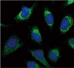
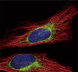
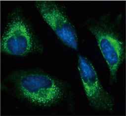
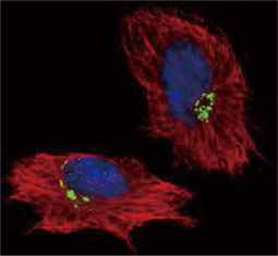
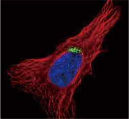
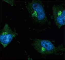
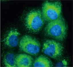
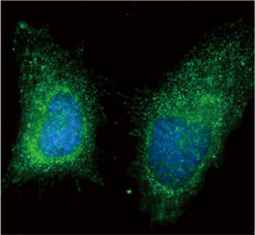
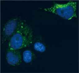
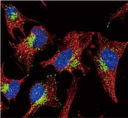
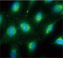
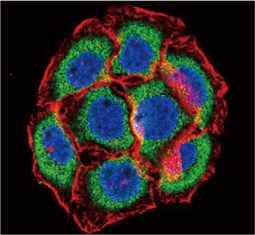
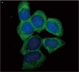
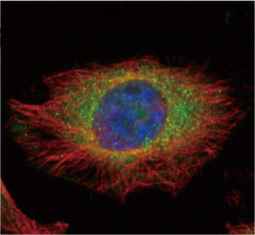
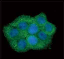
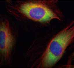
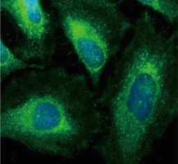
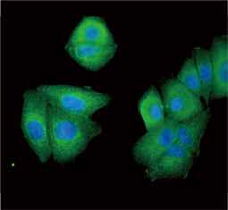
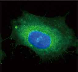
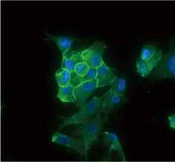
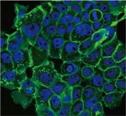
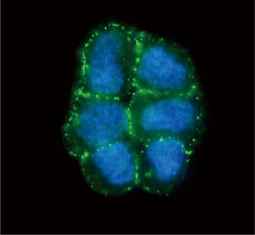
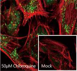
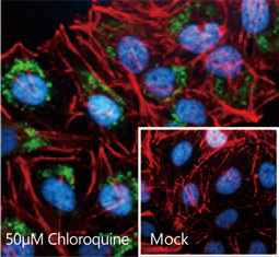
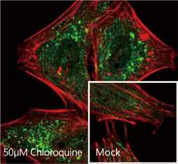
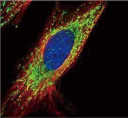
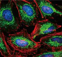
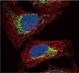
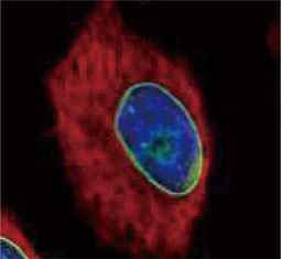
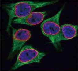
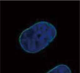
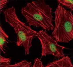
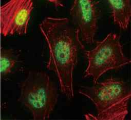
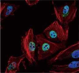
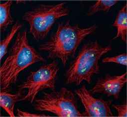
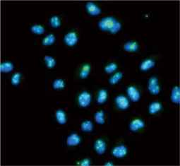
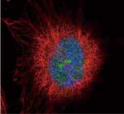
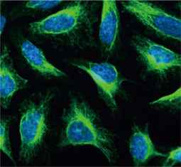
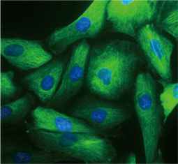
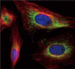
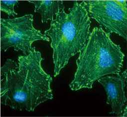
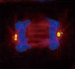
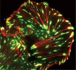
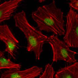
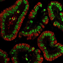
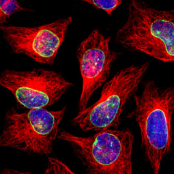
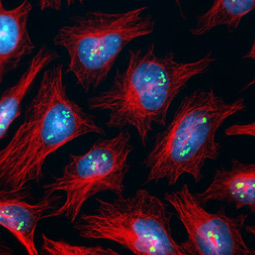
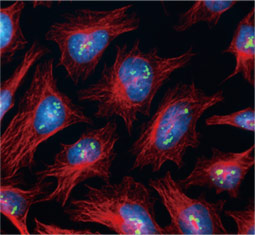
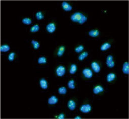
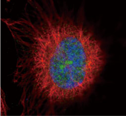
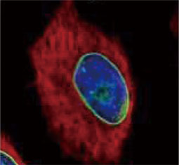
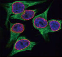
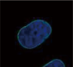
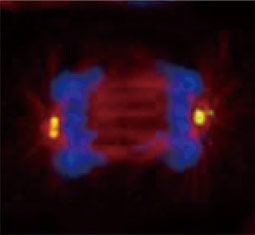
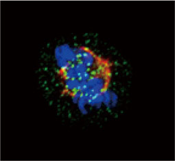
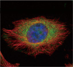
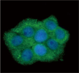
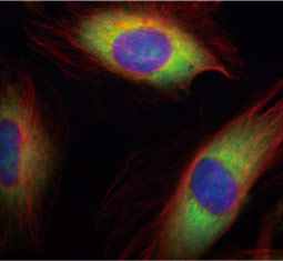
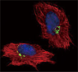
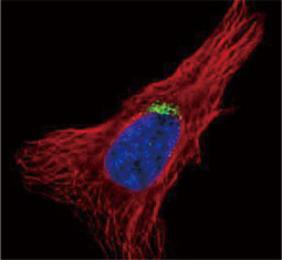
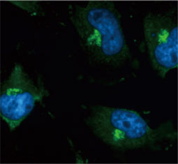
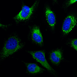
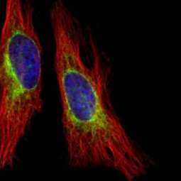
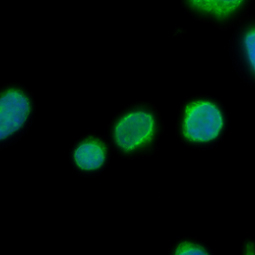
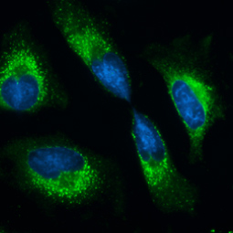
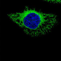
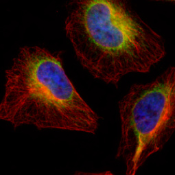
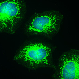
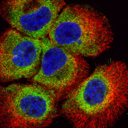
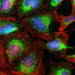
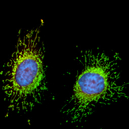
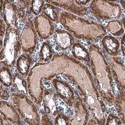
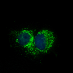
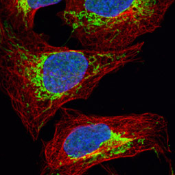
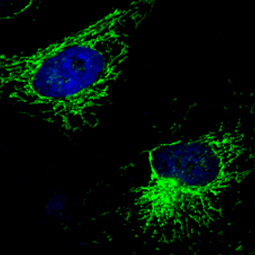
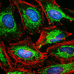
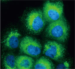
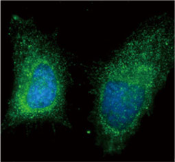
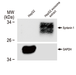
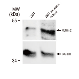
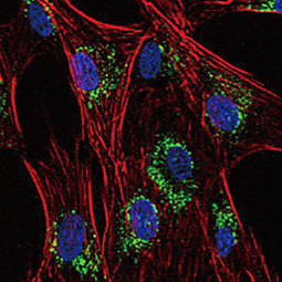
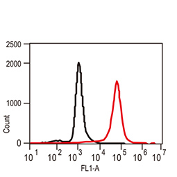
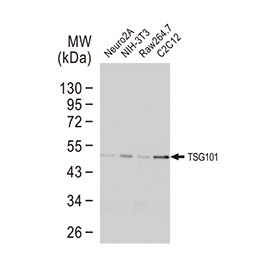
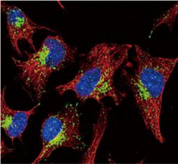
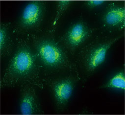
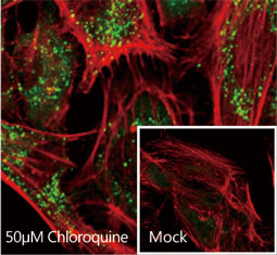
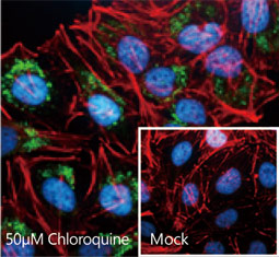
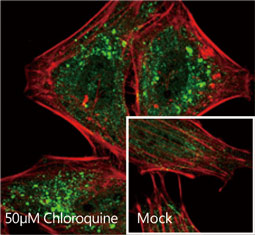
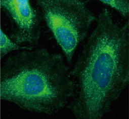
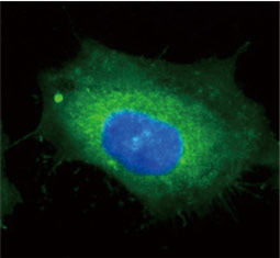
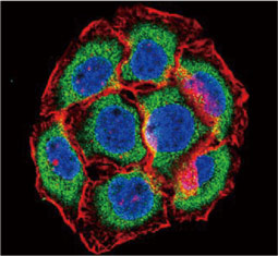
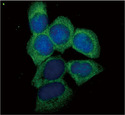
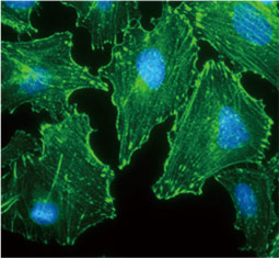
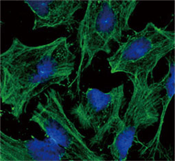
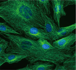
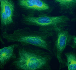
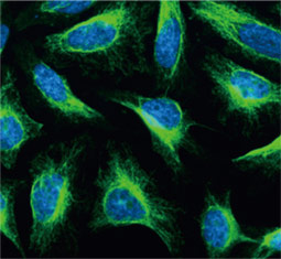
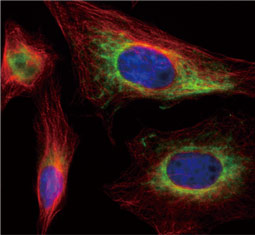
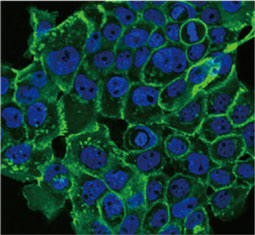
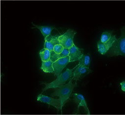
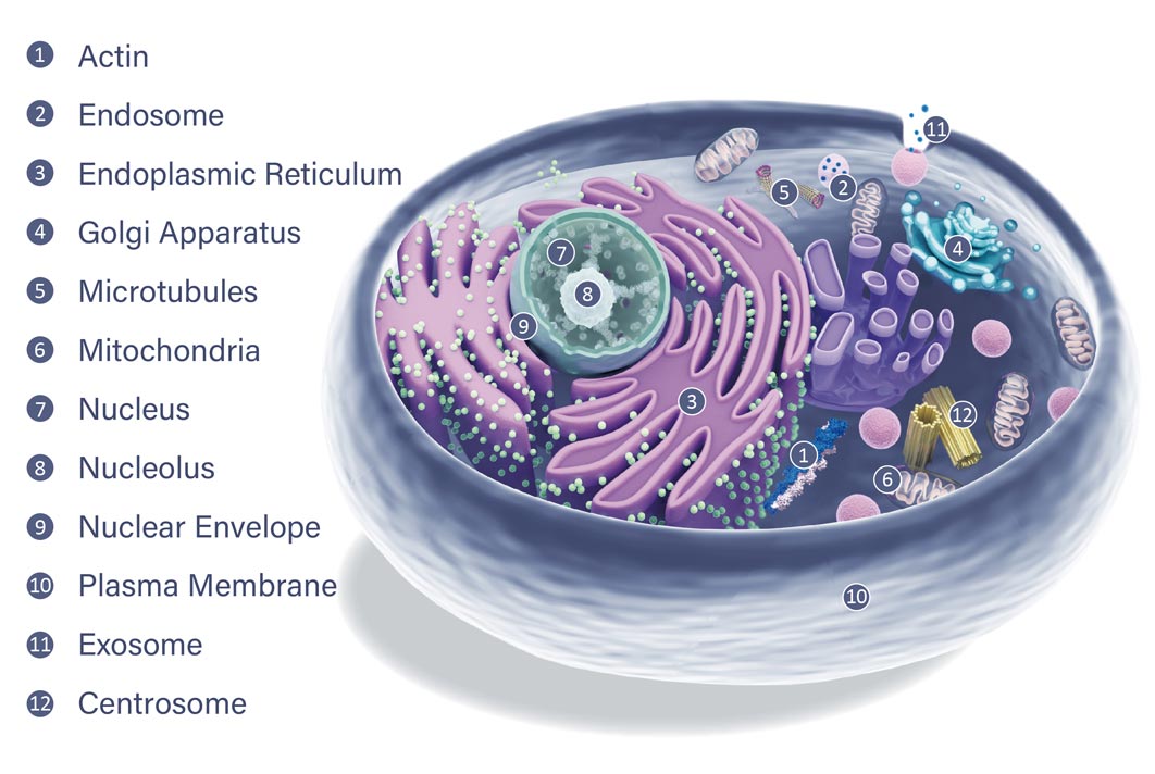
The subcellular location of a protein may suggest potential roles for that factor in one or more cellular processes. Organelle protein-specific antibodies are essential for establishing colocalization of a particular protein of interest with an organelle, thereby contributing crucial insight into its possible function(s). In addition, these organelle marker antibodies can often be used in cell fractionation studies analyzed by western blot alone or after immunoprecipitation.
Organelle Markers
|
|
|||||
| Organelle | Product Name | Clonality | Reactivity | Applications | Cat. No. | |
| Actin | beta Actin antibody | Rb pAb | Hu, Ms, Dm, Rat, Yeast, Cat, Rice, C. albicans | ICC/IF, IHC-P, IP, WB, IB | GTX109639 | |
| Cytoplasm | HSPA1A antibody | Rb pAb | Hu, Ms, Rat | ICC/IF, IHC-P, IP, WB | GTX111088 | |
| Plasma membrane | pan Cadherin antibody [CH-19] | Ms mAb | Hu, Ms, Chk, Dog, Pig, Rat, Cat, Goat, Gpig, Hm, Rb, SRat, Shp, Snk, Bov | ICC/IF, IHC-P, WB | GTX26528 | |
| Endoplasmic Reticulum | GRP94 antibody | Rb pAb | Hu, Ms, Rat | ICC/IF, IHC-P, IP, WB | GTX103232 | |
| Endoplasmic Reticulum | Calnexin antibody [C3], C-term | Rb pAb | Hu, Ms, Rat, Shp | ICC/IF, IHC-P, IP, WB | GTX109669 | |
| Golgi Apparatus | GOLPH2 antibody | Rb pAb | Hu | ICC/IF, IHC-P, WB | GTX116154 | |
| Golgi Apparatus | GOLGA5 antibody [N2C2], Internal | Rb pAb | Hu, Ms | ICC/IF, IHC-P, WB | GTX104255 | |
| Junction (Tight Junction) | ZO-1 antibody [N1N2], N-term | Rb pAb | Hu, Ms | ICC/IF, IHC-P, WB | GTX108613 | |
| Junction (Gap Junction) | Connexin 43 antibody | Rb pAb | Hu, Ms, Rat | IHC-P, WB | GTX50571 | |
| Centromere | gamma Tubulin antibody | Rb pAb | Hu, Ms, Rat, Zfsh | ICC/IF, IHC, IHC-F, IHC-P, IP, WB | GTX113286 | |
| Kinetochores (M phase) | ZWINT antibody | Rb pAb | Hu | ICC/IF, WB | GTX107155 | |
| Kinetochores (M phase) | HEC1 antibody [9G3.23] | Ms mAb | Hu, Ms, Pig, Rat, Hm, Kg, AGMK | FACS, ICC/IF, IHC, IP, WB | GTX70268 | |
| Lysosome | LAMP2 antibody | Rb pAb | Hu | ICC/IF, IHC-P, WB | GTX103214 | |
| Lysosome | GALNS antibody | Rb pAb | Hu, Ms, Rat | ICC/IF, IHC-P, WB | GTX110237 | |
| Endosome | RAB7A antibody | Rb pAb | Hu, Ms, Rat | ICC/IF, IHC-P, WB | GTX132548 | |
| Exosome | CD9 antibody [MM2/57] | Ms mAb | Hu, Dog, Pig, Cat, Frt, Hrs, Mink, Rb, Bov, RMK, Lma | Blocking, FACS, IHC-Fr, WB | GTX76184 | |
| Exosome | CD63 antibody [MEM-259] | Ms mAb | Hu | FACS, ICC/IF, IHC-P, IP, WB | GTX28219 | |
| Exosome | CD81 antibody | Rb pAb | Hu | Blocking, FACS, ICC/IF, IHC-P, WB | GTX101766 | |
| Exosome | TSG101 antibody [4A10] | Ms mAb | Hu, Ms, Mk, Rat, Hm | ELISA, FACS, ICC/IF, IHC, IHC-P, IP, WB | GTX70255 | |
| Autophagosome | LC3B antibody | Rb pAb | Hu, Ms, Pig, Rat | IP, FACS, ICC/IF, IHC-P, WB | GTX127375 | |
| Autophagosome | SQSTM1 antibody [N3C1], Internal | Rb pAb | Hu, Ms, Rat, Zfsh, Bov, Honeybee | FACS, ICC/IF, IHC-P, IP, WB | GTX100685 | |
| Microtubules/ Spindle | alpha Tubulin antibody [GT114] | Ms mAb | Hu, Ms, Dm, Rat, Zfsh | ICC/IF, IHC-Fr, IHC-P, WB | GTX628802 | |
| Mitochondria (Inner Membrane) | COX4 antibody | Rb pAb | Hu, Ms, Rat | ICC/IF, IHC-P, WB | GTX114330 | |
| Mitochondria (Inner Membrane) | Prohibitin antibody | Rb pAb | Hu, Ms, Rat | ICC/IF, IHC-P, IP, WB | GTX101105 | |
| Mitochondria (Outer Membrane) | VDAC2 antibody [C2C3], C-term | Rb pAb | Hu, Ms | ELISA, ICC/IF, IHC-P, IP, WB | GTX104745 | |
| Nuclear Envelope | Lamin A + C antibody | Rb pAb | Hu, Ms, Rat | ICC/IF, IHC-P, IP, WB | GTX101127 | |
| Nuclear Envelope | POM121 antibody [N2N3] | Rb pAb | Hu, Ms | ICC/IF, IHC-P, WB | GTX102128 | |
| Nucleolus | PAF49 antibody | Rb pAb | Hu | ICC/IF, IHC-P, WB | GTX102175 | |
| Nucleolus | RPA70 antibody [C1C3] | Rb pAb | Hu, Ms | ICC/IF, IHC-P, IP, WB | GTX108749 | |
| Nucleus | PCNA antibody | Rb pAb | Hu, Ms, Rat | ICC/IF, IHC, IHC-P, IP, WB | GTX100539 | |
| Nucleus | HDAC1 antibody | Rb pAb | Hu, Ms, Rat | ICC/IF, IHC-P, IP, WB, ChIP assay | GTX100513 | |
| Nucleus | HDAC2 antibody | Rb pAb | Hu, Ms, Rat | ICC/IF, IHC-P, IP, WB, ChIP assay | GTX109642 | |
| Internal Controls | Vinculin antibody [N3C1], Internal | Rb pAb | Hu, Ms, Rat | IHC-P, WB | GTX109749 | |
| Internal Controls | Cofilin 1 antibody | Rb pAb | Hu, Ms, Rat | WB, ICC/IF, IHC-P, IHC-Fr | GTX102156 | |
| Internal Controls | Cyclophilin A antibody | Rb pAb | Hu, Ms, Rat, Hm | WB, ICC/IF, IHC-P | GTX104698 | |
| Internal Controls | Histone H3 antibody | Rb pAb | Hu, Ms, Rat, Dm, Mk, Fungi, Rice | WB, ICC/IF, IHC-P, IP, ChIP assay | GTX122148 | |
| Internal Controls | HSP60 antibody | Rb pAb | Hu, Ms, Rat, Hm | WB, ICC/IF, IHC-P, ELISA | GTX110089 |
Endoplasmic Reticulum (ER) |
|||||
|
|
|||||
Golgi Apparatus |
|||||
|
|
|||||
Endosome |
|||||
|
|||||
Exosome |
|||||
|
|
|||||
Lysosome |
|||||
|
|
|||||
Proteasome |
|||||
|
|
|||||
Ribosome |
|||||
|
|
|||||
Peroxisome
Plasma Membrane |
|||||
|
|
|||||
Adhesion Junction |
|||||
|
|
|||||
Tight Junction |
|||||
|
|
|||||
Desmosome
Autophagosome |
|||||
|
|
|||||
Mitochondria |
|||||
|
|
|||||
Nuclear Envelope |
|||||
|
|
|||||
Nucleus |
|||||
|
|
|||||
Nucleolus |
|||||
|
|
|||||
Intermediate Filament |
|||||
|
|
|||||
Actin |
|||||
|
|
|||||
Tubulin |
|||||
|
|
|||||
Centrosome |
|||||
|
|
|||||
Kinetochore |
|||||
|
|
|||||
Focal Adhesion |
|||||
|
|
|||||
Highlighted Products
| Name | Catalogue Number |
| Fibrillarin antibody | GTX113684 |
| Fibrillarin antibody | GTX101807 |
| PAF49 antibody [GT635] | GTX629076 |
| Fibrillarin antibody [38F3] | GTX24566 |
| PAF49 antibody | GTX102175 |
| Fibrillarin antibody | GTX65859 |
| PAF49 antibody [GT1964] | GTX629115 |
| PAF49 antibody [GT7212] | GTX629075 |
| EIF6 antibody [N1C3-2] | GTX117971 |
| Fibrillarin antibody | GTX32604 |
| EIF6 antibody | GTX54010 |
| Fibrillarin antibody [38F3] | GTX82695 |
Highlighted Products
| Name | Catalogue Number |
| Lamin A + C antibody | Cat No. GTX101127 |
| Lamin A + C antibody [GT1137] | Cat No. GTX00774 |
| Lamin B2 antibody [GT144] | Cat No. GTX628803 |
| Lamin A + C antibody | Cat No. GTX101126 |
| Lamin A + C antibody [GT9712] | Cat No. GTX629404 |
| Lamin A + C antibody | Cat No. GTX111677 |
| Lamin A + C antibody [mab636] | Cat No. GTX80813 |
| Lamin B2 antibody [N3C2], Internal | Cat No. GTX109894 |
| Lamin B2 antibody | Cat No. GTX110309 |
| Lamin B2 antibody | Cat No. GTX33292 |
| Lamin A + C antibody | Cat No. GTX13910 |
| Lamin A antibody [4E7] | Cat No. GTX60478 |
| Lamin A antibody [JOL4] | Cat No. GTX80380 |
| Lamin A + C antibody [JoL3] | Cat No. GTX24789 |
| Lamin A + C antibody [JoL5] | Cat No. GTX25090 |
Highlighted Products
| Name | Catalogue Number |
| gamma Tubulin antibody | Cat No. GTX113286 |
| gamma Tubulin antibody [GT4511] | Cat No. GTX629704 |
| gamma Tubulin antibody [4D11] | Cat No. GTX15794 |
| gamma Tubulin antibody [TU-30] | Cat No. GTX79861 |
| gamma Tubulin antibody [GTU-88] | Cat No. GTX11316 |
| gamma Tubulin antibody [TU-32] | Cat No. GTX79860 |
| gamma Tubulin antibody | Cat No. GTX11317 |
| gamma Tubulin antibody | Cat No. GTX11321 |
| gamma Tubulin antibody (Cy3) | Cat No. GTX11319 |
Highlighted Products
| Name | Catalogue Number |
| Hec1 antibody [9G3.23] | Cat No. GTX70268 |
| Hec1 (phospho Ser 55) antibody | Cat No. GTX70017 |
| Hec1 (phospho Ser 165) antibody | Cat No. GTX70013 |
| Hec1 antibody | Cat No. GTX110735 |
| Hec1 antibody | Cat No. GTX70016 |
| Hec1 (non-phospho Ser 165) antibody | Cat No. GTX70012 |
| Hec1 (non-phospho Ser 76/77) antibody | Cat No. GTX70014 |
| Hec1 (phospho Ser 76/77) antibody | Cat No. GTX70015 |
| Hec1 (phospho Ser 165) blocking peptide | Cat No. GTX70013-PEP |
| Hec1 (non-phospho Ser 165) blocking peptide | Cat No. GTX70012-PEP |
| Hec1 (non-phospho Ser 55) blocking peptide | Cat No. GTX70016-PEP |
| Hec1 (phospho Ser 76/77) blocking peptide | Cat No. GTX70015-PEP |
| Hec1 (phospho Ser 55) blocking peptide | Cat No. GTX70017-PEP |
| Hec1 antibody, C-term | Cat No. GTX23393 |
| Hec1 antibody [EPR5342] | Cat No. GTX63082 |
| Hec1 antibody | Cat No. GTX32648 |
| Hec1 blocking peptide | Cat No. GTX23393-PEP |
Highlighted Products
| Name | Catalogue Number |
| RPS6 antibody | Cat No. GTX113542 |
| RPL17 antibody [N1C3-2] | Cat No. GTX111934 |
| RPS6 (phospho Ser235/236) antibody | Cat No. GTX130430 |
| RPS6 (phospho Ser235) antibody | Cat No. GTX132281 |
| RPS6 (phospho Ser235) antibody [GT4610] | Cat No. GTX633811 |
| RPS6 antibody | Cat No. GTX130450 |
| RPS6 (phospho Ser240/Ser244) antibody | Cat No. GTX133942 |
| RPS6 (phospho Ser235) antibody [GT829] | Cat No. GTX633823 |
| RPL4 antibody | Cat No. GTX112184 |
| RPL17 antibody | Cat No. GTX66472 |
| RPS6 (phospho Ser235/236) antibody | Cat No. GTX12864 |
| RPS6 (phospho Ser240/Ser244) antibody | Cat No. GTX66604 |
| RPL17 antibody [N1C3] | Cat No. GTX101831 |
| RPS6 (phospho Ser244/247) antibody | Cat No. GTX12865 |
| RPS6 (phospho Ser235) antibody | Cat No. GTX78950 |
| RPS6 (phospho Ser236) antibody | Cat No. GTX17944 |
| RPL4 antibody | Cat No. GTX66468 |
| Human RPL17 protein, His tag | Cat No. GTX111934-pro |
| RPS6 (phospho Ser240) antibody | Cat No. GTX54985 |
| RPL17 antibody | Cat No. GTX87817 |
| RPS6 antibody | Cat No. GTX50552 |
| RPS6 (phospho Ser235) antibody | Cat No. GTX50267 |
| RPS6 (phospho Ser235) antibody [9H270] | Cat No. GTX15031 |
| RPL17 antibody, N-term | Cat No. GTX88967 |
| RPL17 blocking peptide | Cat No. GTX88967-PEP |
Highlighted Products
| Name | Catalogue Number |
| PEX19 antibody | Cat No. GTX110721 |
| PEX19 antibody [GT554] | Cat No. GTX628212 |
| ACAA1 antibody | Cat No. GTX114230 |
| ACAA1 antibody | Cat No. GTX114229 |
| PEX19 antibody | Cat No. GTX114959 |
| PEX19 antibody [GT533] | Cat No. GTX628892 |
| PEX26 antibody | Cat No. GTX109551 |
| Human PEX26 protein, His tag | Cat No. GTX109551-pro |
| PEX19 antibody | Cat No. GTX32780 |
| ACAA1 antibody | Cat No. GTX32986 |
| ACAA1 antibody [AT9E5] | Cat No. GTX57678 |
| PEX26 antibody, Internal | Cat No. GTX88069 |
| PEX26 blocking peptide | Cat No. GTX88069-PEP |
| Human PEX19 protein | Cat No. GTX48152-pro |
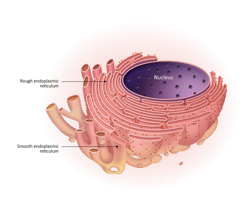

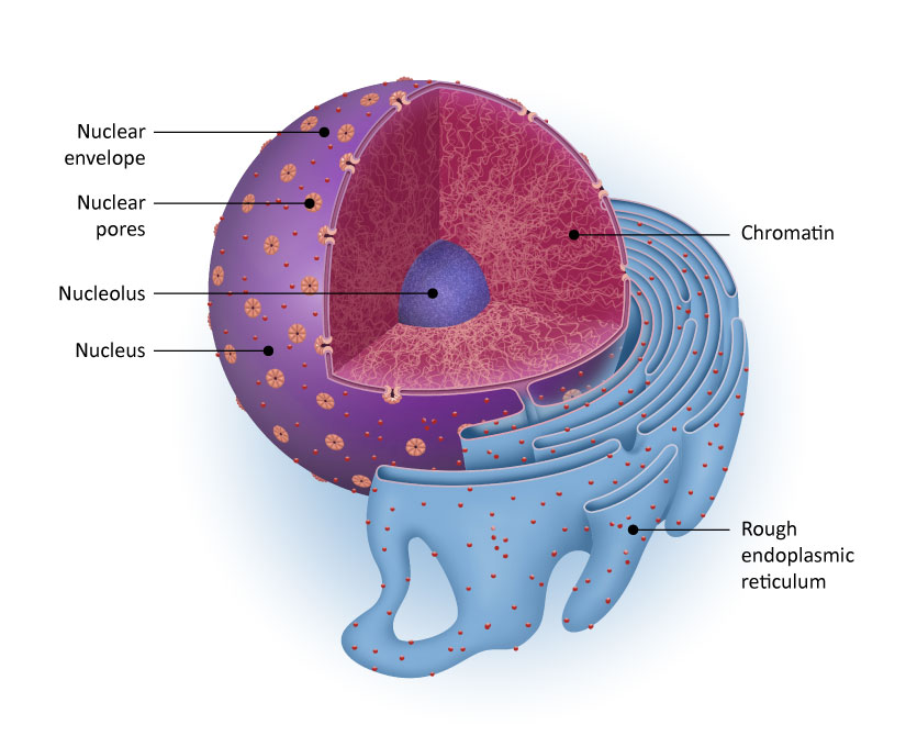
The endoplasmic reticulum (ER) is a eukaryotic membranous organelle consisting of flattened, connected sacs (cisternae) that is responsible for synthesis of membrane and secretory proteins and their subsequent proper folding and posttranslational decoration. It can be divided into two distinct types: rough endoplasmic reticulum (RER) and smooth endoplasmic reticulum (SER). The RER is studded with ribosomes to synthesize membrane and secreted proteins and has some continuity with the nuclear membrane lumen. No ribosomes are associated with the SER, which is involved in lipid metabolism. Soluble proteins are transported from the ER to the Golgi apparatus by way of vesicles for further modification, and are subsequently released from the Golgi in vesicles destined to fuse with the cell membrane. ER quality control mechanisms are triggered when accumulation of unfolded or misfolded proteins is detected by various sensors, including PERK, IRE1, and ATF6. ER stress is associated with a variety of conditions including cancer, diabetes, inflammatory diseases, and neurodegenerative disorders.
GeneTex is proud to offer an outstanding selection of antibodies for endoplasmic reticulum research. These antibodies are validated for various applications and are an important component of our reagent catalog supporting studies of organelle proteins. Please see the highlighted products below.
Highlighted Products
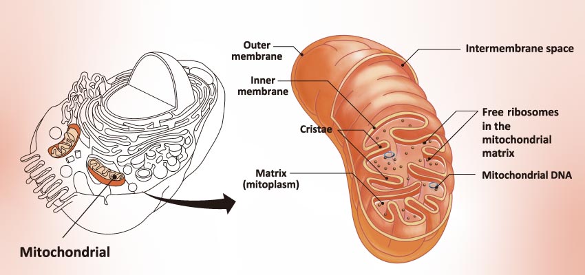

Mitochondria are dual-membrane organelles essential for the production of >90% of the metabolic energy in the eukaryotic cell. Their structural features include inner and outer membranes that bound an intermembrane space. The inner membrane is arranged into folds called cristae, which reach into the central matrix. Mitochondrial dysfunction has been linked to such natural processes as aging in addition to being a key factor in a plethora of neurodegenerative, cardiovascular, metabolic, autoimmune, and neoplastic pathologies that plague humans. GeneTex proudly offers a selection of high quality mitochondrial markers.
Highlighted Products
Highlighted Products
| Name | Catalogue Number |
| RAB7A antibody | Cat No. GTX130847 |
| RAB7A antibody | Cat No. GTX132548 |
| RAB11B antibody [N1C3] | Cat No. GTX119095 |
| RAB7A antibody [Rab7-117] | Cat No. GTX16196 |
| RAB7A antibody | Cat No. GTX02417 |
| Human RAB7A protein, His tag | Cat No. GTX109102-pro |
| RAB7A antibody [AT10E4] | Cat No. GTX57709 |
| Human RAB7A protein, His tag | Cat No. GTX67953-pro |
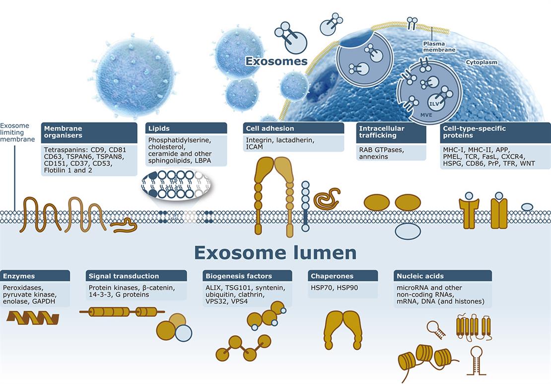

The term “extracellular vesicle” refers to a highly diverse group of membrane-derived, cargo-bearing (e.g., proteins, lipids, nucleic acids) structures that represent an important mode of intercellular communication. Based on the type of membrane from which they originate, extracellular vesicles have been divided into two broad groups: exosomes (derived from inward budding of endosomal membranes in larger structures known as multi-vesicular endosomes (MVEs)) and microvesicles (generated from outward budding of the plasma membrane).
While much is known about microvesicles and exosomes, key aspects of their biogenesis, cargo, release from cells, and targeting to recipient cells remain unclear. Complicating this research is that many features of extracellular vesicles are influenced by cell-type, stimuli from normal or pathological milieus, and the physiological state of the cell. Nevertheless, they are involved in many processes that include coagulation, immune function, and tumorigenesis. Since they are found in biological fluids, there is intense research directed at potentially using extracellular vesicles as biomarkers. Other efforts are being directed at utilizing them to carry compounds or to invoke particular responses in target cells.
GeneTex is proud to offer a set of well-validated antibodies to facilitate your exosome & microvesicle research in various applications and species. See below for the highlighted antibodies.
Highlighted Products
Highlighted Products
| Name | Catalogue Code |
| LAMP1 antibody [GT25212] | Cat No. GTX634336 |
| LAMP2 antibody | Cat No. GTX103214 |
| LAMP2 antibody [GT856] | Cat No. GTX635790 |
| LAMP1 antibody [07] | Cat No. GTX02073 |
| LAMP2A antibody | Cat No. GTX132655 |
| LAMP2 antibody [N2C2], Internal | Cat No. GTX111812 |
| LAMP2 antibody [GL2A7] | Cat No. GTX13524 |
| LAMP1 antibody | Cat No. GTX130356 |
| LAMP1 antibody [Ly1C6] | Cat No. GTX13523 |
| LAMP2 antibody | Cat No. GTX30881 |
| LAMP1 antibody | Cat No. GTX19294 |
| LAMP1 antibody | Cat No. GTX33293 |
| LAMP1 antibody [4E9/11] | Cat No. GTX31229 |
| LAMP2 antibody [M3/84] | Cat No. GTX42498 |
| LAMP2 antibody [M3/84] | Cat No. GTX42497 |
| LAMP2 antibody | Cat No. GTX32699 |
| LAMP2 antibody | Cat No. GTX66812 |
| LAMP1 antibody [1D4B] | Cat No. GTX42501 |
| LAMP2 antibody [AC17] (FITC) | Cat No. GTX43775 |
| LAMP1 antibody [4E9/11] (FITC) | Cat No. GTX43372 |
| LAMP2 blocking peptide | Cat No. GTX30881-PEP |
Highlighted Products
| Name | Catalogue Number |
| PEX19 antibody | Cat No. GTX110721 |
| PEX19 antibody [GT554] | Cat No. GTX628212 |
| ACAA1 antibody | Cat No. GTX114230 |
| ACAA1 antibody | Cat No. GTX114229 |
| PEX19 antibody | Cat No. GTX114959 |
| PEX19 antibody [GT533] | Cat No. GTX628892 |
| PEX26 antibody | Cat No. GTX109551 |
| Human PEX26 protein, His tag | Cat No. GTX109551-pro |
| PEX19 antibody | Cat No. GTX32780 |
| ACAA1 antibody | Cat No. GTX32986 |
| ACAA1 antibody [AT9E5] | Cat No. GTX57678 |
| PEX26 antibody, Internal | Cat No. GTX88069 |
| PEX26 blocking peptide | Cat No. GTX88069-PEP |
| Human PEX19 protein | Cat No. GTX48152-pro |
Highlighted Products
Highlighted Products
| Name | Catalogue Number |
| beta Actin antibody | Cat No. GTX109639 |
| beta Actin antibody [GT5512] | Cat No. GTX629630 |
| beta Actin antibody | Cat No. GTX110564 |
| beta Actin antibody [AC-15] | Cat No. GTX26276 |
| beta Actin antibody | Cat No. GTX100313 |
| alpha Actinin 2 antibody [N1N3] | Cat No. GTX103219 |
| beta Actin antibody | Cat No. GTX124214 |
| alpha Actinin 4 antibody [C2C3], C-term | Cat No. GTX101669 |
| alpha Actinin 4 antibody [N2C1], Internal | Cat No. GTX113115 |
| beta Actin antibody | Cat No. GTX30632 |
| alpha Actinin 2 antibody [N2C1], Internal | Cat No. GTX111167 |
| beta Actin antibody | Cat No. GTX100315 |
| alpha Actinin 3 antibody | Cat No. GTX103216 |
| alpha Actinin 4 antibody [N1N2], N-term | Cat No. GTX113116 |
| alpha Actinin 1 antibody [N2N3] | Cat No. GTX103240 |
| alpha Actinin 4 antibody [7H6] | Cat No. GTX15648 |
| beta Actin antibody [8H10D10] | Cat No. GTX83163 |
| alpha Actinin 2 antibody [GT1253] | Cat No. GTX632361 |
| beta Actin antibody [RM112] | Cat No. GTX33610 |
| beta Actin antibody | Cat No. GTX21801 |
| alpha Actinin 1 antibody | Cat No. GTX55505 |
| beta Actin antibody | Cat No. GTX31732 |
| alpha Actinin 1 antibody [0.T.02] | Cat No. GTX18061 |
| alpha Actinin 1 antibody [AT1D10] | Cat No. GTX57633 |
| beta Actin antibody | Cat No. GTX85117 |
| alpha Actinin 3 antibody | Cat No. GTX54908 |
| beta Actin antibody | Cat No. GTX85119 |
| Fractin antibody | Cat No. GTX82694 |
| beta Actin blocking peptide | Cat No. GTX31732-PEP |
