The conventional approach to commercial antibody production has improved remarkably since its inception well over 40 years ago. The development of reliable antibody reagents remains an essential goal in the quest to advance science and medicine. On the forefront of high-quality antibody manufacturing, GeneTex has solidified its position in the research community and the biotechnology industry.
Our experience and expertise can advance your research.
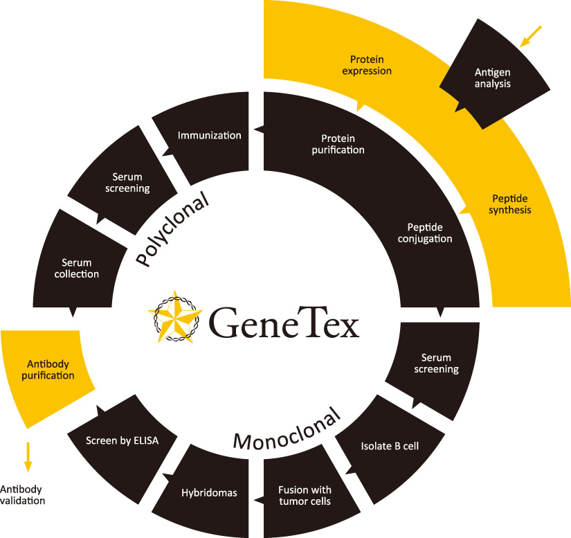
Let our team of antibody production experts help you develop a novel polyclonal or recombinant monoclonal antibody against your target of interest, with superior specificity, sensitivity, and application utility. GeneTex provides exceptional, comprehensive service that spans antigen preparation, animal immunization, antibody screening and purification, and recombinant cloning if desired. Our efficient workflow is guided by multiple production and performance checkpoints that are customized for each project, as are our pricing packages.
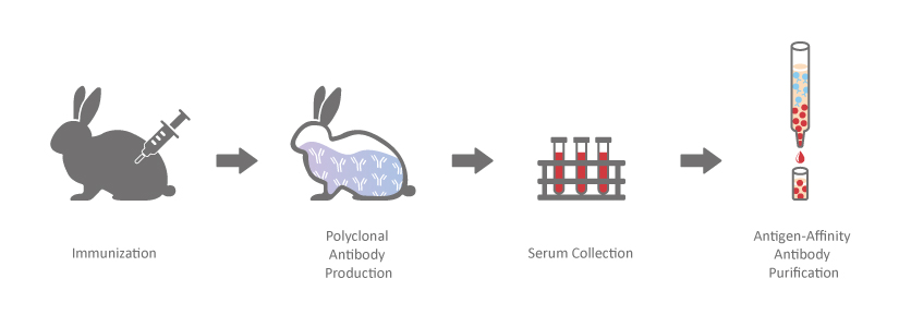
Polyclonal antibodies are a heterogeneous mix of immunoglobulins derived from various B cells that individually target distinct epitopes on an immunogen. GeneTex has produced over ten thousand polyclonal antibodies in its history, resulting in exceptional manufacturing and quality control expertise that translates into reliable antibody reagents. Let GeneTex help you develop the ideal polyclonal antibodies to accelerate your research progress.
Key Design and Production Features
Professional epitope design to optimize immunogen synthesis, solubility and antigenicity properties.
Efficient production of high-quality peptide or protein immunogens.
Enhanced cocktail immunization techniques.
Antigen-affinity chromatography purification of antibodies.
GeneTex’s recombinant antibody protocol involves cloning antibody-coding DNA from the peripheral blood B cells of an immunized rabbit or from a monoclonal hybridoma, employing an innovative technology to screen and identify antibodies that fit your specific application goals. Importantly, our FACS-based method preserves the natural pairing of the heavy and light chains from a single B cell or hybridoma cell, thereby maintaining the matched antibody variable regions. The entire procedure starting with FACS sorting requires only a few weeks. In addition, the generated recombinant antibody can be engineered into various Fc backbones, including rabbit, mouse, rat, or human IgG depending on your needs. And once cloned, your antibody is inexhaustible with exceptional reproducibility.
With a 20-year history of quality results, GeneTex sets itself apart from other antibody companies by taking the necessary steps to properly validate our antibodies. Antibody validation remains an industry-wide challenge with ramifications that can impact progress in the life sciences and medicine.
At GeneTex, product reliability is our primary concern. Stringent validation protocols have been established to test our antibodies for various applications, including WB, IP, IHC, IFA, FACS, and ELISA, using cell lines with known target protein expression levels and a diverse array of tissue samples. We know the importance of replicating results, so quality consistency between lots is maintained confidently through a combination of pre-qualified individual lots, dominant clones, and thorough quality control.
Founded by scientists, for scientists, we at GeneTex understand that researchers expect and demand product performance and fidelity. With GeneTex’s Knockout/Knockdown validation project, we’re committed to demonstrating to our customers that the products they are purchasing are completely reliable, every single time.
Our Commitment
There has been growing distrust of commercially available antibodies with regard to specificity and performance variability. As a result, the research community has demanded that antibody suppliers increase their efforts to more thoroughly validate their products. One approach to address this is through knockout or knockdown of the target protein.
At GeneTex, we continue to provide the research community with the highest quality and most stringently validated antibodies by utilizing CRISPR/Cas9- and shRNA-generated cell lysates for our western blot assays. To date, we have evaluated most of our featured products and will continue to expand these efforts throughout our entire antibody catalogue.
GeneTex primary antibodies have been tested through the use of various analytic strategies to ensure persistent quality and confirm antibody specificity. Based on the guidelines described by the International Working Group on Antibody Validation (IWGAV), GeneTex has been employing multiple approaches to certify antibody performance in our quality assurance process.
See GeneTex’s Validation Methods
GeneTex understands the absolute necessity for reliable antibodies to achieve accurate and reproducible experimental results. To optimize the performance of our reagents, we employ various analytic validation strategies to ensure both consistent quality (see GeneTex’s Approach to Antibody Lot-to-Lot Variability) and specificity. These modalities are in line with guidelines described by the International Working Group on Antibody Validation (IWGAV) and have become fundamental components of our quality assurance process:
Polyclonal antibodies generated by an immunized animal consist of a heterogeneous mix of immunoglobulins derived from many B-cells that individually target distinct epitopes on an antigen. Often representing up to 5% of the total immunoglobulin, these antibodies are generally purified from the immune sera using antigen-affinity chromatography. Polyclonal antibodies are particularly useful for detecting proteins that are expressed at lower levels, and their performance is impacted to a lesser degree by alterations in the antigen (e.g., denaturation). GeneTex offers an extensive catalogue of affinity-purified polyclonal antibodies directed against almost all key targets/proteins in the spectrum of biomedical research.
Monoclonal antibodies are produced by immortal hybridoma cells created from the fusion of antibody-producing splenic B-cells and myeloma cells, or by recombinant protein expression in mammalian cells. These antibodies have monovalent affinity and bind to a single epitope on the antigen. Purification is commonly accomplished through Protein A/G/L affinity chromatography.
GeneTex offers a broad selection of monoclonal antibodies directed against the majority of key targets/proteins in the spectrum of biomedical research.
A recombinant antibody (rAb) is generated from the cloned, in vitro-expressed heavy and light chains of a selected monoclonal antibody obtained through classical hybridoma methodology, display strategies alone or in combination, sorted single B cells from immunized animals, or by one of many other techniques. The fact that it is cloned means the rAb is defined by its primary sequence, which can be engineered to optimize affinity and be expressed in different binder formats. The numerous advantages of rAbs have been well-documented, with consistency of performance being of paramount significance.
GeneTex’s rAb protocol employs a multi-parameter FACS-based approach to isolate antigen-specific IgG+ memory B cells from an immunized animal, with subsequent cloning of the antibody variable-region genes into an IgG backbone and expression in mammalian cells. This protocol is very rapid and can be completed in weeks, and also affords the opportunity to identify antibodies with diverse capabilities in various applications. Importantly, it allows cloning of the heavy and light chains from the same B cell, thereby preserving natural pairing. And once cloned, the supply of a given rAb is inexhaustible with exceptional reproducibility.
One outstanding advantage of this protocol is that the suitability of the individual clones for desired applications can be tested during screening. For example, GeneTex’s Iba1 rabbit recombinant antibody [HL22] (GTX635363) detects Iba1, a protein commonly used as an immunohistochemical marker of both quiescent and activated microglia. In its development and production, both paraffin- and frozen-IHC analyses (IHC-P, IHC-Fr, respectively) were conducted in the first screening process to identify the best clones for these applications, as shown below (Figure 1). The clones were compared to a highly cited, market-leading commercial antibody to gauge their performance. This strategy provides us with valuable perspective on a clone’s market competitiveness during development. In addition, our recombinant antibody team has successfully expressed the antigen-binding regions of this antibody in the context of both mouse and rat IgG backbones, thus extending flexibility for multiple staining.
Figure 1. GeneTex’s recombinant rabbit monoclonal Iba1 antibody [HL22] (GTX635363) is superior to a competitor’s highly cited rabbit polyclonal antibody for both IHC-P (panels A vs. D) and IHC-Fr (panels B, C vs. E, F).
Another new rabbit recombinant product is GeneTex’s Ras (G12D mutant) antibody [HL10] (GTX635362), which specifically detects the G12D mutant by western blot with minimal background (Figure 2). Most importantly, it can also be used for IHC-P, where it demonstrates a robust signal and clear specificity for the G12D mutation in WT and KRAS G12D pancreatic tumor tissue sections (Figure 2A). This Ras (G12D mutant) antibody (GTX635362) is the first commercial recombinant version that shows exceptional specificity by IHC-P for this key tumorigenic mutant protein on sequence-verified human pancreatic tumor samples.
Figure 2. (A) GeneTex’s recombinant rabbit RAS (G12D mutant) antibody [HL10] (GTX635362) is sensitive and specific for the RAS G12D mutation by IHC of a KRAS G12D mutant pancreatic tumor tissue section (top) compared to a wild-type KRAS section (bottom). (B) The antibody is sensitive and specific for the RAS G12D mutation by WB of extracts from wild-type and mutant KRAS-confirmed human pancreatic cell lines. GAPDH for the loading control was detected by GTX100118. Lane1: HPDE. Lane2: HPNE. Lane3: AsPC1 (KRAS G12D). Lane4: BxPC3 (KRAS WT). Lane5: CFPAC1 (KRAS G12V). Lane6: HPAC (KRAS G12D). Lane7: HPAF-II (KRAS G12D). Lane8: MIA PaCa-22 (KRAS G12C). Lane9: PANC1 (KRAS G12D). Lane10: SU86.86 (KRAS G12D).
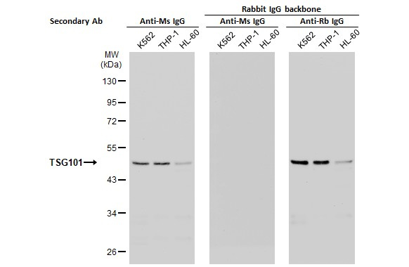
As mentioned above, the antigen-binding regions of a recombinant antibody can be inserted into various host IgG backbones or expressed in different binder formats (e.g., Fab fragments or scFvs). Here, GeneTex’s cloned TSG101 mouse monoclonal antibody (GTX70255) was converted to a rabbit IgG backbone (GTX635396) with preservation of performance. No cross-reaction was observed when the converted TSG101 rabbit IgG recombinant antibody was used in combination with an anti-mouse IgG secondary antibody (Figure 3).
Figure 3. Switch from a cloned mouse monoclonal antibody to a rabbit IgG backbone
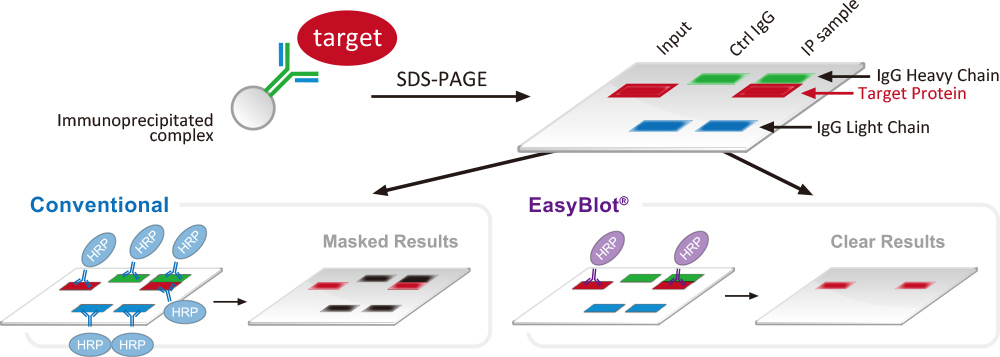
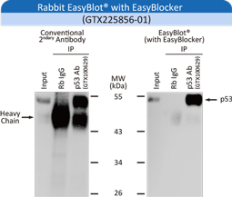
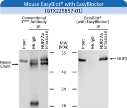
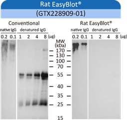
GeneTex offers over 1000 secondary antibodies with different conjugates to give our customers full flexibility in their experimental design. We also offer our specialized EasyBlot® secondary antibodies for the specific recognition of non-denatured primary antibodies used for the western blot assays that commonly follow IP/co-IP protocols.
GeneTex antibody panels are a convenient and economical option for researchers.
Each panel contains carefully selected high-quality and highly cited antibodies in small amounts (25μl) directed against related targets involving signaling pathways, protein modifications, or specific cell markers. In addition, a secondary antibody is included to facilitate western blot analyses.
GeneTex offers validated antibody pairs for ELISA. These ELISA antibody pairs and kits are made for quantification of various cytokines, growth factors, enzymes, immunoglobins, and extracellular matrix components. They are available for human, mouse, rat and other species.
Inclusion of an appropriate isotype control is essential for the proper interpretation of experimental results. Because isotype control antibodies have no relevant specificity, they enable the researcher to distinguish non-specific “background” binding from antigen-specific antibody binding. Non-specific binding of primary antibodies may occur due to either binding of antibody to Fc receptors or non-specific interactions with cellular proteins, lipids, or carbohydrates. Additionally, autofluorescence or endogenous enzyme activity in target cells may give a false positive that could be misinterpreted as antibody binding.
These factors vary by target cell type and the isotype of the primary antibody. Therefore, the primary antibody used should be matched to an isotype control of identical species and isotype (including both heavy and light chains).
GeneTex offers a comprehensive catalog of recombinant proteins and peptides, including growth factors, cytokines, CD antigens, signaling proteins, Fc-fusion proteins and more. These high-quality and high-purity products are guaranteed to assist your various research needs.
Choose from thousands of bioactive proteins for functional testing involving physiological and pathological studies, enzyme assays, and functional ELISAs.
In Parkinson’s disease (PD), alpha-synuclein protein forms aggregates in brain neurons. Termed ‘Lewy bodies’, these deposits are associated with neuronal death. Ongoing research is trying to identify ways to prevent alpha-synuclein accumulation, promote its removal, or trigger its recycling. GeneTex is proud to offer a set of active recombinant proteins for PD research, including preformed fibrils of human alpha-synuclein protein (GTX17669-pro), mouse alpha-synuclein protein (GTX17671-pro), and human tau protein (GTX17675-pro). The activity of these proteins was assessed using various approaches. For example, the fibrillar structures of the human and mouse proteins were visualized by transmission electron microscopy (TEM), as shown below.
When primary rat hippocampal neurons were treated with human alpha-synuclein protein (GTX17669-pro) or mouse alpha-synuclein protein (GTX17671-pro), the subsequent Lewy body inclusions were detected by immunostaining. Furthermore, immunohistochemistry revealed fibril aggregation in rat brain 30 days after injection of the active alpha-synuclein protein.
GeneTex also offers an active human Interferon-gamma (IFN-γ) (GTX00083-pro). IFN-γ is a dimerized soluble cytokine that is the only member of the type II class of interferons. The importance of IFN-γ in the immune system stems in part from its ability to inhibit viral replication directly, and most importantly from its immunostimulatory and immunomodulatory effects. IFN-γ is an important activator of human monocytic THP-1 cells. THP-1 cells treated with IFN-γ acquire a fusiform or polygonal morphology and become more adherent, as shown below.
Another example is tumor necrosis factor (TNF) protein. TNF has marked anti-tumor effects. However, it may also cause anemia and hepatic dysfunction in patients. The effective dose of TNF can be reduced by promoting its activity with actinomycin D. A proliferation assay was performed on murine L929 cells treated with GeneTex’s human TNF-alpha protein (active) (GTX65267-pro) and actinomycin D to examine cell toxicity, as shown below. The results demonstrate the utility of this assay to evaluate the in vitro cytotoxic effect of TNF-alpha in anti-tumor research.
Enzyme Assays
Enzyme activation is achieved through different molecular modifications, including cleavage, phosphorylation and acetylation. These reactions are common during normal and pathological cellular processes. GeneTex provides a series of active proteins to initiate enzymatic signaling. For example, phospho-ERK1/2 induction was observed following addition of GeneTex’s human Progranulin protein (active) (GTX65641-pro) to neuronal-differentiated mouse P19 cells (see below).
Vascular endothelial growth factor 165 (VEGF165) is the most abundant variant of Vascular endothelial growth factor-A. It is a glycosylated mitogen that, coupled with its receptor Vascular Endothelial Growth Factor Receptor 1 (VEGFR1), specifically acts on endothelial cells to mediate vascular permeability, induce angiogenesis, vasculogenesis, and inhibit apoptosis. To demonstrate the binding activity of GeneTex’s Human VEGF165 protein (active) (GTX00110-pro) to its receptor VEGFR1, an ELISA assay was conducted to detect their physical interaction. As shown below, this effect was nicely in a dose dependent manner.
GeneTex offers formalin-fixed paraffin-embedded (FFPE) slides from a wide variety of sources. We have single cell line slides, single tissue slides, cell line arrays, and tissue arrays from different organs. Our human tissue section collection includes products from patients diagnosed with many different diseases.
GeneTex’s goal is to provide researchers a comprehensive cell/tissue sample catalog to accelerate their research.
GeneTex offers lysates from a wide variety of cell lines and tissue sources. This includes products from human, mouse, and rat lines in the form of total protein lysates and subcellular fraction preps (i.e., nucleus, cytosol, or membrane). Our cell and tissue lysate inventory lists products from cancer and/or normal cell lines/tissues, and the tissue lysate samples are from donor tissues and organs of specified age and gender. Our catalog also has human tissue lysates from patients diagnosed with various diseases
GeneTex offers serum and plasma products from a large number of animal species. These are available in convenient sizes to best serve your research needs.
GeneTex carries a broad selection of ancillary products to support your IHC, ICC/IF, and western blot experiments, including buffers, blocking reagents, antigen retrieval solutions, signal detection and enhancement reagents, staining chemicals, and mounting media. Our Trident product line offers a comprehensive collection of reagents for all of your western blotting needs. In addition, GeneTex’s catalog lists high-quality reagents for IHC and ICC/IF, various lectins for glycosylation research, convenient Ready-to-Use chemicals, and a suite of caspase inhibitors
GeneTex offers a series of research kits to accelerate your research. The EasyBlot IP/WB Kit helps you to obtain optimal IP/WB results. The Trident Protein Extraction Kits are filter cartridge-based assay kits for isolating nuclear, membrane or cytosolic fractions, as well as for organelle isolation (i.e., mitochondria and endosomes). GeneTex’s extensive catalog of cellular and biochemical assay kits include products focusing on cell proliferation, apoptosis, cell viability and cytotoxicity, and drug screening, among many other interests related to cell biology.
Your best choice for trouble-free IP-WB experiments
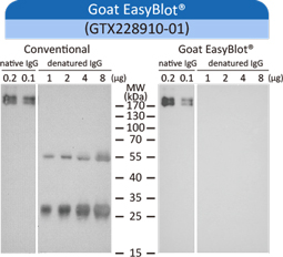
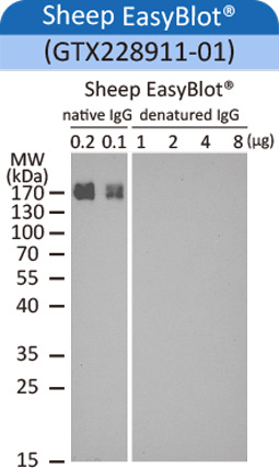
Immunoprecipitation (IP) and co-immunoprecipitation (co-IP) assays often necessitate the use of the same antibody, or antibodies from the same host, during both the IP and western blot (WB) procedures. Denatured immunoglobulin (Ig) in the IP sample can be bound by the secondary antibody used for the WB. Thus, depending on the molecular weight of the target protein, the desired band may be partially or completely obscured on the WB image by signal from denatured heavy (-50 kDa) and/or light (-25 kDa) Ig chains.
GeneTex’s EasyBlot® secondary antibodies are specifically optimized to eliminate this problem. They bind only the native (non-denatured and non-reduced) primary antibody used for WB analysis and not the denatured IP antibody chains. In addition, the specially formulated EasyBlocker reagent will greatly reduce background signal caused by free Protein A or Protein G.
EasyBlot®secondary antibodies are indispensable for achieving the cleanest results for your IP-WB experiments.
EasyBlot®: The easiest way to get the best result!
Clear
Efficiently eliminates denatured IgG masking of your target protein’s signal
Compatible
Suitable for WB following either IP or co-IP analysis using Protein A-, Protein G-, or Agarose-conjugated antibodies
Comprehensive
Broad range of EasyBlot® reagents for detecting various hosts: rabbit, mouse, rat, goat or sheep
Convenient: Simple and user-friendly!
Easy to switch from regular secondary antibodies to EasyBlot® secondary antibodies
Combine with EasyBlocker to further optimize your specific signal
Significantly reduce background generated by free Protein A/G with the EasyBlocker reagent.
Significantly reduce background generated by free Protein A/G with the EasyBlocker reagent.
Significantly reduce background generated by free Protein A/G with the EasyBlocker reagent.
View All Products Immunohistochemistry (IHC) is an indispensable application routinely employed in academic and clinical research as well as in Read More...
Recombinant Monoclonal Antibodies for GPCR Targets
View All Products GeneTex Addresses GPCR Targets with its Recombinant Monoclonal Antibody Production Platform GPCR Flyer GeneTex, a multinational antibody Read More...
Home Liver-specific Loss of OPA1 Results in Mitohormesis and Prevents Paracetamol-induced Hepatotoxicity
Contact UsView All Products Mitochondrial dysfunction is a frequent cause of liver disease, with fission and fusion being key processes Read More...
GeneTex’s New S100 beta Recombinant Monoclonal Antibody
Contact Us View All Products S100 beta (S100B) is a small, acidic homodimeric calcium-binding protein in the S100 protein family, Read More...
Contact UsView All Products Inhibition of the PD-L1/PD-1 interaction has become a major clinical strategy against a number of solid Read More...
Targeting the MMP-2/TYRO3/BRD3 Axis Ameliorates Colorectal Cancer Malignancy
Contact UsView All Products Receptor tyrosine kinases (RTKs) are well known to drive cancer progression and metastasis when mutated or Read More...
HCK Inhibition: Boosting Immunotherapy’s Efficacy and Utility?
Contact Us View All Products An immunologically “cold” tumor microenvironment (TME), even one associated with an otherwise immunogenic tumor, can Read More...
Development
Developmental biology is the study of how organisms grow and develop. It is a very broad field that incorporates a Read More...
GeneTex FAQs
If your questions are not listed below, please contact technical support: technical@stratech.co.uk Technical FAQImmunocytochemistry/ Immunofluorescence (ICC/IF) Troubleshooting TipsWestern Blot (WB) Read More...
Influenza A and B Recombinant Monoclonal Antibodies
Influenza viruses cause disease that can range in severity from relatively mild to lethal. The influenza A and B viruses Read More...
Organelle Markers
The subcellular location of a protein may suggest potential roles for that factor in one or more cellular processes. Organelle Read More...
The ALS-Reproducible Antibody Platform (ALS-RAP) Webinar
When: Tues, June 8th, 2021 (4pm UK Time) 11:00AM - 12:30PM EDT Convert this time to your time zone Highlighted Read More...
Exosome Research
What are exosomes? Extracellular vesicles (EVs) 30 – 200 nm in size were once thought to be natural cellular debris; Read More...
Webinar PD-L1
Thank you for your interest in the seminar “Enhancing anti-cancer immune response by targeting glycosylated PD-L1". Objectives: 1. Immunity and Read More...
Epitope Tags & Reporters
Epitope tags are peptide or protein sequences covalently conjugated, usually to the C- or N-terminus, of a recombinant protein. The Read More...
Loading Controls
Loading controls are used to ensure that equal amounts of each protein sample are loaded into the wells of an Read More...
VetSignal
GeneTex is proud to launch its new VetSignal™ line of products to advance veterinary science research. These antibodies and other Read More...
Zebrafish
As the leading manufacturer of zebrafish antibodies, GeneTex offers an extensive portfolio of reagents validated for use in this model Read More...
Infectious Diseases
The 2020 Nobel Prize in Physiology or Medicine has been awarded to “Harvey J Alter, Michael Houhgton and Charles M Read More...
Signaling Pathways
Cell signaling refers to the various sequences of molecular events that mark a cell’s response to its environment, whether that Read More...


](https://www.genetex.com/upload/media/product/subcategory/Recombinant-Antibodies/img01.jpg "Iba1 antibody [HL22](GTX635363)")
](https://www.genetex.com/upload/media/product/subcategory/Recombinant-Antibodies/img02.jpg "RAS (G12D Mutant) antibody [HL10](GTX635362)")

")